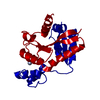
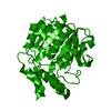
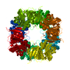
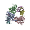
Journal: J Am Chem Soc / Year: 2021 Title: Copper Centers in the Cryo-EM Structure of Particulate Methane Monooxygenase Reveal the Catalytic Machinery of Methane Oxidation. Authors: W-H Chang / H-H Lin / I-K Tsai / S-H Huang / S-C Chung / I-P Tu / S S-F Yu / S I Chan / Abstract: The particulate methane monooxygenase (pMMO) is the first enzyme in the C1 metabolic pathway in methanotrophic bacteria. As this enzyme converts methane into methanol efficiently near room ...The particulate methane monooxygenase (pMMO) is the first enzyme in the C1 metabolic pathway in methanotrophic bacteria. As this enzyme converts methane into methanol efficiently near room temperature, it has become the paradigm for developing an understanding of this difficult C1 chemistry. pMMO is a membrane-bound protein with three subunits (PmoB, PmoA, and PmoC) and 12-14 coppers distributed among different sites. X-ray crystal structures that have revealed only three mononuclear coppers at three sites have neither disclosed the location of the active site nor the catalytic mechanism of the enzyme. Here we report a cyro-EM structure of -pMMO from (Bath) at 2.5 Å, and develop quantitative electrostatic-potential profiling to scrutinize the nonprotein densities for signatures of the copper cofactors. Our results confirm a mononuclear Cu at the site, resolve two Cus at the site, and uncover additional Cu clusters at the PmoA/PmoC interface within the membrane ( site) and in the water-exposed -terminal subdomain of the PmoB ( clusters). These findings complete the minimal set of copper factors required for catalytic turnover of pMMO, offering a glimpse of the catalytic machinery for methane oxidation according to the chemical principles underlying the mechanism proposed earlier.
History
Deposition
May 20, 2021
-
Header (metadata) release
Aug 4, 2021
-
Map release
Aug 4, 2021
-
Update
Aug 4, 2021
-
Current status
Aug 4, 2021
Processing site: PDBj / Status: Released
-
Structure visualization
Movie
Surface view with section colored by density value
Film or detector model: GATAN K2 SUMMIT (4k x 4k) / Detector mode: COUNTING / Digitization - Frames/image: 1-60 / Number real images: 23484 / Average electron dose: 80.0 e/Å2
Electron beam
Acceleration voltage: 300 kV / Electron source: FIELD EMISSION GUN
Type of model: OTHER / Details: cryoSPARC ab-initio Reconstruction
Final reconstruction
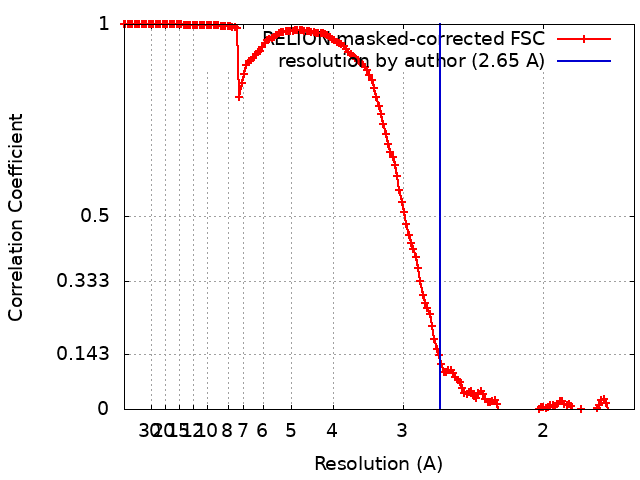
Resolution.type: BY AUTHOR / Resolution: 2.65 Å / Resolution method: FSC 0.143 CUT-OFF / Software - Name: cryoSPARC (ver. 2.15) / Details: Reconstructed with the one of subset from 3DVA / Number images used: 243147
Initial angle assignment
Type: RANDOM ASSIGNMENT / Software - Name: cryoSPARC (ver. 2.15)
In the structure databanks used in Yorodumi, some data are registered as the other names, "COVID-19 virus" and "2019-nCoV". Here are the details of the virus and the list of structure data.
Jan 31, 2019. EMDB accession codes are about to change! (news from PDBe EMDB page)
EMDB accession codes are about to change! (news from PDBe EMDB page)
The allocation of 4 digits for EMDB accession codes will soon come to an end. Whilst these codes will remain in use, new EMDB accession codes will include an additional digit and will expand incrementally as the available range of codes is exhausted. The current 4-digit format prefixed with “EMD-” (i.e. EMD-XXXX) will advance to a 5-digit format (i.e. EMD-XXXXX), and so on. It is currently estimated that the 4-digit codes will be depleted around Spring 2019, at which point the 5-digit format will come into force.
The EM Navigator/Yorodumi systems omit the EMD- prefix.
Related info.:Q: What is EMD? / ID/Accession-code notation in Yorodumi/EM Navigator
Yorodumi is a browser for structure data from EMDB, PDB, SASBDB, etc.
This page is also the successor to EM Navigator detail page, and also detail information page/front-end page for Omokage search.
The word "yorodu" (or yorozu) is an old Japanese word meaning "ten thousand". "mi" (miru) is to see.
Related info.:EMDB / PDB / SASBDB / Comparison of 3 databanks / Yorodumi Search / Aug 31, 2016. New EM Navigator & Yorodumi / Yorodumi Papers / Jmol/JSmol / Function and homology information / Changes in new EM Navigator and Yorodumi
 Movie
Movie Controller

 Yorodumi
 Open data
 Basic information
 Map data
 Sample
 Function and homology information
 Methylococcus capsulatus str. Bath (bacteria)
 Authors
 Citation
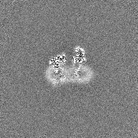
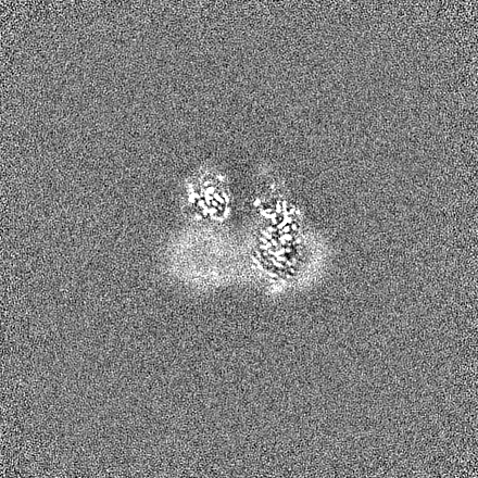
 Structure visualization
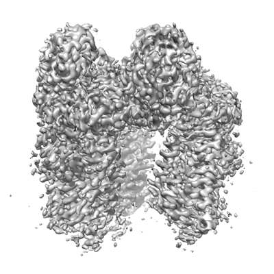
 Downloads & links
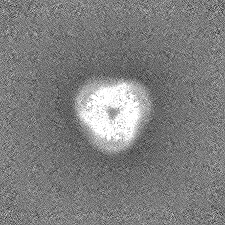
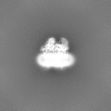
 emd_31327.png
 http://ftp.pdbj.org/pub/emdb/structures/EMD-31327

 Z (Sec.)
 Y (Row.)
 X (Col.)


 Sample components
 Processing
 Electron microscopy
 FIELD EMISSION GUN