 Movie
Movie Controller

[English] 日本語
 Yorodumi
- PDB-7ev9: cryoEM structure of particulate methane monooxygenase associated ... -
+ Open data

- Basic information
| Entry | Database: PDB / ID: 7ev9 | ||||||
|---|---|---|---|---|---|---|---|
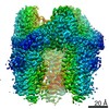
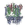
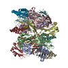
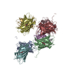
| Title | cryoEM structure of particulate methane monooxygenase associated with Cu(I) | ||||||
 Components Components |
| ||||||
 Keywords Keywords | OXIDOREDUCTASE / particulate methane monooxygenase / copper contained | ||||||
| Function / homology |  Function and homology information Function and homology informationmethane monooxygenase (particulate) / methane monooxygenase (soluble) / methane monooxygenase [NAD(P)H] activity / monooxygenase activity / membrane / metal ion binding Similarity search - Function | ||||||
| Biological species |  Methylococcus capsulatus (bacteria) Methylococcus capsulatus (bacteria) | ||||||
| Method | ELECTRON MICROSCOPY / single particle reconstruction / cryo EM / Resolution: 2.6 Å | ||||||
 Authors Authors | Chang, W.H. / Lin, H.H. / Tsai, I.K. / Huang, S.H. / Chung, S.C. / Tu, I.P. / Yu, S.F. / Chan, S.I. | ||||||
 Citation Citation | Journal: J Am Chem Soc / Year: 2021 Title: Copper Centers in the Cryo-EM Structure of Particulate Methane Monooxygenase Reveal the Catalytic Machinery of Methane Oxidation. Authors: W-H Chang / H-H Lin / I-K Tsai / S-H Huang / S-C Chung / I-P Tu / S S-F Yu / S I Chan /  Abstract: The particulate methane monooxygenase (pMMO) is the first enzyme in the C1 metabolic pathway in methanotrophic bacteria. As this enzyme converts methane into methanol efficiently near room ...The particulate methane monooxygenase (pMMO) is the first enzyme in the C1 metabolic pathway in methanotrophic bacteria. As this enzyme converts methane into methanol efficiently near room temperature, it has become the paradigm for developing an understanding of this difficult C1 chemistry. pMMO is a membrane-bound protein with three subunits (PmoB, PmoA, and PmoC) and 12-14 coppers distributed among different sites. X-ray crystal structures that have revealed only three mononuclear coppers at three sites have neither disclosed the location of the active site nor the catalytic mechanism of the enzyme. Here we report a cyro-EM structure of -pMMO from (Bath) at 2.5 Å, and develop quantitative electrostatic-potential profiling to scrutinize the nonprotein densities for signatures of the copper cofactors. Our results confirm a mononuclear Cu at the site, resolve two Cus at the site, and uncover additional Cu clusters at the PmoA/PmoC interface within the membrane ( site) and in the water-exposed -terminal subdomain of the PmoB ( clusters). These findings complete the minimal set of copper factors required for catalytic turnover of pMMO, offering a glimpse of the catalytic machinery for methane oxidation according to the chemical principles underlying the mechanism proposed earlier. | ||||||
| History |
|
- Structure visualization
| Movie |
Movie viewer |
|---|---|
| Structure viewer | Molecule: MolmilJmol/JSmol |
- Downloads & links
-Download
| PDBx/mmCIF format | 7ev9.cif.gz | 528.1 KB | Display | PDBx/mmCIF format |
|---|---|---|---|---|
| PDB format | pdb7ev9.ent.gz | 400.6 KB | Display | PDB format |
| PDBx/mmJSON format | 7ev9.json.gz | Tree view | PDBx/mmJSON format | |
| Others |  Other downloads Other downloads |
-Validation report
| Arichive directory | https://data.pdbj.org/pub/pdb/validation_reports/ev/7ev9ftp://data.pdbj.org/pub/pdb/validation_reports/ev/7ev9 | HTTPS FTP |
|---|
-Related structure data
| Related structure data | 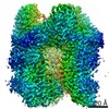 31325MC M: map data used to model this data C: citing same article ( |
|---|---|
| Similar structure data |


-Links
 PDBj
PDBj- Assembly
| Deposited unit | 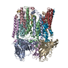
|
|---|---|
| 1 |
|
-Components
| #1: Protein | Mass: 46129.746 Da / Num. of mol.: 3 / Source method: isolated from a natural source Source: (natural) Methylococcus capsulatus (strain ATCC 33009 / NCIMB 11132 / Bath) (bacteria)Strain: ATCC 33009 / NCIMB 11132 / Bath References: UniProt: G1UBD1, methane monooxygenase (particulate) #2: Protein | Mass: 28445.098 Da / Num. of mol.: 3 / Source method: isolated from a natural source Source: (natural) Methylococcus capsulatus (strain ATCC 33009 / NCIMB 11132 / Bath) (bacteria)Strain: ATCC 33009 / NCIMB 11132 / Bath References: UniProt: Q607G3, methane monooxygenase (particulate) #3: Protein | Mass: 29839.309 Da / Num. of mol.: 3 / Source method: isolated from a natural source Source: (natural) Methylococcus capsulatus (strain ATCC 33009 / NCIMB 11132 / Bath) (bacteria)Strain: ATCC 33009 / NCIMB 11132 / Bath References: UniProt: Q603F1, methane monooxygenase (soluble) #4: Chemical | ChemComp-CU1 /   Mass: 63.546 Da / Num. of mol.: 24 / Source method: obtained synthetically / Formula: Cu / Feature type: SUBJECT OF INVESTIGATION Mass: 63.546 Da / Num. of mol.: 24 / Source method: obtained synthetically / Formula: Cu / Feature type: SUBJECT OF INVESTIGATIONHas ligand of interest | Y | |
|---|
-Experimental details
-Experiment
| Experiment | Method: ELECTRON MICROSCOPY |
|---|---|
| EM experiment | Aggregation state: PARTICLE / 3D reconstruction method: single particle reconstruction |
- Sample preparation
| Component | Name: particulate methane monooxygenase (pMMO) / Type: COMPLEX / Entity ID: #1-#3 / Source: NATURAL |
|---|---|
| Molecular weight | Value: 0.312 MDa / Experimental value: NO |
| Source (natural) | Organism: Methylococcus capsulatus str. Bath (bacteria) / Cellular location: membrane |
| Buffer solution | pH: 7.2 |
| Specimen | Embedding applied: YES / Shadowing applied: NO / Staining applied: NO / Vitrification applied: YES |
| EM embedding | Material: vitrified ice |
| Vitrification | Cryogen name: ETHANE / Humidity: 98 % / Chamber temperature: 278 K |
- Electron microscopy imaging
| Experimental equipment |  Model: Titan Krios / Image courtesy: FEI Company |
|---|---|
| Microscopy | Model: FEI TITAN KRIOS |
| Electron gun | Electron source:  FIELD EMISSION GUN / Accelerating voltage: 300 kV / Illumination mode: FLOOD BEAM FIELD EMISSION GUN / Accelerating voltage: 300 kV / Illumination mode: FLOOD BEAM |
| Electron lens | Mode: BRIGHT FIELD / Nominal magnification: 165000 X / Nominal defocus max: 2200 nm / Nominal defocus min: 1000 nm / Cs: 2.7 mm |
| Specimen holder | Cryogen: NITROGEN |
| Image recording | Electron dose: 80 e/Å2 / Detector mode: COUNTING / Film or detector model: GATAN K2 SUMMIT (4k x 4k) / Num. of real images: 23484 |
| Image scans | Movie frames/image: 80 / Used frames/image: 1-60 |
- Processing
| EM software |
| ||||||||||||||||||||||||
|---|---|---|---|---|---|---|---|---|---|---|---|---|---|---|---|---|---|---|---|---|---|---|---|---|---|
| CTF correction | Type: PHASE FLIPPING AND AMPLITUDE CORRECTION | ||||||||||||||||||||||||
| Symmetry | Point symmetry: C3 (3 fold cyclic) | ||||||||||||||||||||||||
| 3D reconstruction | Resolution: 2.6 Å / Resolution method: FSC 0.143 CUT-OFF / Num. of particles: 128116 / Symmetry type: POINT |
