Movie
Movie Controller

[English] 日本語
 Yorodumi
- EMDB-31184: In situ structure of capping enzyme lambda2, penetration protein ... -
+ Open data

- Basic information
| Entry | Database: EMDB / ID: EMD-31184 | |||||||||||||||||||||||||||
|---|---|---|---|---|---|---|---|---|---|---|---|---|---|---|---|---|---|---|---|---|---|---|---|---|---|---|---|---|
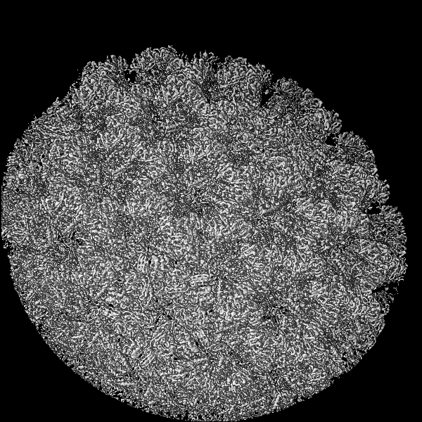
| Title | In situ structure of capping enzyme lambda2, penetration protein mu1 of mammalian reovirus capsid asymmetric unit. | |||||||||||||||||||||||||||
 Map data Map data | ||||||||||||||||||||||||||||
 Sample Sample |
| |||||||||||||||||||||||||||
 Keywords Keywords | mammalian reovirus 3 / capping enzyme lambda2 / penetration protein mu1 / VIRUS / VIRAL PROTEIN-TRANSFERASE complex | |||||||||||||||||||||||||||
| Function / homology |  Function and homology information Function and homology informationhost cell surface binding / viral outer capsid / symbiont entry into host cell via permeabilization of host membrane / mRNA guanylyltransferase / mRNA guanylyltransferase activity / mRNA (guanine-N7)-methyltransferase / mRNA 5'-cap (guanine-N7-)-methyltransferase activity / GTP binding / ATP binding Similarity search - Function | |||||||||||||||||||||||||||
| Biological species |  Mammalian orthoreovirus 3 / Mammalian orthoreovirus 3 Dearing Mammalian orthoreovirus 3 / Mammalian orthoreovirus 3 Dearing | |||||||||||||||||||||||||||
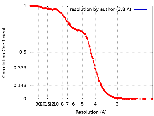
| Method | single particle reconstruction / cryo EM / Resolution: 3.8 Å | |||||||||||||||||||||||||||
 Authors Authors | Zhou ZH / Pan M | |||||||||||||||||||||||||||
| Funding support |  United States, 8 items United States, 8 items
| |||||||||||||||||||||||||||
 Citation Citation | Journal: Nat Commun / Year: 2021 Title: Asymmetric reconstruction of mammalian reovirus reveals interactions among RNA, transcriptional factor µ2 and capsid proteins. Authors: Muchen Pan / Ana L Alvarez-Cabrera / Joon S Kang / Lihua Wang / Chunhai Fan / Z Hong Zhou /  Abstract: Mammalian reovirus (MRV) is the prototypical member of genus Orthoreovirus of family Reoviridae. However, lacking high-resolution structures of its RNA polymerase cofactor μ2 and infectious ...Mammalian reovirus (MRV) is the prototypical member of genus Orthoreovirus of family Reoviridae. However, lacking high-resolution structures of its RNA polymerase cofactor μ2 and infectious particle, limits understanding of molecular interactions among proteins and RNA, and their contributions to virion assembly and RNA transcription. Here, we report the 3.3 Å-resolution asymmetric reconstruction of transcribing MRV and in situ atomic models of its capsid proteins, the asymmetrically attached RNA-dependent RNA polymerase (RdRp) λ3, and RdRp-bound nucleoside triphosphatase μ2 with a unique RNA-binding domain. We reveal molecular interactions among virion proteins and genomic and messenger RNA. Polymerase complexes in three Spinoreovirinae subfamily members are organized with different pseudo-D symmetries to engage their highly diversified genomes. The above interactions and those between symmetry-mismatched receptor-binding σ1 trimers and RNA-capping λ2 pentamers balance competing needs of capsid assembly, external protein removal, and allosteric triggering of endogenous RNA transcription, before, during and after infection, respectively. | |||||||||||||||||||||||||||
| History |
|
- Structure visualization
| Movie |
Movie viewer |
|---|---|
| Structure viewer | EM map: SurfViewMolmilJmol/JSmol |
| Supplemental images |

- Downloads & links
-EMDB archive
| Map data | emd_31184.map.gz | 663 MB | EMDB map data format | |
|---|---|---|---|---|
| Header (meta data) | emd-31184-v30.xmlemd-31184.xml | 14.5 KB 14.5 KB | Display Display | EMDB header |
| FSC (resolution estimation) | emd_31184_fsc.xml | 27.3 KB | Display | FSC data file |
| Images | 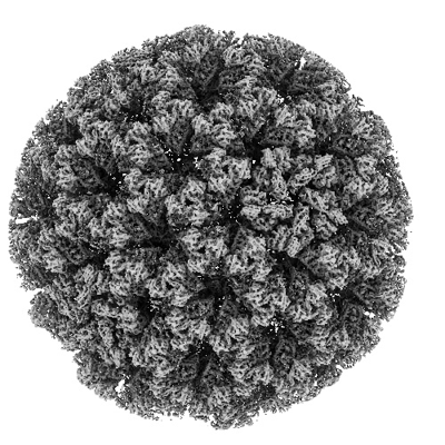 emd_31184.png emd_31184.png | 205.3 KB | ||
| Filedesc metadata | emd-31184.cif.gz | 6.4 KB | ||
| Archive directory |  http://ftp.pdbj.org/pub/emdb/structures/EMD-31184ftp://ftp.pdbj.org/pub/emdb/structures/EMD-31184 http://ftp.pdbj.org/pub/emdb/structures/EMD-31184ftp://ftp.pdbj.org/pub/emdb/structures/EMD-31184 | HTTPS FTP |
-Related structure data
| Related structure data |  7ellMC  7elhC M: atomic model generated by this map C: citing same article ( |
|---|---|
| Similar structure data |


-Links
| EMDB pages | EMDB (EBI/PDBe) / EMDataResource |
|---|---|
| Related items in Molecule of the Month |


-Map
| File | Download / File: emd_31184.map.gz / Format: CCP4 / Size: 824 MB / Type: IMAGE STORED AS FLOATING POINT NUMBER (4 BYTES) | ||||||||||||||||||||||||||||||||||||||||||||||||||||||||||||||||||||
|---|---|---|---|---|---|---|---|---|---|---|---|---|---|---|---|---|---|---|---|---|---|---|---|---|---|---|---|---|---|---|---|---|---|---|---|---|---|---|---|---|---|---|---|---|---|---|---|---|---|---|---|---|---|---|---|---|---|---|---|---|---|---|---|---|---|---|---|---|---|
| Projections & slices | Image control
Images are generated by Spider. | ||||||||||||||||||||||||||||||||||||||||||||||||||||||||||||||||||||
| Voxel size | X=Y=Z: 1.07 Å | ||||||||||||||||||||||||||||||||||||||||||||||||||||||||||||||||||||
| Density |
| ||||||||||||||||||||||||||||||||||||||||||||||||||||||||||||||||||||
| Symmetry | Space group: 1 | ||||||||||||||||||||||||||||||||||||||||||||||||||||||||||||||||||||
| Details | EMDB XML:
CCP4 map header:
| ||||||||||||||||||||||||||||||||||||||||||||||||||||||||||||||||||||
 Z (Sec.)
 Y (Row.)
 X (Col.)


-Supplemental data
- Sample components
-Entire : Mammalian orthoreovirus 3 Dearing
| Entire | Name: Mammalian orthoreovirus 3 Dearing |
|---|---|
| Components |
|
-Supramolecule #1: Mammalian orthoreovirus 3 Dearing
| Supramolecule | Name: Mammalian orthoreovirus 3 Dearing / type: virus / ID: 1 / Parent: 0 / Macromolecule list: #3 / NCBI-ID: 10886 / Sci species name: Mammalian orthoreovirus 3 Dearing / Virus type: VIRION / Virus isolate: SEROTYPE / Virus enveloped: No / Virus empty: No |
|---|
-Macromolecule #1: Mu1
| Macromolecule | Name: Mu1 / type: protein_or_peptide / ID: 1 / Number of copies: 10 / Enantiomer: LEVO |
|---|---|
| Source (natural) | Organism: Mammalian orthoreovirus 3 |
| Molecular weight | Theoretical: 4.050441 KDa |
| Sequence | String: GNASSIVQTI NVTGDGNVFK PSAETSSTAV PSLSLSPGML N UniProtKB: Mu1 |
-Macromolecule #2: Mu1
| Macromolecule | Name: Mu1 / type: protein_or_peptide / ID: 2 / Number of copies: 10 / Enantiomer: LEVO |
|---|---|
| Source (natural) | Organism: Mammalian orthoreovirus 3 |
| Molecular weight | Theoretical: 72.228719 KDa |
| Sequence | String: PGGVPWIAVG DETSVTSPGA LRRMTSKDIP ETAIINTDNS SGAVPSESAL VPYIDEPLVV VTEHAITNFT KAEMALEFNR EFLDKMRVL SVSPKYSDLL TYVDCYVGVS ARQALNNFQK QVPVITPTRQ TMYVDSIQAA LKALEKWEID LRVAQTLLPT N VPIGEVSC ...String: PGGVPWIAVG DETSVTSPGA LRRMTSKDIP ETAIINTDNS SGAVPSESAL VPYIDEPLVV VTEHAITNFT KAEMALEFNR EFLDKMRVL SVSPKYSDLL TYVDCYVGVS ARQALNNFQK QVPVITPTRQ TMYVDSIQAA LKALEKWEID LRVAQTLLPT N VPIGEVSC PMQSVVKLLD DQLPDDSLIR RYPKEAAVAL AKRNGGIQWM DVSEGTVMNE AVNAVAASAL APSASAPPLE EK SKLTEQA MDLVTAAEPE IIASLVPVPA PVFAIPPKPA DYNVRTLRID EATWLRMIPK SMNTPFQIQV TDNTGTNWHL NLR GGTRVV NLDQIAPMRF VLDLGGKSYK ETSWDPNGKK VGFIVFQSKI PFELWTAASQ IGQATVVNYV QLYAEDSSFT AQSI IATTS LAYNYEPEQL NKTDPEMNYY LLATFIDSAA ITPTNMTQPD VWDALLTMSP LSAGEVTVKG AVVSEVVPAD LIGSY TPES LNTSLPNDAA RCMIDRASKI AEAIKIDDDA GPDEYSPNSV PIQGQLAISQ LETGYGVRIF NPKGILSKIA SRAMQA FIG DPSTIITQAA PVLSDKNNWI ALAQGVKTSL RTKSLSAGVK TAVSKLSSSE SIQNWTQGFL DKVSAHFPAP KPDCPTS GD SGESSNRRVK RDSYAGVVKR GYTR UniProtKB: Mu1 |
-Macromolecule #3: mRNA (guanine-N(7)-)-methyltransferase
| Macromolecule | Name: mRNA (guanine-N(7)-)-methyltransferase / type: protein_or_peptide / ID: 3 / Number of copies: 1 / Enantiomer: LEVO / EC number: mRNA (guanine-N7)-methyltransferase |
|---|---|
| Source (natural) | Organism: Mammalian orthoreovirus 3 |
| Molecular weight | Theoretical: 143.998625 KDa |
| Sequence | String: MANVWGVRLA DSLSSPTIET RTRQYTLHDL CSDLDANPGR EPWKPLRNQR TNNIVAVQLF RPLQGLVLDT QLYGFPGAFD DWERFMREK LRVLKYEVLR IYPISNYSNE HVNVFVANAL VGAFLSNQAF YDLLPLLIIN DTMIGDLLGT GASLSQFFQS H GDVLEVAA ...String: MANVWGVRLA DSLSSPTIET RTRQYTLHDL CSDLDANPGR EPWKPLRNQR TNNIVAVQLF RPLQGLVLDT QLYGFPGAFD DWERFMREK LRVLKYEVLR IYPISNYSNE HVNVFVANAL VGAFLSNQAF YDLLPLLIIN DTMIGDLLGT GASLSQFFQS H GDVLEVAA GRKYLQMENY SNDDDDPPLF AKDLSDYAKA FYSDTYEVLD RFFWTHDSSA GVLVHYDKPT NGHHYLLGTL TQ MVSAPPY IINATDAMLL ESCLEQFSAN VRARPAQPVT RLDQCYHLRW GAQYVGEDSL TYRLGVLSLL ATNGYQLARP IPR QLTNRW LSSFVSQIMS DGVNETPLWP QERYVQIAYD SPSVVDGATQ YGYVRKNQLR LGMRISALQS LSDTPSPVQW LPQY TIDQA AMDEGDLMVS RLTQLPLRPD YGNIWVGDAL SYYVDYNRSH RVVLSSELPQ LPDTYFDGDE QYGRSLFSLA RKIGD RSLV KDTAVLKHAY QAIDPNTGKE YLRSGQSVAY FGASAGHSGA DQPLVIEPWI QGKISGVPPP SSVRQFGYDV ARGAIV DLA RPFPSGDYQF VYSDVDQVVD GHDDLSISSG LVESLLSSCM HATAPGGSFV VKINFPTRPV WHYIEQKILP NITSYML IK PFVTNNVELF FVAFGVHQHS SLTWTSGVYF FLVDHFYRYE TLSTISRQLP SFGYVDDGSS VTGIETISIE NPGFSNMT Q AARIGISGLC ANVGNARKSI AIYESHGARV LTITSRRSPA SARRKSRLRY LPLIDPRSLE VQARTILPAD PVLFENVSG ASPHVCLTMM YNFEVSSAVY DGDVVLDLGT GPEAKILELI PATSPVTCVD IRPTAQPSGC WNVRTTFLEL DYLSDGWITG VRGDIVTCM LSLGAAAAGK SMTFDAAFQQ LIKVLSKSTA NVVLVQVNCP TDVVRSIKGY LEIDSTNKRY RFPKFGRDEP Y SDMDALEK ICRTAWPNCS ITWVPLSYDL RWTRLALLES TTLSSASIRI AELMYKYMPI MRIDIHGLPM EKRGNFIVGQ NC SLVIPGF NAQDVFNCYF NSALAFSTED VNAAMIPQVS AQFDATKGEW TLDMVFSDAG IYTMQALVGS NANPVSLGSF VVD SPDVDI TDAWPAQLDF TIAGTDVDIT VNPYYRLMTF VRIDGQWQIA NPDKFQFFSS ASGTLVMNVK LDIADKYLLY YIRD VQSRD VGFYIQHPLQ LLNTITLPTN EDLFLSAPDM REWAVKESGN TICILNSQGF VLPQDWDVLT DTISWSPSIP TYIVP PGDY TLTPL UniProtKB: Outer capsid protein lambda-2 |
-Macromolecule #4: MYRISTIC ACID
| Macromolecule | Name: MYRISTIC ACID / type: ligand / ID: 4 / Number of copies: 10 / Formula: MYR |
|---|---|
| Molecular weight | Theoretical: 228.371 Da |
| Chemical component information |  ChemComp-MYR: |
-Experimental details
-Structure determination
| Method | cryo EM |
|---|---|
 Processing Processing | single particle reconstruction |
| Aggregation state | particle |
-Sample preparation
| Buffer | pH: 7.4 |
|---|---|
| Vitrification | Cryogen name: ETHANE |
- Electron microscopy
| Microscope | FEI TITAN KRIOS |
|---|---|
| Image recording | Film or detector model: GATAN K2 SUMMIT (4k x 4k) / Average electron dose: 56.0 e/Å2 |
| Electron beam | Acceleration voltage: 300 kV / Electron source:  FIELD EMISSION GUN FIELD EMISSION GUN |
| Electron optics | Illumination mode: FLOOD BEAM / Imaging mode: BRIGHT FIELD |
| Experimental equipment |  Model: Titan Krios / Image courtesy: FEI Company |