 Movie
Movie Controller

[English] 日本語
 Yorodumi
- PDB-7elh: In situ structure of transcriptional enzyme complex and capsid sh... -
+ Open data

- Basic information
| Entry | Database: PDB / ID: 7elh | ||||||||||||||||||
|---|---|---|---|---|---|---|---|---|---|---|---|---|---|---|---|---|---|---|---|
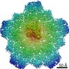
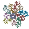
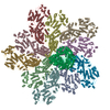
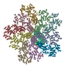
| Title | In situ structure of transcriptional enzyme complex and capsid shell protein of mammalian reovirus at initiation state | ||||||||||||||||||
 Components Components |
| ||||||||||||||||||
 Keywords Keywords | VIRAL PROTEIN/TRANSFERASE/RNA / asymmetric / mu2 / lambda3 / lambda1 / VIRUS / VIRAL PROTEIN-TRANSFERASE-RNA complex | ||||||||||||||||||
| Function / homology |  Function and homology information Function and homology informationT=2 icosahedral viral capsid / host cytoskeleton / viral inner capsid / 7-methylguanosine mRNA capping / viral genome replication / viral capsid / viral nucleocapsid / RNA helicase activity / RNA helicase / RNA-directed RNA polymerase ...T=2 icosahedral viral capsid / host cytoskeleton / viral inner capsid / 7-methylguanosine mRNA capping / viral genome replication / viral capsid / viral nucleocapsid / RNA helicase activity / RNA helicase / RNA-directed RNA polymerase / nucleotide binding / RNA-directed RNA polymerase activity / structural molecule activity / ATP hydrolysis activity / RNA binding / zinc ion binding / ATP binding Similarity search - Function | ||||||||||||||||||
| Biological species |  Mammalian orthoreovirus 3Mammalian orthoreovirus 3 Dearing Mammalian orthoreovirus 3Mammalian orthoreovirus 3 Dearing | ||||||||||||||||||
| Method | ELECTRON MICROSCOPY / single particle reconstruction / cryo EM / Resolution: 3.3 Å | ||||||||||||||||||
 Authors Authors | Zhou, Z.H. / Pan, M. | ||||||||||||||||||
| Funding support |  United States, 5items United States, 5items
| ||||||||||||||||||
 Citation Citation | Journal: Nat Commun / Year: 2021 Title: Asymmetric reconstruction of mammalian reovirus reveals interactions among RNA, transcriptional factor µ2 and capsid proteins. Authors: Muchen Pan / Ana L Alvarez-Cabrera / Joon S Kang / Lihua Wang / Chunhai Fan / Z Hong Zhou /  Abstract: Mammalian reovirus (MRV) is the prototypical member of genus Orthoreovirus of family Reoviridae. However, lacking high-resolution structures of its RNA polymerase cofactor μ2 and infectious ...Mammalian reovirus (MRV) is the prototypical member of genus Orthoreovirus of family Reoviridae. However, lacking high-resolution structures of its RNA polymerase cofactor μ2 and infectious particle, limits understanding of molecular interactions among proteins and RNA, and their contributions to virion assembly and RNA transcription. Here, we report the 3.3 Å-resolution asymmetric reconstruction of transcribing MRV and in situ atomic models of its capsid proteins, the asymmetrically attached RNA-dependent RNA polymerase (RdRp) λ3, and RdRp-bound nucleoside triphosphatase μ2 with a unique RNA-binding domain. We reveal molecular interactions among virion proteins and genomic and messenger RNA. Polymerase complexes in three Spinoreovirinae subfamily members are organized with different pseudo-D symmetries to engage their highly diversified genomes. The above interactions and those between symmetry-mismatched receptor-binding σ1 trimers and RNA-capping λ2 pentamers balance competing needs of capsid assembly, external protein removal, and allosteric triggering of endogenous RNA transcription, before, during and after infection, respectively. | ||||||||||||||||||
| History |
|
- Structure visualization
| Movie |
Movie viewer |
|---|---|
| Structure viewer | Molecule: MolmilJmol/JSmol |
- Downloads & links
-Download
| PDBx/mmCIF format | 7elh.cif.gz | 2.4 MB | Display | PDBx/mmCIF format |
|---|---|---|---|---|
| PDB format | pdb7elh.ent.gz | Display | PDB format | |
| PDBx/mmJSON format | 7elh.json.gz | Tree view | PDBx/mmJSON format | |
| Others |  Other downloads Other downloads |
-Validation report
| Arichive directory | https://data.pdbj.org/pub/pdb/validation_reports/el/7elhftp://data.pdbj.org/pub/pdb/validation_reports/el/7elh | HTTPS FTP |
|---|
-Related structure data
| Related structure data |  31183MC  7ellC M: map data used to model this data C: citing same article ( |
|---|---|
| Similar structure data |


-Links
 PDBj
PDBj


- Assembly
| Deposited unit | 
|
|---|---|
| 1 |
|
-Components
-Protein , 4 types, 17 molecules ABDEFGHIJKLMegikm
| #1: Protein | Mass: 83331.289 Da / Num. of mol.: 1 / Source method: isolated from a natural source / Source: (natural) Mammalian orthoreovirus 3 / References: UniProt: Q6EDZ8 | ||
|---|---|---|---|
| #2: Protein | Mass: 142449.016 Da / Num. of mol.: 1 / Source method: isolated from a natural source / Source: (natural) Mammalian orthoreovirus 3References: UniProt: A0A0B5CSU4, RNA-directed RNA polymerase | ||
| #4: Protein | Mass: 122842.859 Da / Num. of mol.: 10 / Source method: isolated from a natural source / Source: (natural) Mammalian orthoreovirus 3 / References: UniProt: F1ARN3#5: Protein | Mass: 19112.762 Da / Num. of mol.: 5 / Source method: isolated from a natural source / Source: (natural) Mammalian orthoreovirus 3 / References: UniProt: F1ARN3 |
-RNA chain , 2 types, 9 molecules CNOPQRSTU
| #3: RNA chain | Mass: 1263.842 Da / Num. of mol.: 1 / Source method: obtained synthetically / Source: (synth.) Mammalian orthoreovirus 3 Dearing |
|---|---|
| #6: RNA chain | Mass: 19016.188 Da / Num. of mol.: 8 / Source method: isolated from a natural source / Source: (natural) Mammalian orthoreovirus 3 Dearing |
-Non-polymers , 1 types, 1 molecules 
| #7: Chemical | ChemComp-PO4 /  Mass: 94.971 Da / Num. of mol.: 1 / Source method: obtained synthetically / Formula: PO4 / Feature type: SUBJECT OF INVESTIGATION Mass: 94.971 Da / Num. of mol.: 1 / Source method: obtained synthetically / Formula: PO4 / Feature type: SUBJECT OF INVESTIGATION |
|---|
-Details
| Has ligand of interest | Y |
|---|
-Experimental details
-Experiment
| Experiment | Method: ELECTRON MICROSCOPY |
|---|---|
| EM experiment | Aggregation state: PARTICLE / 3D reconstruction method: single particle reconstruction |
- Sample preparation
| Component | Name: Mammalian orthoreovirus 3 Dearing / Type: VIRUS / Entity ID: #1-#3, #6 / Source: NATURAL |
|---|---|
| Source (natural) | Organism: Mammalian orthoreovirus 3 Dearing |
| Details of virus | Empty: NO / Enveloped: NO / Isolate: OTHER / Type: VIRION |
| Buffer solution | pH: 7.4 |
| Specimen | Embedding applied: NO / Shadowing applied: NO / Staining applied: NO / Vitrification applied: YES |
| Vitrification | Cryogen name: ETHANE |
- Electron microscopy imaging
| Experimental equipment |  Model: Titan Krios / Image courtesy: FEI Company |
|---|---|
| Microscopy | Model: FEI TITAN KRIOS |
| Electron gun | Electron source:  FIELD EMISSION GUN / Accelerating voltage: 300 kV / Illumination mode: FLOOD BEAM FIELD EMISSION GUN / Accelerating voltage: 300 kV / Illumination mode: FLOOD BEAM |
| Electron lens | Mode: BRIGHT FIELD |
| Image recording | Electron dose: 56 e/Å2 / Film or detector model: GATAN K2 SUMMIT (4k x 4k) |
- Processing
| CTF correction | Type: PHASE FLIPPING AND AMPLITUDE CORRECTION |
|---|---|
| 3D reconstruction | Resolution: 3.3 Å / Resolution method: FSC 0.143 CUT-OFF / Num. of particles: 102966 / Symmetry type: POINT |
| Refinement | Highest resolution: 3.3 Å Details: The distance between Residue H HIS 230 and Residue H ASN 241 is 48.65 Angstrom. Authors state that the map density of this large gaps is poor. For now the large gap could not be explained. ...Details: The distance between Residue H HIS 230 and Residue H ASN 241 is 48.65 Angstrom. Authors state that the map density of this large gaps is poor. For now the large gap could not be explained. Maybe there could have been some broken points in the large gap, but no evidence has been found. However, they could make sure that the solved coordinates are correct. The cryo-EM and the X-ray (PDB-1EJ6) results could support each other. |
