 ムービー
ムービー コントローラー


+ データを開く

- 基本情報
基本情報
| 登録情報 | データベース: EMDB / ID: EMD-3090 | |||||||||
|---|---|---|---|---|---|---|---|---|---|---|
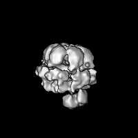
| タイトル | Structural basis for DNA strand separation by a hexameric replicative helicase | |||||||||
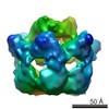
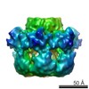
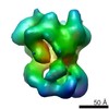
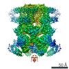
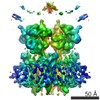
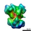
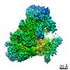
 マップデータ マップデータ | Reconstruction of full-length E1 helicase assembled on the DNA, labelled with monovalent tetrameric streptavidin (MTS) on the dsDNA | |||||||||
 試料 試料 |
| |||||||||
 キーワード キーワード | papillomavirus / helicase / DNA replication fork / electron microscopy / structural analysis | |||||||||
| 機能・相同性 |  機能・相同性情報 機能・相同性情報DNA 3'-5' helicase / biotin binding / DNA helicase activity / DNA replication / host cell nucleus / ATP hydrolysis activity / DNA binding / extracellular region / ATP binding 類似検索 - 分子機能 | |||||||||
| 生物種 |  Bovine papillomavirus (パピローマウイルス) / Bovine papillomavirus (パピローマウイルス) /  Streptomyces avidinii (バクテリア) Streptomyces avidinii (バクテリア) | |||||||||
| 手法 | 単粒子再構成法 / ネガティブ染色法 / 解像度: 18.0 Å | |||||||||
 データ登録者 データ登録者 | Chaban Y / Stead JA / Ryzhenkova K / Whelan F / Lamber K / Antson A / Sanders CM / Orlova EV | |||||||||
 引用 引用 | ジャーナル: Nucleic Acids Res / 年: 2015 タイトル: Structural basis for DNA strand separation by a hexameric replicative helicase. 著者: Yuriy Chaban / Jonathan A Stead / Ksenia Ryzhenkova / Fiona Whelan / Ekaterina P Lamber / Alfred Antson / Cyril M Sanders / Elena V Orlova /  要旨: Hexameric helicases are processive DNA unwinding machines but how they engage with a replication fork during unwinding is unknown. Using electron microscopy and single particle analysis we determined ...Hexameric helicases are processive DNA unwinding machines but how they engage with a replication fork during unwinding is unknown. Using electron microscopy and single particle analysis we determined structures of the intact hexameric helicase E1 from papillomavirus and two complexes of E1 bound to a DNA replication fork end-labelled with protein tags. By labelling a DNA replication fork with streptavidin (dsDNA end) and Fab (5' ssDNA) we located the positions of these labels on the helicase surface, showing that at least 10 bp of dsDNA enter the E1 helicase via a side tunnel. In the currently accepted 'steric exclusion' model for dsDNA unwinding, the active 3' ssDNA strand is pulled through a central tunnel of the helicase motor domain as the dsDNA strands are wedged apart outside the protein assembly. Our structural observations together with nuclease footprinting assays indicate otherwise: strand separation is taking place inside E1 in a chamber above the helicase domain and the 5' passive ssDNA strands exits the assembly through a separate tunnel opposite to the dsDNA entry point. Our data therefore suggest an alternative to the current general model for DNA unwinding by hexameric helicases. | |||||||||
| 履歴 |
|
- 構造の表示
構造の表示
| ムービー |
ムービービューア |
|---|---|
| 構造ビューア | EMマップ: SurfViewMolmilJmol/JSmol |
| 添付画像 |

- ダウンロードとリンク
-EMDBアーカイブ
| マップデータ | emd_3090.map.gz | 1.8 MB | EMDBマップデータ形式 | |
|---|---|---|---|---|
| ヘッダ (付随情報) | emd-3090-v30.xmlemd-3090.xml | 16 KB 16 KB | 表示 表示 | EMDBヘッダ |
| 画像 |  emd_3090.png emd_3090.png | 160.4 KB | ||
| アーカイブディレクトリ |  http://ftp.pdbj.org/pub/emdb/structures/EMD-3090ftp://ftp.pdbj.org/pub/emdb/structures/EMD-3090 http://ftp.pdbj.org/pub/emdb/structures/EMD-3090ftp://ftp.pdbj.org/pub/emdb/structures/EMD-3090 | HTTPS FTP |
-検証レポート
| 文書・要旨 | emd_3090_validation.pdf.gz | 195.3 KB | 表示 | EMDB検証レポート |
|---|---|---|---|---|
| 文書・詳細版 | emd_3090_full_validation.pdf.gz | 194.5 KB | 表示 | |
| XML形式データ | emd_3090_validation.xml.gz | 5.8 KB | 表示 | |
| アーカイブディレクトリ | https://ftp.pdbj.org/pub/emdb/validation_reports/EMD-3090ftp://ftp.pdbj.org/pub/emdb/validation_reports/EMD-3090 | HTTPS FTP |
-関連構造データ


-リンク
| EMDBのページ | EMDB (EBI/PDBe) / EMDataResource |
|---|---|
| 「今月の分子」の関連する項目 |


-マップ
| ファイル | ダウンロード / ファイル: emd_3090.map.gz / 形式: CCP4 / 大きさ: 29.8 MB / タイプ: IMAGE STORED AS FLOATING POINT NUMBER (4 BYTES) | ||||||||||||||||||||||||||||||||||||||||||||||||||||||||||||
|---|---|---|---|---|---|---|---|---|---|---|---|---|---|---|---|---|---|---|---|---|---|---|---|---|---|---|---|---|---|---|---|---|---|---|---|---|---|---|---|---|---|---|---|---|---|---|---|---|---|---|---|---|---|---|---|---|---|---|---|---|---|
| 注釈 | Reconstruction of full-length E1 helicase assembled on the DNA, labelled with monovalent tetrameric streptavidin (MTS) on the dsDNA | ||||||||||||||||||||||||||||||||||||||||||||||||||||||||||||
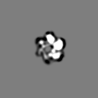
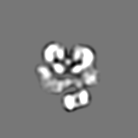
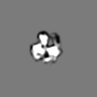
| 投影像・断面図 | 画像のコントロール
画像は Spider により作成 | ||||||||||||||||||||||||||||||||||||||||||||||||||||||||||||
| ボクセルのサイズ | X=Y=Z: 1.6 Å | ||||||||||||||||||||||||||||||||||||||||||||||||||||||||||||
| 密度 |
| ||||||||||||||||||||||||||||||||||||||||||||||||||||||||||||
| 対称性 | 空間群: 1 | ||||||||||||||||||||||||||||||||||||||||||||||||||||||||||||
| 詳細 | EMDB XML:
CCP4マップ ヘッダ情報:
| ||||||||||||||||||||||||||||||||||||||||||||||||||||||||||||
 Z (Sec.)
 Y (Row.)
 X (Col.)


-添付データ
- 試料の構成要素
-全体 : Full-length E1 helicase-DNA complex labelled with monovalent tetr...
| 全体 | 名称: Full-length E1 helicase-DNA complex labelled with monovalent tetrameric streptavidin (MTS) on the dsDNA end of the DNA fork |
|---|---|
| 要素 |
|
-超分子 #1000: Full-length E1 helicase-DNA complex labelled with monovalent tetr...
| 超分子 | 名称: Full-length E1 helicase-DNA complex labelled with monovalent tetrameric streptavidin (MTS) on the dsDNA end of the DNA fork タイプ: sample / ID: 1000 集合状態: one MTS binds to one hexamer of full-length E1 helices via dsDNA Number unique components: 3 |
|---|---|
| 分子量 | 実験値: 460 KDa / 理論値: 460 KDa / 手法: Theoretical calculation |
-分子 #1: Full-length hexameric E1 helicase
| 分子 | 名称: Full-length hexameric E1 helicase / タイプ: protein_or_peptide / ID: 1 / Name.synonym: FLE1 詳細: Full-length E1 helicase-DNA complex was labelled with monovalent tetrameric streptavidin (MTS) on the dsDNA end of the DNA fork コピー数: 1 / 集合状態: hexamer / 組換発現: Yes |
|---|---|
| 由来(天然) | 生物種: Bovine papillomavirus (パピローマウイルス) 別称: Bovine papillomavirus / Organelle: Nucleus / 細胞中の位置: Nucleus |
| 分子量 | 実験値: 410 KDa / 理論値: 410 KDa |
| 組換発現 | 生物種: |
| 配列 | UniProtKB: Replication protein E1 |
-分子 #2: Monovalent tetrameric streptavidin
| 分子 | 名称: Monovalent tetrameric streptavidin / タイプ: protein_or_peptide / ID: 2 / Name.synonym: MTS / コピー数: 1 / 集合状態: tetramer / 組換発現: Yes |
|---|---|
| 由来(天然) | 生物種: Streptomyces avidinii (バクテリア) / 別称: Streptomyces avidinii |
| 分子量 | 実験値: 53 KDa / 理論値: 53 KDa |
| 組換発現 | 生物種: |
| 配列 | UniProtKB: Streptavidin |
-実験情報
-構造解析
| 手法 | ネガティブ染色法 |
|---|---|
 解析 解析 | 単粒子再構成法 |
| 試料の集合状態 | particle |
-試料調製
| 濃度 | 3 mg/mL |
|---|---|
| 緩衝液 | pH: 8 詳細: 10 mM Tris-Cl pH 8.0, 225 mM NaCl, 2 mM DTT, 0.1 mM PMSF, 0.1 mM EDTA |
| 染色 | タイプ: NEGATIVE / 詳細: Sample was stained with 2% uranyl acetate |
| グリッド | 詳細: Sample was applied on to carbon-coated copper grids (400 mesh, freshly glow-discharged in air) |
| 凍結 | 凍結剤: NONE / 装置: OTHER |
- 電子顕微鏡法
| 顕微鏡 | FEI TECNAI F20 |
|---|---|
| 温度 | 最低: 291 K / 最高: 296 K / 平均: 293 K |
| アライメント法 | Legacy - 非点収差: Objective lens astigmatism was corrected at 100,000 times magnification |
| 日付 | 2012年12月14日 |
| 撮影 | カテゴリ: CCD フィルム・検出器のモデル: GATAN ULTRASCAN 4000 (4k x 4k) デジタル化 - サンプリング間隔: 1.6 µm / 実像数: 210 / 平均電子線量: 20 e/Å2 / カメラ長: 1000 / 詳細: No subframe averaging was used. / ビット/ピクセル: 16 |
| 電子線 | 加速電圧: 200 kV / 電子線源:  FIELD EMISSION GUN FIELD EMISSION GUN |
| 電子光学系 | 倍率(補正後): 67000 / 照射モード: FLOOD BEAM / 撮影モード: BRIGHT FIELD / Cs: 2.1 mm / 最大 デフォーカス(公称値): 1.5 µm / 最小 デフォーカス(公称値): 0.5 µm / 倍率(公称値): 62000 |
| 試料ステージ | 試料ホルダー: Negative stain holder / 試料ホルダーモデル: OTHER |
| 実験機器 |  モデル: Tecnai F20 / 画像提供: FEI Company |
+画像解析
-原子モデル構築 1
| 初期モデル | PDB ID: Chain - #0 - Chain ID: A / Chain - #1 - Chain ID: B / Chain - #2 - Chain ID: C / Chain - #3 - Chain ID: D / Chain - #4 - Chain ID: E / Chain - #5 - Chain ID: F |
|---|---|
| ソフトウェア | 名称: Chimera, Veda |
| 詳細 | The domains were separately fitted by manual docking using Chimera |
| 精密化 | 空間: REAL / プロトコル: RIGID BODY FIT / 当てはまり具合の基準: correlation coefficient |
| 得られたモデル | 
PDB-5a9m: |

-原子モデル構築 2
| 初期モデル | PDB ID: Chain - Chain ID: A |
|---|---|
| ソフトウェア | 名称: Chimera |
| 詳細 | The domains were separately fitted by manual docking using Chimera based on the local correlation. |
| 精密化 | 空間: REAL / プロトコル: RIGID BODY FIT 当てはまり具合の基準: local correlation coefficient |
| 得られたモデル |
PDB-5a9m: |

-原子モデル構築 3
| 初期モデル | PDB ID: Chain - #0 - Chain ID: A / Chain - #1 - Chain ID: B / Chain - #2 - Chain ID: C / Chain - #3 - Chain ID: D |
|---|---|
| ソフトウェア | 名称: Chimera |
| 詳細 | The tetramer structure was fitted in Chimera using the local correlation criterion. |
| 精密化 | 空間: REAL / プロトコル: RIGID BODY FIT / 当てはまり具合の基準: correlation coefficient |
| 得られたモデル |
PDB-5a9m: |

