 Movie
Movie Controller

[English] 日本語
 Yorodumi
- PDB-1ksx: Crystal Structures of Two Intermediates in the Assembly of the Pa... -
+ Open data

- Basic information
| Entry | Database: PDB / ID: 1ksx | ||||||
|---|---|---|---|---|---|---|---|
| Title | Crystal Structures of Two Intermediates in the Assembly of the Papillomavirus Replication Initiation Complex | ||||||
 Components Components |
| ||||||
 Keywords Keywords | REPLICATION/DNA / PAPILLOMAVIRUS / DNA-BINDING DOMAIN / REPLICATION / INITIATOR PROTEIN / HELICASE / REPLICATION-DNA COMPLEX | ||||||
| Function / homology |  Function and homology information Function and homology information3'-5' DNA helicase activity / DNA 3'-5' helicase / DNA replication / host cell nucleus / ATP hydrolysis activity / DNA binding / ATP binding Similarity search - Function | ||||||
| Biological species |  Bovine papillomavirus Bovine papillomavirus | ||||||
| Method |  X-RAY DIFFRACTION / SYNCHROTRON / MOLECULAR REPLACEMENT / Resolution: 3.2 Å X-RAY DIFFRACTION / SYNCHROTRON / MOLECULAR REPLACEMENT / Resolution: 3.2 Å | ||||||
 Authors Authors | Enemark, E.J. / Stenlund, A. / Joshua-Tor, L. | ||||||
 Citation Citation | Journal: EMBO J. / Year: 2002 Title: Crystal structures of two intermediates in the assembly of the papillomavirus replication initiation complex. Authors: Enemark, E.J. / Stenlund, A. / Joshua-Tor, L. | ||||||
| History |
|
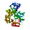
- Structure visualization
| Structure viewer | Molecule: MolmilJmol/JSmol |
|---|
- Downloads & links
-Download
| PDBx/mmCIF format | 1ksx.cif.gz | 256.3 KB | Display | PDBx/mmCIF format |
|---|---|---|---|---|
| PDB format | pdb1ksx.ent.gz | 206.8 KB | Display | PDB format |
| PDBx/mmJSON format | 1ksx.json.gz | Tree view | PDBx/mmJSON format | |
| Others |  Other downloads Other downloads |
-Validation report
| Arichive directory | https://data.pdbj.org/pub/pdb/validation_reports/ks/1ksxftp://data.pdbj.org/pub/pdb/validation_reports/ks/1ksx | HTTPS FTP |
|---|
-Related structure data
| Related structure data |  1ksyC  1f08S C: citing same article ( S: Starting model for refinement |
|---|---|
| Similar structure data |


-Links
 PDBj
PDBj


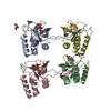
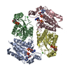
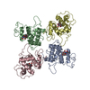
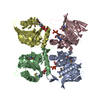
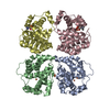
- Assembly
| Deposited unit | 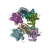
| ||||||||
|---|---|---|---|---|---|---|---|---|---|
| 1 | 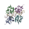
| ||||||||
| 2 | 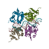
| ||||||||
| Unit cell |
| ||||||||
| Details | E1 multimerization occurs upon binding to the adjacent sites of the target DNA sequence. |
-Components
| #1: DNA chain | Mass: 6444.222 Da / Num. of mol.: 4 / Source method: obtained synthetically #2: Protein | Mass: 16877.770 Da / Num. of mol.: 8 / Fragment: DNA-binding domain Source method: isolated from a genetically manipulated source Source: (gene. exp.) Bovine papillomavirus / Plasmid: PET11CGST / Species (production host): Escherichia coli / Production host:  #3: Water | ChemComp-HOH / |  Mass: 18.015 Da / Num. of mol.: 20 / Source method: isolated from a natural source / Formula: H2O Mass: 18.015 Da / Num. of mol.: 20 / Source method: isolated from a natural source / Formula: H2O |
|---|
-Experimental details
-Experiment
| Experiment | Method: X-RAY DIFFRACTION / Number of used crystals: 1 |
|---|
- Sample preparation
| Crystal | Density Matthews: 3.33 Å3/Da / Density % sol: 63.05 % | ||||||||||||||||||||||||
|---|---|---|---|---|---|---|---|---|---|---|---|---|---|---|---|---|---|---|---|---|---|---|---|---|---|
| Crystal grow | Temperature: 290 K / Method: vapor diffusion, hanging drop / pH: 7.5 Details: Ca(CF3COO)2, DTT, pH 7.5, VAPOR DIFFUSION, HANGING DROP, temperature 290K | ||||||||||||||||||||||||
| Components of the solutions |
| ||||||||||||||||||||||||
| Crystal grow | *PLUS | ||||||||||||||||||||||||
| Components of the solutions | *PLUS
|
-Data collection
| Diffraction | Mean temperature: 100 K |
|---|---|
| Diffraction source | Source: SYNCHROTRON / Site: NSLS  / Beamline: X26C / Wavelength: 1.1 / Beamline: X26C / Wavelength: 1.1 |
| Detector | Type: ADSC QUANTUM 4 / Detector: CCD / Date: Nov 4, 2000 / Details: mirrors |
| Radiation | Monochromator: Silicon / Protocol: SINGLE WAVELENGTH / Monochromatic (M) / Laue (L): M / Scattering type: x-ray |
| Radiation wavelength | Wavelength: 1.1 Å / Relative weight: 1 |
| Reflection | Resolution: 3.2→50 Å / Num. all: 35201 / Num. obs: 35038 / % possible obs: 99.7 % / Observed criterion σ(F): 0 / Observed criterion σ(I): -3 / Redundancy: 3.6 % / Rmerge(I) obs: 0.195 / Net I/σ(I): 9.14 |
| Reflection shell | Resolution: 3.2→3.31 Å / Redundancy: 3.5 % / Rmerge(I) obs: 0.584 / Mean I/σ(I) obs: 2.4 / Num. unique all: 3474 / % possible all: 99.3 |
| Reflection | *PLUS Lowest resolution: 50 Å / Num. measured all: 125960 |
| Reflection shell | *PLUS % possible obs: 99.3 % / Num. unique obs: 3474 / Num. measured obs: 12242 |
- Processing
| Software |
| ||||||||||||||||||||||||||||||||||||||||||||||||||||||||||||||||||||||||||||||||
|---|---|---|---|---|---|---|---|---|---|---|---|---|---|---|---|---|---|---|---|---|---|---|---|---|---|---|---|---|---|---|---|---|---|---|---|---|---|---|---|---|---|---|---|---|---|---|---|---|---|---|---|---|---|---|---|---|---|---|---|---|---|---|---|---|---|---|---|---|---|---|---|---|---|---|---|---|---|---|---|---|---|
| Refinement | Method to determine structure: MOLECULAR REPLACEMENT Starting model: PDB ENTRY 1F08 Resolution: 3.2→42.62 Å / Rfactor Rfree error: 0.007 / Data cutoff high absF: 130431.95 / Data cutoff low absF: 0 / Isotropic thermal model: RESTRAINED / Cross valid method: THROUGHOUT / σ(F): 0 / Stereochemistry target values: Engh & Huber Details: THE NUMBER OF NON-HYDROGEN ATOMS USED IN REFINEMENT IS LESS THAN SPECIFIED IN REMARK 3. THE VALUES LISTED IN REMARK 3 ARE 4-FOLD GREATER (REFLECTING THE 4-FOLD STRICT NCS). 2328 PROTEIN ...Details: THE NUMBER OF NON-HYDROGEN ATOMS USED IN REFINEMENT IS LESS THAN SPECIFIED IN REMARK 3. THE VALUES LISTED IN REMARK 3 ARE 4-FOLD GREATER (REFLECTING THE 4-FOLD STRICT NCS). 2328 PROTEIN ATOMS, 428 NUCLEIC ACID ATOMS, AND 5 SOLVENT ATOMS WERE USED IN REFINEMENT.
| ||||||||||||||||||||||||||||||||||||||||||||||||||||||||||||||||||||||||||||||||
| Solvent computation | Solvent model: FLAT MODEL / Bsol: 23.0378 Å2 / ksol: 0.240909 e/Å3 | ||||||||||||||||||||||||||||||||||||||||||||||||||||||||||||||||||||||||||||||||
| Displacement parameters | Biso mean: 59.5 Å2
| ||||||||||||||||||||||||||||||||||||||||||||||||||||||||||||||||||||||||||||||||
| Refine analyze |
| ||||||||||||||||||||||||||||||||||||||||||||||||||||||||||||||||||||||||||||||||
| Refinement step | Cycle: LAST / Resolution: 3.2→42.62 Å
| ||||||||||||||||||||||||||||||||||||||||||||||||||||||||||||||||||||||||||||||||
| Refine LS restraints |
| ||||||||||||||||||||||||||||||||||||||||||||||||||||||||||||||||||||||||||||||||
| LS refinement shell | Resolution: 3.2→3.4 Å / Rfactor Rfree error: 0.028 / Total num. of bins used: 6
| ||||||||||||||||||||||||||||||||||||||||||||||||||||||||||||||||||||||||||||||||
| Xplor file |
| ||||||||||||||||||||||||||||||||||||||||||||||||||||||||||||||||||||||||||||||||
| Refinement | *PLUS Highest resolution: 3.2 Å / Lowest resolution: 50 Å / Rfactor obs: 0.2634 / Rfactor Rfree: 0.2844 | ||||||||||||||||||||||||||||||||||||||||||||||||||||||||||||||||||||||||||||||||
| Solvent computation | *PLUS | ||||||||||||||||||||||||||||||||||||||||||||||||||||||||||||||||||||||||||||||||
| Displacement parameters | *PLUS | ||||||||||||||||||||||||||||||||||||||||||||||||||||||||||||||||||||||||||||||||
| Refine LS restraints | *PLUS
|
