Japan Agency for Medical Research and Development (AMED)
JP19am0101001 (support number 0053)
Japan
Japan Society for the Promotion of Science (JSPS)
15K21608
Japan
Japan Science and Technology
JPMJCR14M1
Japan
Japan Science and Technology
JPMJCR13M7
Japan
Japan Society for the Promotion of Science (JSPS)
25111004
Japan
Japan Agency for Medical Research and Development (AMED)
JP19am0101079 (support number 0739)
Japan
Japan Society for the Promotion of Science (JSPS)
19H05707
Japan
Japan Society for the Promotion of Science (JSPS)
18H03989
Japan
Citation
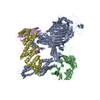
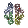
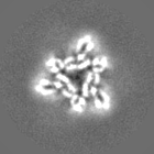
Journal: Nat Struct Mol Biol / Year: 2020 Title: Atg9 is a lipid scramblase that mediates autophagosomal membrane expansion. Authors: Kazuaki Matoba / Tetsuya Kotani / Akihisa Tsutsumi / Takuma Tsuji / Takaharu Mori / Daisuke Noshiro / Yuji Sugita / Norimichi Nomura / So Iwata / Yoshinori Ohsumi / Toyoshi Fujimoto / ...Authors: Kazuaki Matoba / Tetsuya Kotani / Akihisa Tsutsumi / Takuma Tsuji / Takaharu Mori / Daisuke Noshiro / Yuji Sugita / Norimichi Nomura / So Iwata / Yoshinori Ohsumi / Toyoshi Fujimoto / Hitoshi Nakatogawa / Masahide Kikkawa / Nobuo N Noda / Abstract: The molecular function of Atg9, the sole transmembrane protein in the autophagosome-forming machinery, remains unknown. Atg9 colocalizes with Atg2 at the expanding edge of the isolation membrane (IM) ...The molecular function of Atg9, the sole transmembrane protein in the autophagosome-forming machinery, remains unknown. Atg9 colocalizes with Atg2 at the expanding edge of the isolation membrane (IM), where Atg2 receives phospholipids from the endoplasmic reticulum (ER). Here we report that yeast and human Atg9 are lipid scramblases that translocate phospholipids between outer and inner leaflets of liposomes in vitro. Cryo-EM of fission yeast Atg9 reveals a homotrimer, with two connected pores forming a path between the two membrane leaflets: one pore, located at a protomer, opens laterally to the cytoplasmic leaflet; the other, at the trimer center, traverses the membrane vertically. Mutation of residues lining the pores impaired IM expansion and autophagy activity in yeast and abolished Atg9's ability to transport phospholipids between liposome leaflets. These results suggest that phospholipids delivered by Atg2 are translocated from the cytoplasmic to the luminal leaflet by Atg9, thereby driving autophagosomal membrane expansion.
History
Deposition
Sep 14, 2020
-
Header (metadata) release
Oct 28, 2020
-
Map release
Oct 28, 2020
-
Update
Dec 16, 2020
-
Current status
Dec 16, 2020
Processing site: PDBj / Status: Released
-
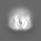
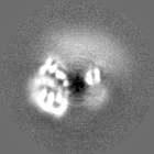
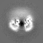
Structure visualization
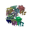
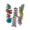
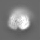
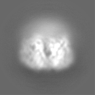
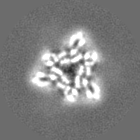
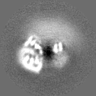
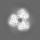
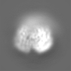
Movie
Surface view with section colored by density value
In the structure databanks used in Yorodumi, some data are registered as the other names, "COVID-19 virus" and "2019-nCoV". Here are the details of the virus and the list of structure data.
Jan 31, 2019. EMDB accession codes are about to change! (news from PDBe EMDB page)
EMDB accession codes are about to change! (news from PDBe EMDB page)
The allocation of 4 digits for EMDB accession codes will soon come to an end. Whilst these codes will remain in use, new EMDB accession codes will include an additional digit and will expand incrementally as the available range of codes is exhausted. The current 4-digit format prefixed with “EMD-” (i.e. EMD-XXXX) will advance to a 5-digit format (i.e. EMD-XXXXX), and so on. It is currently estimated that the 4-digit codes will be depleted around Spring 2019, at which point the 5-digit format will come into force.
The EM Navigator/Yorodumi systems omit the EMD- prefix.
Related info.:Q: What is EMD? / ID/Accession-code notation in Yorodumi/EM Navigator
Yorodumi is a browser for structure data from EMDB, PDB, SASBDB, etc.
This page is also the successor to EM Navigator detail page, and also detail information page/front-end page for Omokage search.
The word "yorodu" (or yorozu) is an old Japanese word meaning "ten thousand". "mi" (miru) is to see.
Related info.:EMDB / PDB / SASBDB / Comparison of 3 databanks / Yorodumi Search / Aug 31, 2016. New EM Navigator & Yorodumi / Yorodumi Papers / Jmol/JSmol / Function and homology information / Changes in new EM Navigator and Yorodumi
 Movie
Movie Controller


 Open data
 Basic information
 Map data
 Sample
 Function and homology information
 Authors
 Japan, 9 items
  Citation
 Structure visualization
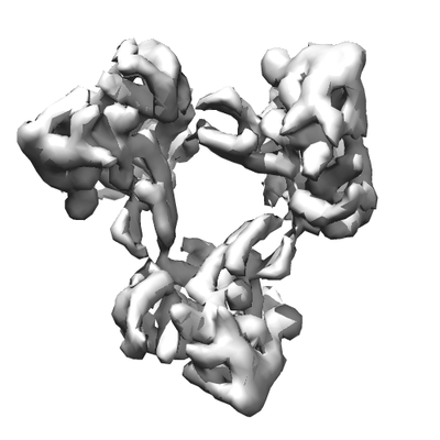
 Downloads & links
 emd_30545.png
 http://ftp.pdbj.org/pub/emdb/structures/EMD-30545


 Z (Sec.)
 Y (Row.)
 X (Col.)


 Sample components
 Processing
 Electron microscopy
 FIELD EMISSION GUN