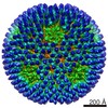
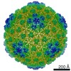
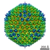
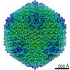
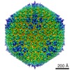
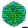
Journal: J Virol / Year: 2015 Title: Structures of Adenovirus Incomplete Particles Clarify Capsid Architecture and Show Maturation Changes of Packaging Protein L1 52/55k. Authors: Gabriela N Condezo / Roberto Marabini / Silvia Ayora / José M Carazo / Raúl Alba / Miguel Chillón / Carmen San Martín / Abstract: Adenovirus is one of the most complex icosahedral, nonenveloped viruses. Even after its structure was solved at near-atomic resolution by both cryo-electron microscopy and X-ray crystallography, the ...Adenovirus is one of the most complex icosahedral, nonenveloped viruses. Even after its structure was solved at near-atomic resolution by both cryo-electron microscopy and X-ray crystallography, the location of minor coat proteins is still a subject of debate. The elaborated capsid architecture is the product of a correspondingly complex assembly process, about which many aspects remain unknown. Genome encapsidation involves the concerted action of five virus proteins, and proteolytic processing by the virus protease is needed to prime the virion for sequential uncoating. Protein L1 52/55k is required for packaging, and multiple cleavages by the maturation protease facilitate its release from the nascent virion. Light-density particles are routinely produced in adenovirus infections and are thought to represent assembly intermediates. Here, we present the molecular and structural characterization of two different types of human adenovirus light particles produced by a mutant with delayed packaging. We show that these particles lack core polypeptide V but do not lack the density corresponding to this protein in the X-ray structure, thereby adding support to the adenovirus cryo-electron microscopy model. The two types of light particles present different degrees of proteolytic processing. Their structures provide the first glimpse of the organization of L1 52/55k protein inside the capsid shell and of how this organization changes upon partial maturation. Immature, full-length L1 52/55k is poised beneath the vertices to engage the virus genome. Upon proteolytic processing, L1 52/55k disengages from the capsid shell, facilitating genome release during uncoating. IMPORTANCE: Adenoviruses have been extensively characterized as experimental systems in molecular biology, as human pathogens, and as therapeutic vectors. However, a clear picture of many aspects of ...IMPORTANCE: Adenoviruses have been extensively characterized as experimental systems in molecular biology, as human pathogens, and as therapeutic vectors. However, a clear picture of many aspects of their basic biology is still lacking. Two of these aspects are the location of minor coat proteins in the capsid and the molecular details of capsid assembly. Here, we provide evidence supporting one of the two current models for capsid architecture. We also show for the first time the location of the packaging protein L1 52/55k in particles lacking the virus genome and how this location changes during maturation. Our results contribute to clarifying standing questions in adenovirus capsid architecture and provide new details on the role of L1 52/55k protein in assembly.
History
Deposition
May 8, 2015
-
Header (metadata) release
Jun 17, 2015
-
Map release
Jul 29, 2015
-
Update
Sep 9, 2015
-
Current status
Sep 9, 2015
Processing site: PDBe / Status: Released
-
Structure visualization
Movie
Surface view with section colored by density value
Category: CCD / Film or detector model: FEI EAGLE (4k x 4k) / Digitization - Sampling interval: 14 µm / Number real images: 889 / Average electron dose: 12 e/Å2
Electron beam
Acceleration voltage: 200 kV / Electron source: FIELD EMISSION GUN
CTF detection, phase flip, particle selection, extraction and normalization with Xmipp. 2D and 3D classification, 3D refinement and postprocessing with Relion.
CTF correction
Details: micrograph
Final reconstruction
Applied symmetry - Point group: I (icosahedral) / Algorithm: OTHER / Resolution.type: BY AUTHOR / Resolution: 12.3 Å / Resolution method: OTHER / Software - Name: Xmipp, Relion / Number images used: 6743
In the structure databanks used in Yorodumi, some data are registered as the other names, "COVID-19 virus" and "2019-nCoV". Here are the details of the virus and the list of structure data.
Jan 31, 2019. EMDB accession codes are about to change! (news from PDBe EMDB page)
EMDB accession codes are about to change! (news from PDBe EMDB page)
The allocation of 4 digits for EMDB accession codes will soon come to an end. Whilst these codes will remain in use, new EMDB accession codes will include an additional digit and will expand incrementally as the available range of codes is exhausted. The current 4-digit format prefixed with “EMD-” (i.e. EMD-XXXX) will advance to a 5-digit format (i.e. EMD-XXXXX), and so on. It is currently estimated that the 4-digit codes will be depleted around Spring 2019, at which point the 5-digit format will come into force.
The EM Navigator/Yorodumi systems omit the EMD- prefix.
Related info.:Q: What is EMD? / ID/Accession-code notation in Yorodumi/EM Navigator
Yorodumi is a browser for structure data from EMDB, PDB, SASBDB, etc.
This page is also the successor to EM Navigator detail page, and also detail information page/front-end page for Omokage search.
The word "yorodu" (or yorozu) is an old Japanese word meaning "ten thousand". "mi" (miru) is to see.
Related info.:EMDB / PDB / SASBDB / Comparison of 3 databanks / Yorodumi Search / Aug 31, 2016. New EM Navigator & Yorodumi / Yorodumi Papers / Jmol/JSmol / Function and homology information / Changes in new EM Navigator and Yorodumi
 Movie
Movie Controller


 Open data
 Basic information
 Map data
 Sample
 Keywords
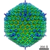
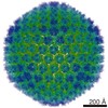
 Human adenovirus 5
 Authors
 Citation
 Structure visualization
 Movie viewer
 Downloads & links
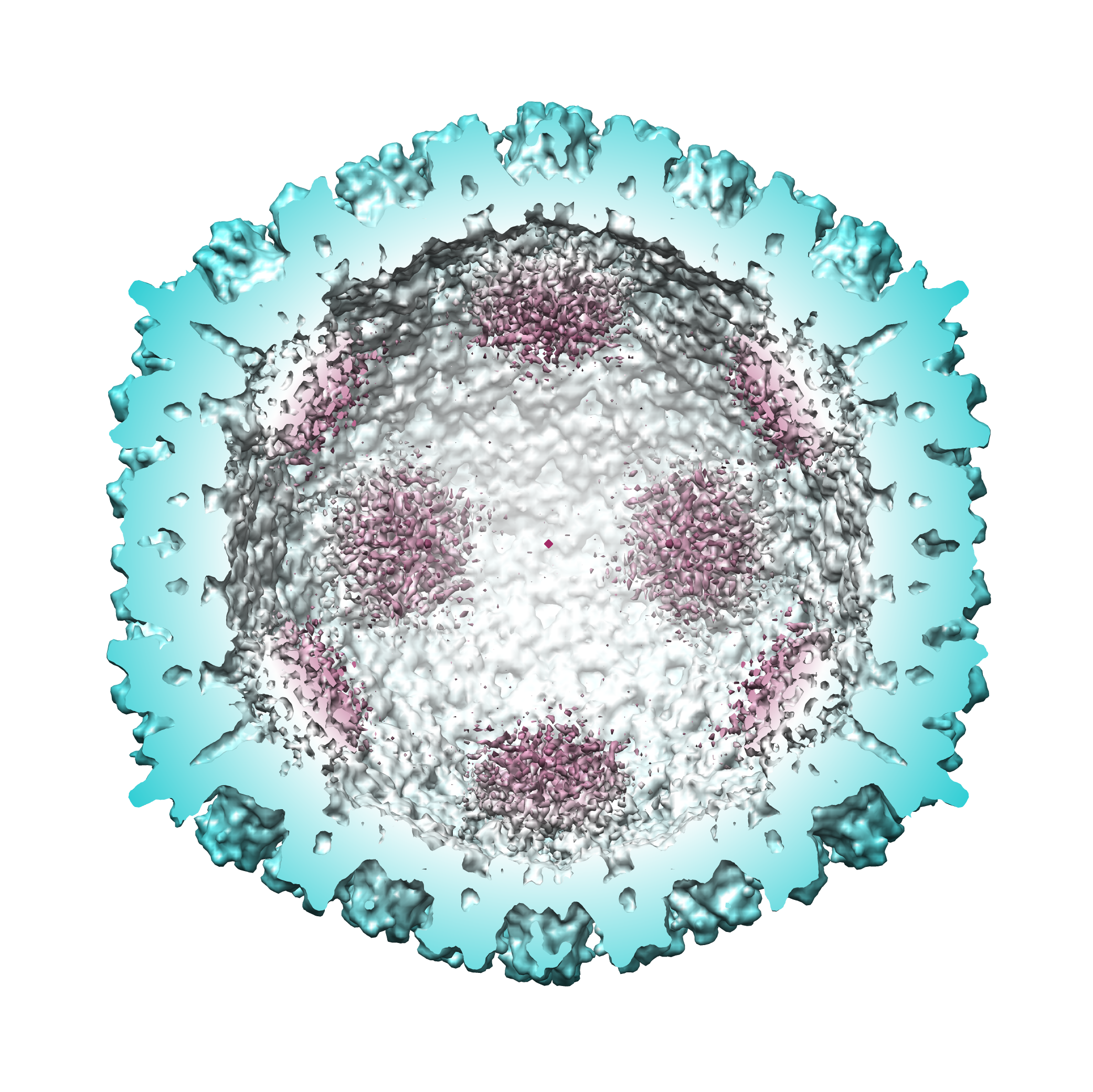 B2.png
 http://ftp.pdbj.org/pub/emdb/structures/EMD-3003
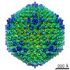

 Z (Sec.)
 Y (Row.)
 X (Col.)


 Sample components
 Homo sapiens (human) / synonym: VERTEBRATES
 Processing
 Electron microscopy
 FIELD EMISSION GUN

