Movie
Movie Controller


+ Open data

- Basic information
| Entry |  | |||||||||
|---|---|---|---|---|---|---|---|---|---|---|
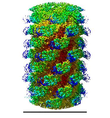
| Title | Pseudomonas phage E217 extended sheath and tail tube | |||||||||
 Map data Map data | ||||||||||
 Sample Sample |
| |||||||||
 Keywords Keywords | Pseudomonas / phage / E217 / VIRUS | |||||||||
| Function / homology | : / Tail fiber protein gp32 / Protein of unknown function DUF3383 / Protein of unknown function (DUF3383) / Tail fiber protein / Tail sheath protein Function and homology information Function and homology information | |||||||||
| Biological species |  Pseudomonas phage vB_PaeM_E217 (virus) Pseudomonas phage vB_PaeM_E217 (virus) | |||||||||
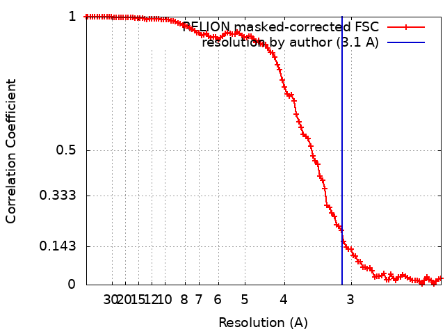
| Method | single particle reconstruction / cryo EM / Resolution: 3.1 Å | |||||||||
 Authors Authors | Li F / Cingolani G / Hou C | |||||||||
| Funding support |  United States, 1 items United States, 1 items
| |||||||||
 Citation Citation | Journal: Nat Commun / Year: 2023 Title: High-resolution cryo-EM structure of the Pseudomonas bacteriophage E217. Authors: Fenglin Li / Chun-Feng David Hou / Ravi K Lokareddy / Ruoyu Yang / Francesca Forti / Federica Briani / Gino Cingolani /  Abstract: E217 is a Pseudomonas phage used in an experimental cocktail to eradicate cystic fibrosis-associated Pseudomonas aeruginosa. Here, we describe the structure of the whole E217 virion before and after ...E217 is a Pseudomonas phage used in an experimental cocktail to eradicate cystic fibrosis-associated Pseudomonas aeruginosa. Here, we describe the structure of the whole E217 virion before and after DNA ejection at 3.1 Å and 4.5 Å resolution, respectively, determined using cryogenic electron microscopy (cryo-EM). We identify and build de novo structures for 19 unique E217 gene products, resolve the tail genome-ejection machine in both extended and contracted states, and decipher the complete architecture of the baseplate formed by 66 polypeptide chains. We also determine that E217 recognizes the host O-antigen as a receptor, and we resolve the N-terminal portion of the O-antigen-binding tail fiber. We propose that E217 design principles presented in this paper are conserved across PB1-like Myoviridae phages of the Pbunavirus genus that encode a ~1.4 MDa baseplate, dramatically smaller than the coliphage T4. | |||||||||
| History |
|
- Structure visualization
| Supplemental images |
|---|
- Downloads & links
-EMDB archive
| Map data | emd_29481.map.gz | 79.1 MB | EMDB map data format | |
|---|---|---|---|---|
| Header (meta data) | emd-29481-v30.xmlemd-29481.xml | 17.6 KB 17.6 KB | Display Display | EMDB header |
| FSC (resolution estimation) | emd_29481_fsc.xml | 10.6 KB | Display | FSC data file |
| Images |  emd_29481.png emd_29481.png | 180.7 KB | ||
| Filedesc metadata | emd-29481.cif.gz | 5.7 KB | ||
| Others | emd_29481_half_map_1.map.gzemd_29481_half_map_2.map.gz | 79.4 MB 79.4 MB | ||
| Archive directory |  http://ftp.pdbj.org/pub/emdb/structures/EMD-29481ftp://ftp.pdbj.org/pub/emdb/structures/EMD-29481 http://ftp.pdbj.org/pub/emdb/structures/EMD-29481ftp://ftp.pdbj.org/pub/emdb/structures/EMD-29481 | HTTPS FTP |
-Related structure data
| Related structure data |  8fuvMC  8envC 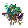 8eonC 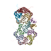 8frsC  8fvgC 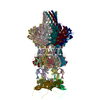 8fvhC C: citing same article ( M: atomic model generated by this map |
|---|---|
| Similar structure data |


-Links
| EMDB pages | EMDB (EBI/PDBe) / EMDataResource |
|---|
-Map
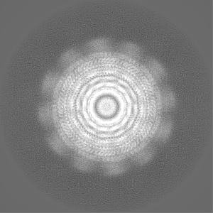
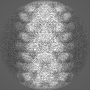
| File | Download / File: emd_29481.map.gz / Format: CCP4 / Size: 103 MB / Type: IMAGE STORED AS FLOATING POINT NUMBER (4 BYTES) | ||||||||||||||||||||||||||||||||||||
|---|---|---|---|---|---|---|---|---|---|---|---|---|---|---|---|---|---|---|---|---|---|---|---|---|---|---|---|---|---|---|---|---|---|---|---|---|---|
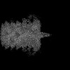
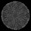
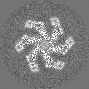
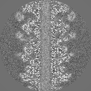
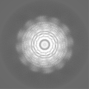
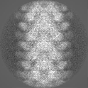
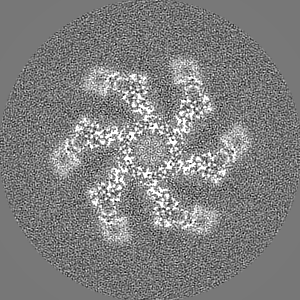
| Projections & slices | Image control
Images are generated by Spider. | ||||||||||||||||||||||||||||||||||||
| Voxel size | X=Y=Z: 1.12 Å | ||||||||||||||||||||||||||||||||||||
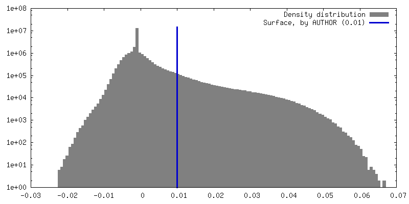
| Density |
| ||||||||||||||||||||||||||||||||||||
| Symmetry | Space group: 1 | ||||||||||||||||||||||||||||||||||||
| Details | EMDB XML:
|
 Z (Sec.)
 Y (Row.)
 X (Col.)


-Supplemental data
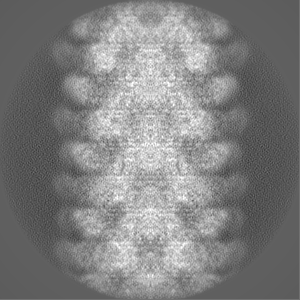
-Half map: #1
| File | emd_29481_half_map_1.map | ||||||||||||
|---|---|---|---|---|---|---|---|---|---|---|---|---|---|
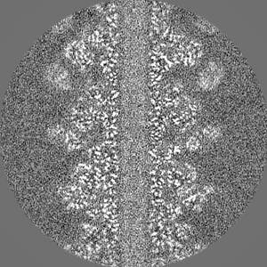
| Projections & Slices |
| ||||||||||||
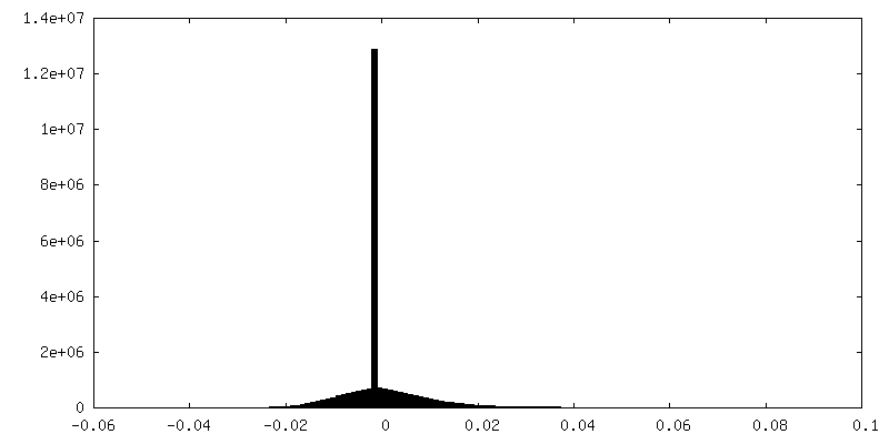
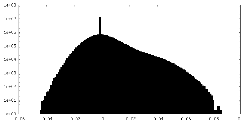
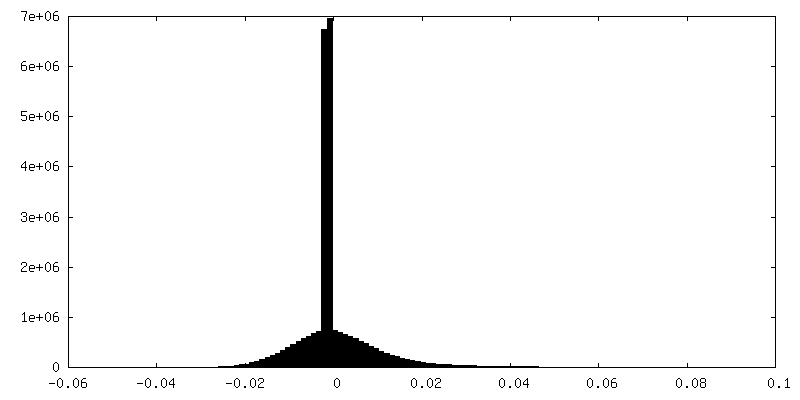
| Density Histograms |

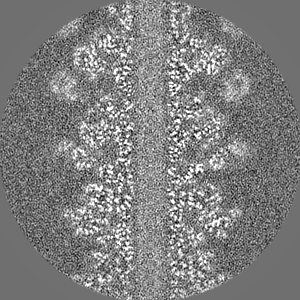
-Half map: #2
| File | emd_29481_half_map_2.map | ||||||||||||
|---|---|---|---|---|---|---|---|---|---|---|---|---|---|
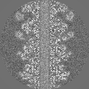
| Projections & Slices |
| ||||||||||||
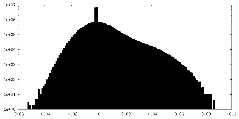
| Density Histograms |

- Sample components
-Entire : Pseudomonas phage vB_PaeM_E217
| Entire | Name: Pseudomonas phage vB_PaeM_E217 (virus) |
|---|---|
| Components |
|
-Supramolecule #1: Pseudomonas phage vB_PaeM_E217
| Supramolecule | Name: Pseudomonas phage vB_PaeM_E217 / type: virus / ID: 1 / Parent: 0 / Macromolecule list: all / NCBI-ID: 2034346 / Sci species name: Pseudomonas phage vB_PaeM_E217 / Virus type: VIRION / Virus isolate: OTHER / Virus enveloped: No / Virus empty: No |
|---|
-Macromolecule #1: Sheath protein gp31
| Macromolecule | Name: Sheath protein gp31 / type: protein_or_peptide / ID: 1 / Details: Extended state / Number of copies: 1 / Enantiomer: LEVO |
|---|---|
| Source (natural) | Organism: Pseudomonas phage vB_PaeM_E217 (virus) |
| Molecular weight | Theoretical: 53.69475 KDa |
| Sequence | String: MISQSRYIRI ISGVGAAAPV AGRKLILRVM TTNNVIPPGI VIEFDNANAV LSYFGAQSEE YQRAAAYFKF ISKSVNSPSS ISFARWVNT AIAPMVVGDN LPKTIADFAG FSAGVLTIMV GAAEQNITAI DTSAATSMDN VASIIQTEIR KNADPQLAQA T VTWNQNTN ...String: MISQSRYIRI ISGVGAAAPV AGRKLILRVM TTNNVIPPGI VIEFDNANAV LSYFGAQSEE YQRAAAYFKF ISKSVNSPSS ISFARWVNT AIAPMVVGDN LPKTIADFAG FSAGVLTIMV GAAEQNITAI DTSAATSMDN VASIIQTEIR KNADPQLAQA T VTWNQNTN QFTLVGATIG TGVLAVAKSA DPQDMSTALG WSTSNVVNVA GQSADLPDAA VAKSTNVSNN FGSFLFAGAP LD NDQIKAV SAWNAAQNNQ FIYTVATSLA NLGTLFTLVN GNAGTALNVL SATAANDFVE QCPSEILAAT NYDEPGASQN YMY YQFPGR NITVSDDTVA NTVDKSRGNY IGVTQANGQQ LAFYQRGILC GGPTDAVDMN VYANEIWLKS AIAQALLDLF LNVN AVPAS STGEAMTLAV LQPVLDKATA NGTFTYGKEI SAVQQQYITQ VTGDRRAWRQ VQTLGYWINI TFSSYTNSNT GLTEW KANY TLIYSKGDAI RFVEGSDVMI UniProtKB: Tail sheath protein |
-Macromolecule #2: Tail fiber protein gp32
| Macromolecule | Name: Tail fiber protein gp32 / type: protein_or_peptide / ID: 2 / Number of copies: 6 / Enantiomer: LEVO |
|---|---|
| Source (natural) | Organism: Pseudomonas phage vB_PaeM_E217 (virus) |
| Molecular weight | Theoretical: 15.174168 KDa |
| Sequence | String: FGSICAFTAS RTFPNGFTVT EEFADADPID SPPFAAADTG AGLNGDMVVW NRANILEVVV NVIPNTEGER NLAVLLDANR TGKDKSGAR DVVGLVVAMP DGSKITCTNG TPIDGVLINA VASVGRLKTK PYRFRFEKVI KAGTS UniProtKB: Tail fiber protein |
-Experimental details
-Structure determination
| Method | cryo EM |
|---|---|
 Processing Processing | single particle reconstruction |
| Aggregation state | particle |
-Sample preparation
| Buffer | pH: 8 |
|---|---|
| Vitrification | Cryogen name: ETHANE / Chamber humidity: 100 % |
- Electron microscopy
| Microscope | TFS KRIOS |
|---|---|
| Image recording | Film or detector model: OTHER / Average electron dose: 1.25 e/Å2 |
| Electron beam | Acceleration voltage: 300 kV / Electron source:  FIELD EMISSION GUN FIELD EMISSION GUN |
| Electron optics | Illumination mode: OTHER / Imaging mode: OTHER / Nominal defocus max: 5.0 µm / Nominal defocus min: 1.2 µm |
| Experimental equipment |  Model: Titan Krios / Image courtesy: FEI Company |