Movie
Movie Controller


+ Open data

- Basic information
| Entry |  | ||||||||||||
|---|---|---|---|---|---|---|---|---|---|---|---|---|---|
| Title | XPC release from Core7-XPA-DNA (AP) | ||||||||||||
 Map data Map data | |||||||||||||
 Sample Sample |
| ||||||||||||
 Keywords Keywords | protein-DNA complex / DNA BINDING PROTEIN-DNA complex | ||||||||||||
| Function / homology |  Function and homology information Function and homology informationnucleotide-excision repair factor 1 complex / nucleotide-excision repair involved in interstrand cross-link repair / nucleotide-excision repair, DNA damage recognition / MMXD complex / core TFIIH complex portion of holo TFIIH complex / Cytosolic iron-sulfur cluster assembly / positive regulation of mitotic recombination / hair cell differentiation / nucleotide-excision repair factor 3 complex / nucleotide-excision repair, preincision complex assembly ...nucleotide-excision repair factor 1 complex / nucleotide-excision repair involved in interstrand cross-link repair / nucleotide-excision repair, DNA damage recognition / MMXD complex / core TFIIH complex portion of holo TFIIH complex / Cytosolic iron-sulfur cluster assembly / positive regulation of mitotic recombination / hair cell differentiation / nucleotide-excision repair factor 3 complex / nucleotide-excision repair, preincision complex assembly / UV protection / CAK-ERCC2 complex / regulation of cyclin-dependent protein serine/threonine kinase activity / transcription factor TFIIH core complex / transcription factor TFIIH holo complex / G protein-coupled receptor internalization / DNA 5'-3' helicase / nuclear thyroid hormone receptor binding / transcription preinitiation complex / RNA Polymerase I Transcription Termination / embryonic organ development / UV-damage excision repair / transcription factor TFIID complex / RNA polymerase II general transcription initiation factor activity / regulation of mitotic cell cycle phase transition / RNA Pol II CTD phosphorylation and interaction with CE during HIV infection / RNA Pol II CTD phosphorylation and interaction with CE / Formation of the Early Elongation Complex / Formation of the HIV-1 Early Elongation Complex / mRNA Capping / HIV Transcription Initiation / RNA Polymerase II HIV Promoter Escape / Transcription of the HIV genome / RNA Polymerase II Promoter Escape / RNA Polymerase II Transcription Pre-Initiation And Promoter Opening / RNA Polymerase II Transcription Initiation / RNA Polymerase II Transcription Initiation And Promoter Clearance / ATPase activator activity / DNA topological change / RNA Polymerase I Transcription Initiation / response to UV / DNA 3'-5' helicase / protein localization to nucleus / 3'-5' DNA helicase activity / Tat-mediated elongation of the HIV-1 transcript / Formation of HIV-1 elongation complex containing HIV-1 Tat / Formation of HIV elongation complex in the absence of HIV Tat / hormone-mediated signaling pathway / RNA Polymerase II Transcription Elongation / Formation of RNA Pol II elongation complex / transcription by RNA polymerase I / RNA Polymerase II Pre-transcription Events / transcription-coupled nucleotide-excision repair / DNA helicase activity / TP53 Regulates Transcription of DNA Repair Genes / chromosome segregation / promoter-specific chromatin binding / RNA Polymerase I Promoter Escape / transcription initiation at RNA polymerase II promoter / nucleotide-excision repair / transcription elongation by RNA polymerase II / base-excision repair / NoRC negatively regulates rRNA expression / transcription by RNA polymerase II / intracellular protein localization / spindle / Dual Incision in GG-NER / Transcription-Coupled Nucleotide Excision Repair (TC-NER) / Formation of TC-NER Pre-Incision Complex / Formation of Incision Complex in GG-NER / sequence-specific double-stranded DNA binding / Dual incision in TC-NER / Gap-filling DNA repair synthesis and ligation in TC-NER / 4 iron, 4 sulfur cluster binding / response to oxidative stress / double-stranded DNA binding / 5'-3' DNA helicase activity / damaged DNA binding / nuclear body / protein-macromolecule adaptor activity / nuclear speck / positive regulation of apoptotic process / protein domain specific binding / DNA repair / apoptotic process / chromatin binding / regulation of transcription by RNA polymerase II / nucleolus / regulation of DNA-templated transcription / positive regulation of DNA-templated transcription / protein homodimerization activity / ATP hydrolysis activity / DNA binding / nucleoplasm / zinc ion binding / ATP binding / metal ion binding / nucleus / cytoplasm Similarity search - Function | ||||||||||||
| Biological species |  Homo sapiens (human) / synthetic construct (others) Homo sapiens (human) / synthetic construct (others) | ||||||||||||
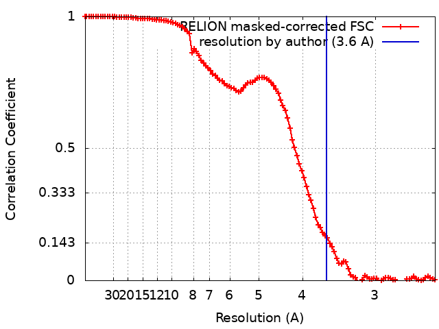
| Method | single particle reconstruction / cryo EM / Resolution: 3.6 Å | ||||||||||||
 Authors Authors | Kim J / Yang W | ||||||||||||
| Funding support |  United States, United States,  Japan, 3 items Japan, 3 items
| ||||||||||||
 Citation Citation | Journal: Nature / Year: 2023 Title: Lesion recognition by XPC, TFIIH and XPA in DNA excision repair. Authors: Jinseok Kim / Chia-Lung Li / Xuemin Chen / Yanxiang Cui / Filip M Golebiowski / Huaibin Wang / Fumio Hanaoka / Kaoru Sugasawa / Wei Yang /   Abstract: Nucleotide excision repair removes DNA lesions caused by ultraviolet light, cisplatin-like compounds and bulky adducts. After initial recognition by XPC in global genome repair or a stalled RNA ...Nucleotide excision repair removes DNA lesions caused by ultraviolet light, cisplatin-like compounds and bulky adducts. After initial recognition by XPC in global genome repair or a stalled RNA polymerase in transcription-coupled repair, damaged DNA is transferred to the seven-subunit TFIIH core complex (Core7) for verification and dual incisions by the XPF and XPG nucleases. Structures capturing lesion recognition by the yeast XPC homologue Rad4 and TFIIH in transcription initiation or DNA repair have been separately reported. How two different lesion recognition pathways converge and how the XPB and XPD helicases of Core7 move the DNA lesion for verification are unclear. Here we report on structures revealing DNA lesion recognition by human XPC and DNA lesion hand-off from XPC to Core7 and XPA. XPA, which binds between XPB and XPD, kinks the DNA duplex and shifts XPC and the DNA lesion by nearly a helical turn relative to Core7. The DNA lesion is thus positioned outside of Core7, as would occur with RNA polymerase. XPB and XPD, which track the lesion-containing strand but translocate DNA in opposite directions, push and pull the lesion-containing strand into XPD for verification. | ||||||||||||
| History |
|
- Structure visualization
| Supplemental images |
|---|
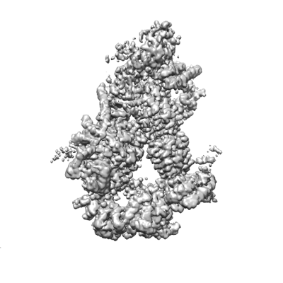
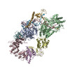
- Downloads & links
-EMDB archive
| Map data | emd_28002.map.gz | 89.7 MB | EMDB map data format | |
|---|---|---|---|---|
| Header (meta data) | emd-28002-v30.xmlemd-28002.xml | 42.1 KB 42.1 KB | Display Display | EMDB header |
| FSC (resolution estimation) | emd_28002_fsc.xml | 10.7 KB | Display | FSC data file |
| Images | 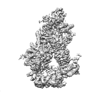 emd_28002.png emd_28002.png | 83.4 KB | ||
| Filedesc metadata | emd-28002.cif.gz | 10.9 KB | ||
| Others | emd_28002_half_map_1.map.gzemd_28002_half_map_2.map.gz | 80.9 MB 80.9 MB | ||
| Archive directory |  http://ftp.pdbj.org/pub/emdb/structures/EMD-28002ftp://ftp.pdbj.org/pub/emdb/structures/EMD-28002 http://ftp.pdbj.org/pub/emdb/structures/EMD-28002ftp://ftp.pdbj.org/pub/emdb/structures/EMD-28002 | HTTPS FTP |
-Related structure data
| Related structure data | 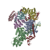 8ebyMC 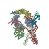 8ebsC 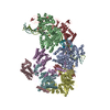 8ebtC 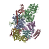 8ebuC 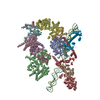 8ebvC 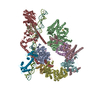 8ebwC 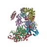 8ebxC C: citing same article ( M: atomic model generated by this map |
|---|---|
| Similar structure data |
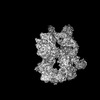
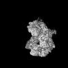
-Links
| EMDB pages | EMDB (EBI/PDBe) / EMDataResource |
|---|---|
| Related items in Molecule of the Month |








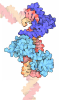
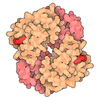



-Map
| File | Download / File: emd_28002.map.gz / Format: CCP4 / Size: 103 MB / Type: IMAGE STORED AS FLOATING POINT NUMBER (4 BYTES) | ||||||||||||||||||||||||||||||||||||
|---|---|---|---|---|---|---|---|---|---|---|---|---|---|---|---|---|---|---|---|---|---|---|---|---|---|---|---|---|---|---|---|---|---|---|---|---|---|
| Projections & slices | Image control
Images are generated by Spider. | ||||||||||||||||||||||||||||||||||||
| Voxel size | X=Y=Z: 1.245 Å | ||||||||||||||||||||||||||||||||||||
| Density |
| ||||||||||||||||||||||||||||||||||||
| Symmetry | Space group: 1 | ||||||||||||||||||||||||||||||||||||
| Details | EMDB XML:
|
 Z (Sec.)
 Y (Row.)
 X (Col.)


-Supplemental data
-Half map: #1
| File | emd_28002_half_map_1.map | ||||||||||||
|---|---|---|---|---|---|---|---|---|---|---|---|---|---|
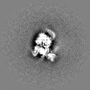
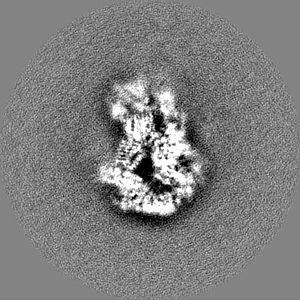
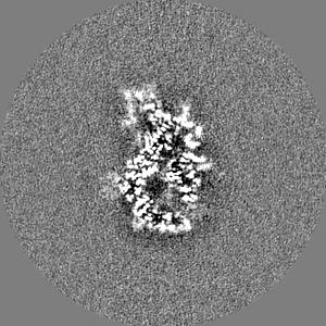
| Projections & Slices |
| ||||||||||||
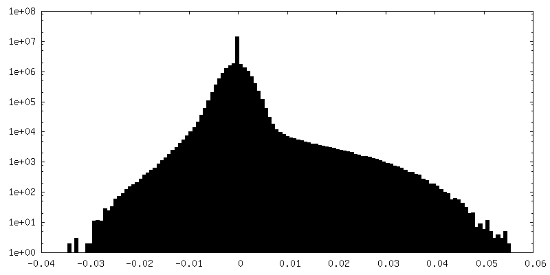
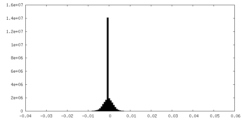
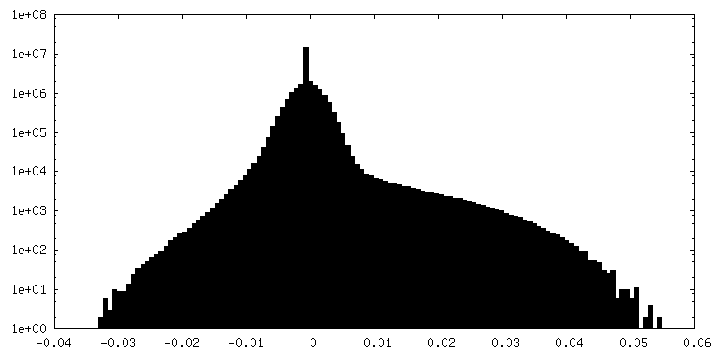
| Density Histograms |

-Half map: #2
| File | emd_28002_half_map_2.map | ||||||||||||
|---|---|---|---|---|---|---|---|---|---|---|---|---|---|
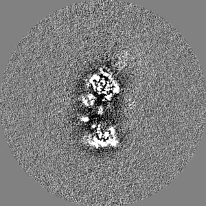
| Projections & Slices |
| ||||||||||||
| Density Histograms |


- Sample components
+Entire : small DNA lesion recognition complex4
+Supramolecule #1: small DNA lesion recognition complex4
+Macromolecule #1: TFIIH basal transcription factor complex helicase XPB subunit

 Spodoptera frugiperda (fall armyworm)
+Macromolecule #2: General transcription and DNA repair factor IIH helicase subunit XPD
+Macromolecule #3: General transcription factor IIH subunit 1
+Macromolecule #4: General transcription factor IIH subunit 4, p52
+Macromolecule #5: General transcription factor IIH subunit 2
+Macromolecule #6: General transcription factor IIH subunit 3
+Macromolecule #7: General transcription factor IIH subunit 5
+Macromolecule #8: DNA repair protein complementing XP-C cells
+Macromolecule #9: DNA repair protein complementing XP-A cells

 Escherichia coli (E. coli)
+Macromolecule #10: DNA
+Macromolecule #11: DNA
+Macromolecule #12: IRON/SULFUR CLUSTER
+Macromolecule #13: ZINC ION
-Experimental details
-Structure determination
| Method | cryo EM |
|---|---|
 Processing Processing | single particle reconstruction |
| Aggregation state | particle |
-Sample preparation
| Concentration | 0.4 mg/mL | ||||||||||||||||||
|---|---|---|---|---|---|---|---|---|---|---|---|---|---|---|---|---|---|---|---|
| Buffer | pH: 7.9 Component:
| ||||||||||||||||||
| Grid | Model: Quantifoil R1.2/1.3 / Material: COPPER / Support film - Material: CARBON / Support film - topology: HOLEY / Pretreatment - Type: GLOW DISCHARGE / Pretreatment - Time: 30 sec. | ||||||||||||||||||
| Vitrification | Cryogen name: ETHANE / Chamber humidity: 100 % / Chamber temperature: 277.15 K / Instrument: FEI VITROBOT MARK IV |
- Electron microscopy
| Microscope | FEI TITAN KRIOS |
|---|---|
| Image recording | Film or detector model: GATAN K3 (6k x 4k) / Number grids imaged: 1 / Number real images: 3883 / Average exposure time: 2.5 sec. / Average electron dose: 54.1 e/Å2 |
| Electron beam | Acceleration voltage: 300 kV / Electron source:  FIELD EMISSION GUN FIELD EMISSION GUN |
| Electron optics | C2 aperture diameter: 70.0 µm / Illumination mode: FLOOD BEAM / Imaging mode: BRIGHT FIELD / Cs: 2.7 mm / Nominal defocus max: 3.0 µm / Nominal defocus min: 2.0 µm / Nominal magnification: 105000 |
| Sample stage | Specimen holder model: FEI TITAN KRIOS AUTOGRID HOLDER / Cooling holder cryogen: NITROGEN |
| Experimental equipment |  Model: Titan Krios / Image courtesy: FEI Company |