 Movie
Movie Controller

[English] 日本語
 Yorodumi
- EMDB-2519: Stalled mammalian ribosome bound to canine pancreatic microsomes -
+ Open data

- Basic information
| Entry | Database: EMDB / ID: EMD-2519 | |||||||||
|---|---|---|---|---|---|---|---|---|---|---|
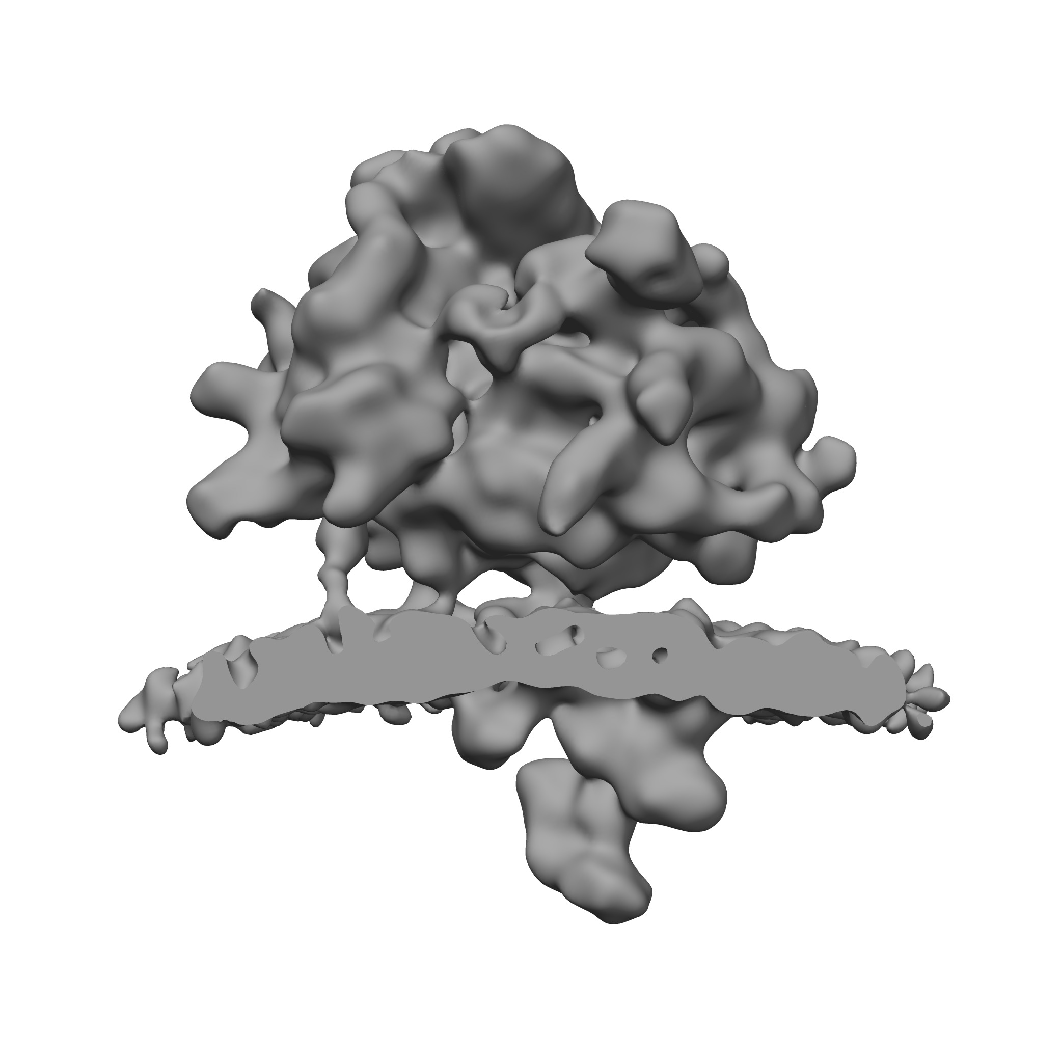
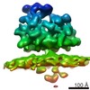
| Title | Stalled mammalian ribosome bound to canine pancreatic microsomes | |||||||||
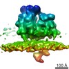
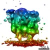
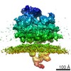
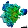
 Map data Map data | Ribosome-stripped canine pancreatic microsomes were populated with rabbit ribosomes, stalled on the CMV regulatory peptide. | |||||||||
 Sample Sample |
| |||||||||
 Keywords Keywords | endoplasmic reticulum / translocon / ribosome / oligosaccharyltransferase / OST / protein-conducting channel / Sec61 / translocon-associated protein complex / TRAP | |||||||||
| Biological species |  | |||||||||
| Method | subtomogram averaging / cryo EM / Resolution: 22.0 Å | |||||||||
 Authors Authors | Pfeffer S / Dudek J / Gogala M / Schorr S / Linxweiler J / Lang S / Becker T / Beckmann R / Zimmermann R / Foerster F | |||||||||
 Citation Citation | Journal: Nat Commun / Year: 2014 Title: Structure of the mammalian oligosaccharyl-transferase complex in the native ER protein translocon. Authors: Stefan Pfeffer / Johanna Dudek / Marko Gogala / Stefan Schorr / Johannes Linxweiler / Sven Lang / Thomas Becker / Roland Beckmann / Richard Zimmermann / Friedrich Förster /  Abstract: In mammalian cells, proteins are typically translocated across the endoplasmic reticulum (ER) membrane in a co-translational mode by the ER protein translocon, comprising the protein-conducting ...In mammalian cells, proteins are typically translocated across the endoplasmic reticulum (ER) membrane in a co-translational mode by the ER protein translocon, comprising the protein-conducting channel Sec61 and additional complexes involved in nascent chain processing and translocation. As an integral component of the translocon, the oligosaccharyl-transferase complex (OST) catalyses co-translational N-glycosylation, one of the most common protein modifications in eukaryotic cells. Here we use cryoelectron tomography, cryoelectron microscopy single-particle analysis and small interfering RNA-mediated gene silencing to determine the overall structure, oligomeric state and position of OST in the native ER protein translocon of mammalian cells in unprecedented detail. The observed positioning of OST in close proximity to Sec61 provides a basis for understanding how protein translocation into the ER and glycosylation of nascent proteins are structurally coupled. The overall spatial organization of the native translocon, as determined here, serves as a reliable framework for further hypothesis-driven studies. | |||||||||
| History |
|
- Structure visualization
| Movie |
 Movie viewer Movie viewer |
|---|---|
| Structure viewer | EM map: SurfViewMolmilJmol/JSmol |
| Supplemental images |
- Downloads & links
-EMDB archive
| Map data | emd_2519.map.gz | 28.3 MB | EMDB map data format | |
|---|---|---|---|---|
| Header (meta data) | emd-2519-v30.xmlemd-2519.xml | 9.7 KB 9.7 KB | Display Display | EMDB header |
| Images | emd_2519.tif | 919 KB | ||
| Archive directory |  http://ftp.pdbj.org/pub/emdb/structures/EMD-2519ftp://ftp.pdbj.org/pub/emdb/structures/EMD-2519 http://ftp.pdbj.org/pub/emdb/structures/EMD-2519ftp://ftp.pdbj.org/pub/emdb/structures/EMD-2519 | HTTPS FTP |
-Validation report
| Summary document | emd_2519_validation.pdf.gz | 242.4 KB | Display | EMDB validaton report |
|---|---|---|---|---|
| Full document | emd_2519_full_validation.pdf.gz | 241.5 KB | Display | |
| Data in XML | emd_2519_validation.xml.gz | 6 KB | Display | |
| Arichive directory | https://ftp.pdbj.org/pub/emdb/validation_reports/EMD-2519ftp://ftp.pdbj.org/pub/emdb/validation_reports/EMD-2519 | HTTPS FTP |
-Related structure data
| Related structure data | 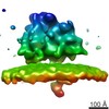 2514C 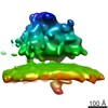 2515C 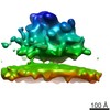 2516C 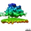 2517C 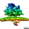 2518C 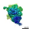 2523C C: citing same article ( |
|---|---|
| Similar structure data |

-Links
| EMDB pages | EMDB (EBI/PDBe) / EMDataResource |
|---|---|
| Related items in Molecule of the Month |

-Map
| File | Download / File: emd_2519.map.gz / Format: CCP4 / Size: 29.8 MB / Type: IMAGE STORED AS FLOATING POINT NUMBER (4 BYTES) | ||||||||||||||||||||||||||||||||||||||||||||||||||||||||||||||||||||
|---|---|---|---|---|---|---|---|---|---|---|---|---|---|---|---|---|---|---|---|---|---|---|---|---|---|---|---|---|---|---|---|---|---|---|---|---|---|---|---|---|---|---|---|---|---|---|---|---|---|---|---|---|---|---|---|---|---|---|---|---|---|---|---|---|---|---|---|---|---|
| Annotation | Ribosome-stripped canine pancreatic microsomes were populated with rabbit ribosomes, stalled on the CMV regulatory peptide. | ||||||||||||||||||||||||||||||||||||||||||||||||||||||||||||||||||||
| Projections & slices | Image control
Images are generated by Spider. | ||||||||||||||||||||||||||||||||||||||||||||||||||||||||||||||||||||
| Voxel size | X=Y=Z: 2.88 Å | ||||||||||||||||||||||||||||||||||||||||||||||||||||||||||||||||||||
| Density |
| ||||||||||||||||||||||||||||||||||||||||||||||||||||||||||||||||||||
| Symmetry | Space group: 1 | ||||||||||||||||||||||||||||||||||||||||||||||||||||||||||||||||||||
| Details | EMDB XML:
CCP4 map header:
| ||||||||||||||||||||||||||||||||||||||||||||||||||||||||||||||||||||
 Z (Sec.)
 Y (Row.)
 X (Col.)


-Supplemental data
- Sample components
-Entire : Ribosome-stripped canine pancreatic microsomes were populated wit...
| Entire | Name: Ribosome-stripped canine pancreatic microsomes were populated with rabbit ribosomes, stalled on the CMV regulatory peptide. |
|---|---|
| Components |
|
-Supramolecule #1000: Ribosome-stripped canine pancreatic microsomes were populated wit...
| Supramolecule | Name: Ribosome-stripped canine pancreatic microsomes were populated with rabbit ribosomes, stalled on the CMV regulatory peptide. type: sample / ID: 1000 / Number unique components: 2 |
|---|
-Supramolecule #1: ER membrane-associated 80S ribosome
| Supramolecule | Name: ER membrane-associated 80S ribosome / type: complex / ID: 1 / Recombinant expression: No / Ribosome-details: ribosome-eukaryote: ALL |
|---|---|
| Source (natural) | Organism: |
-Macromolecule #1: ER protein translocon
| Macromolecule | Name: ER protein translocon / type: protein_or_peptide / ID: 1 / Recombinant expression: No |
|---|---|
| Source (natural) | Organism: |
-Experimental details
-Structure determination
| Method | cryo EM |
|---|---|
 Processing Processing | subtomogram averaging |
| Aggregation state | particle |
-Sample preparation
| Concentration | 2 mg/mL |
|---|---|
| Grid | Details: Lacey carbon molybdenum grid |
| Vitrification | Cryogen name: ETHANE-PROPANE MIXTURE / Chamber humidity: 70 % / Instrument: FEI VITROBOT MARK IV / Method: Blot 3 seconds before plunging. |
- Electron microscopy
| Microscope | FEI TITAN KRIOS |
|---|---|
| Date | Aug 15, 2012 |
| Image recording | Category: CCD / Film or detector model: FEI FALCON I (4k x 4k) / Average electron dose: 60 e/Å2 |
| Electron beam | Acceleration voltage: 300 kV / Electron source:  FIELD EMISSION GUN FIELD EMISSION GUN |
| Electron optics | Illumination mode: FLOOD BEAM / Imaging mode: BRIGHT FIELD / Nominal defocus max: 5.0 µm / Nominal defocus min: 3.0 µm / Nominal magnification: 29000 |
| Sample stage | Specimen holder model: FEI TITAN KRIOS AUTOGRID HOLDER / Tilt series - Axis1 - Min angle: -60 ° / Tilt series - Axis1 - Max angle: 60 ° |
| Experimental equipment |  Model: Titan Krios / Image courtesy: FEI Company |
-Image processing
| Details | Candidate particles were localized using template matching and classified using constrained principal component analysis focusing on the large ribosomal subunit and the ER membrane. At the selected coordinates, unbinned subtomograms were reconstructed individually from the weighted back-projections and aligned to a template. |
|---|---|
| Final reconstruction | Applied symmetry - Point group: C1 (asymmetric) / Resolution.type: BY AUTHOR / Resolution: 22.0 Å / Resolution method: OTHER / Software - Name: av3, tom, PyTom / Number subtomograms used: 2896 |
| CTF correction | Details: micrograph |
