Movie
Movie Controller

[English] 日本語
 Yorodumi
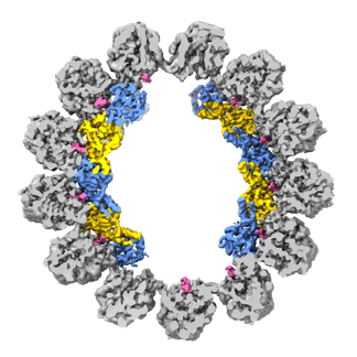
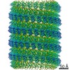
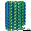
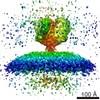
- EMDB-23870: Cryo-EM structure of cortical microtubule from Toxoplasma gondii ... -
+ Open data

- Basic information
| Entry | Database: EMDB / ID: EMD-23870 | |||||||||
|---|---|---|---|---|---|---|---|---|---|---|
| Title | Cryo-EM structure of cortical microtubule from Toxoplasma gondii (without TrxL2) | |||||||||
 Map data Map data | Composite map of T. gondii cortical microtubule (without TrxL2) | |||||||||
 Sample Sample |
| |||||||||
| Function / homology |  Function and homology information Function and homology informationthioredoxin-disulfide reductase (NADPH) activity / negative regulation of Wnt signaling pathway / microtubule-based process / negative regulation of protein ubiquitination / structural constituent of cytoskeleton / microtubule binding / microtubule / Hydrolases; Acting on acid anhydrides; Acting on GTP to facilitate cellular and subcellular movement / cytoskeleton / hydrolase activity ...thioredoxin-disulfide reductase (NADPH) activity / negative regulation of Wnt signaling pathway / microtubule-based process / negative regulation of protein ubiquitination / structural constituent of cytoskeleton / microtubule binding / microtubule / Hydrolases; Acting on acid anhydrides; Acting on GTP to facilitate cellular and subcellular movement / cytoskeleton / hydrolase activity / GTPase activity / GTP binding / metal ion binding / nucleus Similarity search - Function | |||||||||
| Biological species |  | |||||||||
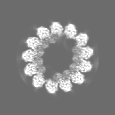
| Method | single particle reconstruction / cryo EM / Resolution: 3.4 Å | |||||||||
 Authors Authors | Wang X / Brown A / Sibley LD / Zhang R | |||||||||
| Funding support |  United States, 1 items United States, 1 items
| |||||||||
 Citation Citation | Journal: Nat Commun / Year: 2021 Title: Cryo-EM structure of cortical microtubules from human parasite Toxoplasma gondii identifies their microtubule inner proteins. Authors: Xiangli Wang / Yong Fu / Wandy L Beatty / Meisheng Ma / Alan Brown / L David Sibley / Rui Zhang / Abstract: In living cells, microtubules (MTs) play pleiotropic roles, which require very different mechanical properties. Unlike the dynamic MTs found in the cytoplasm of metazoan cells, the specialized ...In living cells, microtubules (MTs) play pleiotropic roles, which require very different mechanical properties. Unlike the dynamic MTs found in the cytoplasm of metazoan cells, the specialized cortical MTs from Toxoplasma gondii, a prevalent human pathogen, are extraordinarily stable and resistant to detergent and cold treatments. Using single-particle cryo-EM, we determine their ex vivo structure and identify three proteins (TrxL1, TrxL2 and SPM1) as bona fide microtubule inner proteins (MIPs). These three MIPs form a mesh on the luminal surface and simultaneously stabilize the tubulin lattice in both longitudinal and lateral directions. Consistent with previous observations, deletion of the identified MIPs compromises MT stability and integrity under challenges by chemical treatments. We also visualize a small molecule like density at the Taxol-binding site of β-tubulin. Our results provide the structural basis to understand the stability of cortical MTs and suggest an evolutionarily conserved mechanism of MT stabilization from the inside. | |||||||||
| History |
|
- Structure visualization
| Movie |
Movie viewer |
|---|---|
| Structure viewer | EM map: SurfViewMolmilJmol/JSmol |
| Supplemental images |
- Downloads & links
-EMDB archive
| Map data | emd_23870.map.gz | 64.4 MB | EMDB map data format | |
|---|---|---|---|---|
| Header (meta data) | emd-23870-v30.xmlemd-23870.xml | 22.6 KB 22.6 KB | Display Display | EMDB header |
| Images |  emd_23870.png emd_23870.png | 97.2 KB | ||
| Others | emd_23870_additional_1.map.gzemd_23870_additional_2.map.gz | 50.2 MB 229.3 MB | ||
| Archive directory |  http://ftp.pdbj.org/pub/emdb/structures/EMD-23870ftp://ftp.pdbj.org/pub/emdb/structures/EMD-23870 http://ftp.pdbj.org/pub/emdb/structures/EMD-23870ftp://ftp.pdbj.org/pub/emdb/structures/EMD-23870 | HTTPS FTP |
-Related structure data


-Links
| EMDB pages | EMDB (EBI/PDBe) / EMDataResource |
|---|---|
| Related items in Molecule of the Month |


-Map
| File | Download / File: emd_23870.map.gz / Format: CCP4 / Size: 244.1 MB / Type: IMAGE STORED AS FLOATING POINT NUMBER (4 BYTES) | ||||||||||||||||||||||||||||||||||||||||||||||||||||||||||||||||||||
|---|---|---|---|---|---|---|---|---|---|---|---|---|---|---|---|---|---|---|---|---|---|---|---|---|---|---|---|---|---|---|---|---|---|---|---|---|---|---|---|---|---|---|---|---|---|---|---|---|---|---|---|---|---|---|---|---|---|---|---|---|---|---|---|---|---|---|---|---|---|
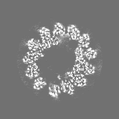
| Annotation | Composite map of T. gondii cortical microtubule (without TrxL2) | ||||||||||||||||||||||||||||||||||||||||||||||||||||||||||||||||||||
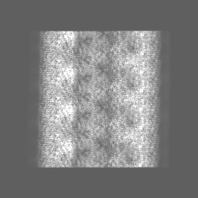
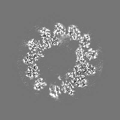
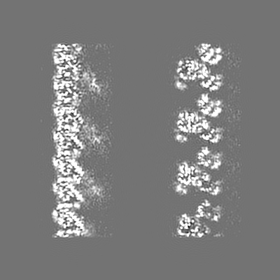
| Projections & slices | Image control
Images are generated by Spider. | ||||||||||||||||||||||||||||||||||||||||||||||||||||||||||||||||||||
| Voxel size | X=Y=Z: 1.096 Å | ||||||||||||||||||||||||||||||||||||||||||||||||||||||||||||||||||||
| Density |
| ||||||||||||||||||||||||||||||||||||||||||||||||||||||||||||||||||||
| Symmetry | Space group: 1 | ||||||||||||||||||||||||||||||||||||||||||||||||||||||||||||||||||||
| Details | EMDB XML:
CCP4 map header:
| ||||||||||||||||||||||||||||||||||||||||||||||||||||||||||||||||||||
 Z (Sec.)
 Y (Row.)
 X (Col.)


-Supplemental data
-Additional map: Unsharpened composite map of T. gondii cortical microtubule...
| File | emd_23870_additional_1.map | ||||||||||||
|---|---|---|---|---|---|---|---|---|---|---|---|---|---|
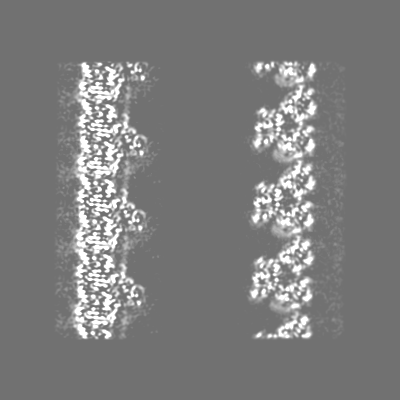
| Annotation | Unsharpened composite map of T. gondii cortical microtubule (without TrxL2) | ||||||||||||
| Projections & Slices |
| ||||||||||||
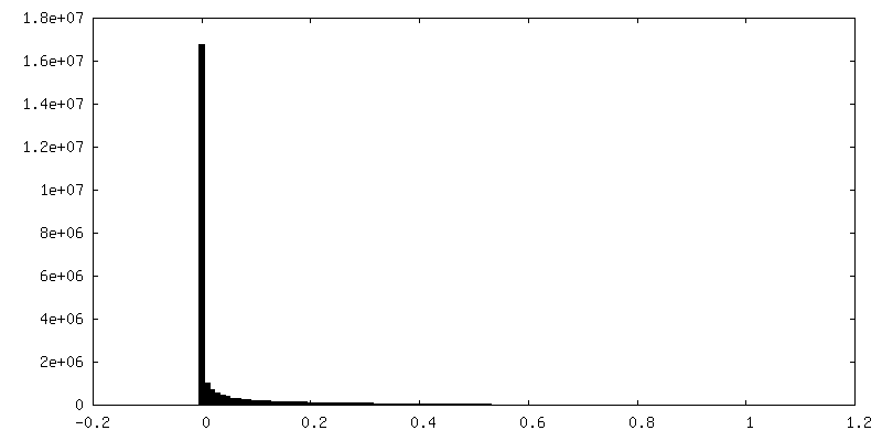
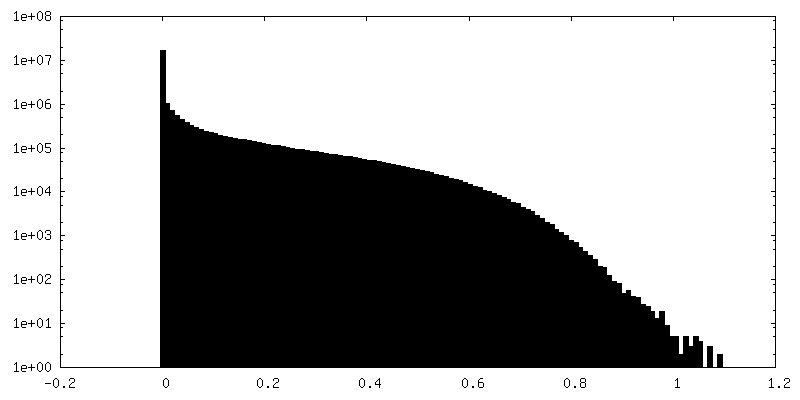
| Density Histograms |


-Additional map: B-factor sharpened composite map of T. gondii cortical...
| File | emd_23870_additional_2.map | ||||||||||||
|---|---|---|---|---|---|---|---|---|---|---|---|---|---|
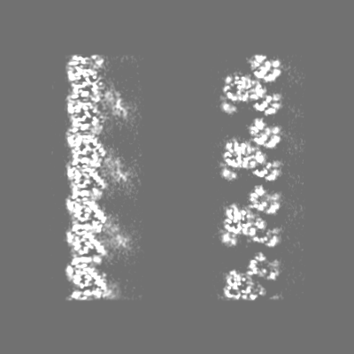
| Annotation | B-factor sharpened composite map of T. gondii cortical microtubule (without TrxL2) | ||||||||||||
| Projections & Slices |
| ||||||||||||
| Density Histograms |


- Sample components
-Entire : cortical microtubule with associated MIPs
| Entire | Name: cortical microtubule with associated MIPs |
|---|---|
| Components |
|
-Supramolecule #1: cortical microtubule with associated MIPs
| Supramolecule | Name: cortical microtubule with associated MIPs / type: complex / ID: 1 / Parent: 0 / Macromolecule list: all |
|---|---|
| Source (natural) | Organism: |
-Macromolecule #1: SPM1
| Macromolecule | Name: SPM1 / type: protein_or_peptide / ID: 1 / Enantiomer: LEVO |
|---|---|
| Source (natural) | Organism: |
| Sequence | String: MSGGNSNTPK KLPSEEGSDY GYPQKPQKYL PKSEQAEPDY SACCKGNDAY KGASHGTVQF SHPEEAQKYA GAAAGAETIQ RGRERVAAD RQPRAAGDVP ARRLHLSDVD EAHRGQSPSR HPGYCVEELC TCGMHKCIPS RAPVPFTGST QYRQEFVPKP L PPPTQVSQ ...String: MSGGNSNTPK KLPSEEGSDY GYPQKPQKYL PKSEQAEPDY SACCKGNDAY KGASHGTVQF SHPEEAQKYA GAAAGAETIQ RGRERVAAD RQPRAAGDVP ARRLHLSDVD EAHRGQSPSR HPGYCVEELC TCGMHKCIPS RAPVPFTGST QYRQEFVPKP L PPPTQVSQ VTLPPSLPFE AESSYRTEFV AKPLPPPAKF SEVKLPPTLP FHGESAYRTD YVPKPLPEVA KPVEVKLPPT LP FNAQSCY RSEYVAKPLP PPVQTVEVKL PPSLPFEGST HYRDEFQVKP LPPATKVTEV KLPPSLPFDA TSMYRSDYVA KSN PICPVS KLPQYPAATY PQNHVFWDPD TKQWY |
-Macromolecule #2: alpha-tubulin
| Macromolecule | Name: alpha-tubulin / type: protein_or_peptide / ID: 2 / Enantiomer: LEVO |
|---|---|
| Source (natural) | Organism: |
| Sequence | String: MREVISIHVG QAGIQIGNAC WELFCLEHGI QPDGQMPSDK TIGGGDDAFN TFFSETGAGK HVPRCVFLDL EPTVVDEVRT GTYRHLFHP EQLISGKEDA ANNFARGHYT IGKEIVDLSL DRIRKLADNC TGLQGFLMFN AVGGGTGSGL GCLLLERLSV D YGKKSKLN ...String: MREVISIHVG QAGIQIGNAC WELFCLEHGI QPDGQMPSDK TIGGGDDAFN TFFSETGAGK HVPRCVFLDL EPTVVDEVRT GTYRHLFHP EQLISGKEDA ANNFARGHYT IGKEIVDLSL DRIRKLADNC TGLQGFLMFN AVGGGTGSGL GCLLLERLSV D YGKKSKLN FCSWPSPQVS TAVVEPYNSV LSTHSLLEHT DVAVMLDNEA IYDICRRNLD IERPTYTNLN RLIAQVISSL TA SLRFDGA LNVDVTEFQT NLVPYPRIHF MLSSYAPIIS AEKAYHEQLS VAEITNSAFE PASMMAKCDP RHGKYMACCL MYR GDVVPK DVNAAVATIK TKRTIQFVDW CPTGFKCGIN YQPPTVVPGG DLAKVMRAVC MISNSTAIAE VFSRMDHKFD LMYA KRAFV HWYVGEGMEE GEFSEAREDL AALEKDYEEV GIETAEGEGE EEGYGDEY |
-Macromolecule #3: beta-tubulin
| Macromolecule | Name: beta-tubulin / type: protein_or_peptide / ID: 3 / Enantiomer: LEVO |
|---|---|
| Source (natural) | Organism: |
| Sequence | String: MREIVHVQGG QCGNQIGAKF WEVISDEHGI DPTGTYCGDS DLQLERINVF YNEATGGRFV PRAILMDLEP GTMDSVRAGP FGQLFRPDN FVFGQTGAGN NWAKGHYTEG AELIDSVLDV VRKEAEGCDC LQGFQITHSL GGGTGSGMGT LLISKVREEY P DRIMETFS ...String: MREIVHVQGG QCGNQIGAKF WEVISDEHGI DPTGTYCGDS DLQLERINVF YNEATGGRFV PRAILMDLEP GTMDSVRAGP FGQLFRPDN FVFGQTGAGN NWAKGHYTEG AELIDSVLDV VRKEAEGCDC LQGFQITHSL GGGTGSGMGT LLISKVREEY P DRIMETFS VFPSPKVSDT VVEPYNATLS VHQLVENADE VQVIDNEALY DICFRTLKLT TPTYGDLNHL VSAAMSGVTC CL RFPGQLN SDLRKLAVNL IPFPRLHFFL IGFAPLTSRG SQQYRALSVP ELTQQMFDAK NMMCASDPRH GRYLTASAMF RGR MSTKEV DEQMLNVQNK NSSYFVEWIP NNMKSSVCDI PPKGLKMSVT FVGNSTAIQE MFKRVSDQFT AMFRRKAFLH WYTG EGMDE MEFTEAESNM NDLVSEYQQY QDATAEEEGE FDEEEGEMGA EEGA |
-Macromolecule #4: TrxL1
| Macromolecule | Name: TrxL1 / type: protein_or_peptide / ID: 4 / Enantiomer: LEVO |
|---|---|
| Source (natural) | Organism: |
| Sequence | String: MSQPVFASPL NVEKRRLNEE RALMQAQKAG GEGVNIQLPP NYGDMDLILF PEGSLKNSNN TVIPQSHLKG KSVALYFADG ADPKCASLL PFLLNYYRTM NEGGANQKIE IIFVSLDRDR EAFESHRAHM PWLSIDLENP LTEILKRHFR VMKEYEVPTY G YGSRTGVP ...String: MSQPVFASPL NVEKRRLNEE RALMQAQKAG GEGVNIQLPP NYGDMDLILF PEGSLKNSNN TVIPQSHLKG KSVALYFADG ADPKCASLL PFLLNYYRTM NEGGANQKIE IIFVSLDRDR EAFESHRAHM PWLSIDLENP LTEILKRHFR VMKEYEVPTY G YGSRTGVP SVIVIGSDGR EAQFLPICSG LEEGDRALLR WDWRNTKFAS DQFHVRPTLL EQ |
-Experimental details
-Structure determination
| Method | cryo EM |
|---|---|
 Processing Processing | single particle reconstruction |
| Aggregation state | filament |
-Sample preparation
| Buffer | pH: 7.4 |
|---|---|
| Grid | Model: Quantifoil R2/2 / Material: COPPER / Mesh: 300 / Pretreatment - Type: GLOW DISCHARGE / Pretreatment - Atmosphere: AIR |
| Vitrification | Cryogen name: ETHANE / Chamber humidity: 95 % / Chamber temperature: 289 K / Instrument: FEI VITROBOT MARK IV / Details: blot for 4 seconds with a blot force of -15. |
- Electron microscopy
| Microscope | FEI TITAN KRIOS |
|---|---|
| Image recording | Film or detector model: GATAN K3 BIOQUANTUM (6k x 4k) / Digitization - Dimensions - Width: 3838 pixel / Digitization - Dimensions - Height: 3710 pixel / Number grids imaged: 2 / Number real images: 9231 / Average exposure time: 9.0 sec. / Average electron dose: 63.7 e/Å2 Details: Images were collected in counting mode, with an exposure rate of 8.5 electrons/pixel/s on the detector camera. |
| Electron beam | Acceleration voltage: 300 kV / Electron source:  FIELD EMISSION GUN FIELD EMISSION GUN |
| Electron optics | Calibrated defocus min: 1.0 µm / Illumination mode: FLOOD BEAM / Imaging mode: BRIGHT FIELD / Cs: 0.01 mm / Nominal defocus max: 3.5 µm / Nominal magnification: 105000 |
| Sample stage | Specimen holder model: FEI TITAN KRIOS AUTOGRID HOLDER / Cooling holder cryogen: NITROGEN |
| Experimental equipment |  Model: Titan Krios / Image courtesy: FEI Company |