Movie
Movie Controller


+ Open data

- Basic information
| Entry | Database: EMDB / ID: EMD-0522 | |||||||||
|---|---|---|---|---|---|---|---|---|---|---|
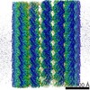
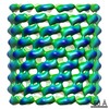
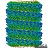
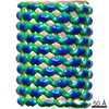
| Title | Ebola virus nucleoprotein - RNA complex | |||||||||
 Map data Map data | Ebola virus nucleoprotein - RNA complex, full map | |||||||||
 Sample Sample |
| |||||||||
 Keywords Keywords | RNA-binding / nucleoprotein / nucleocapsid / capsid / VIRAL PROTEIN-RNA complex | |||||||||
| Function / homology | Ebola nucleoprotein / Ebola nucleoprotein / viral RNA genome packaging / helical viral capsid / viral nucleocapsid / host cell cytoplasm / ribonucleoprotein complex / RNA binding / Nucleoprotein Function and homology information Function and homology information | |||||||||
| Biological species |   Ebola virus - Mayinga, Zaire, 1976 / Zaire ebolavirus (strain Mayinga-76) / Ebola virus - Mayinga, Zaire, 1976 / Zaire ebolavirus (strain Mayinga-76) /  Homo sapiens (human) Homo sapiens (human) | |||||||||
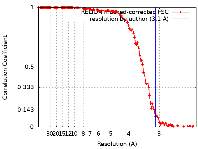
| Method | helical reconstruction / cryo EM / Resolution: 3.1 Å | |||||||||
 Authors Authors | Kirchdoerfer RN / Ward AB | |||||||||
| Funding support |  United States, 2 items United States, 2 items
| |||||||||
 Citation Citation | Journal: Acta Crystallogr F Struct Biol Commun / Year: 2019 Title: Cryo-EM structure of the Ebola virus nucleoprotein-RNA complex. Authors: Robert N Kirchdoerfer / Erica Ollmann Saphire / Andrew B Ward / Abstract: Ebola virus is an emerging virus that is capable of causing a deadly disease in humans. Replication, transcription and packaging of the viral genome are carried out by the viral nucleocapsid. The ...Ebola virus is an emerging virus that is capable of causing a deadly disease in humans. Replication, transcription and packaging of the viral genome are carried out by the viral nucleocapsid. The nucleocapsid is a complex of the viral nucleoprotein, RNA and several other viral proteins. The nucleoprotein forms large, RNA-bound, helical filaments and acts as a scaffold for additional viral proteins. The 3.1 Å resolution single-particle cryo-electron microscopy structure of the nucleoprotein-RNA helical filament presented here resembles previous structures determined at lower resolution, while providing improved molecular details of protein-protein and protein-RNA interactions. The higher resolution of the structure presented here will facilitate the design and characterization of novel and specific Ebola virus therapeutics targeting the nucleocapsid. | |||||||||
| History |
|
- Structure visualization
| Movie |
Movie viewer |
|---|---|
| Structure viewer | EM map: SurfViewMolmilJmol/JSmol |
| Supplemental images |

- Downloads & links
-EMDB archive
| Map data | emd_0522.map.gz | 163.9 MB | EMDB map data format | |
|---|---|---|---|---|
| Header (meta data) | emd-0522-v30.xmlemd-0522.xml | 19.9 KB 19.9 KB | Display Display | EMDB header |
| FSC (resolution estimation) | emd_0522_fsc.xml | 12.8 KB | Display | FSC data file |
| Images | 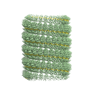 emd_0522.png emd_0522.png | 159.4 KB | ||
| Masks | emd_0522_msk_1.map | 178 MB | Mask map | |
| Filedesc metadata | emd-0522.cif.gz | 6.8 KB | ||
| Others | emd_0522_half_map_1.map.gzemd_0522_half_map_2.map.gz | 140.4 MB 140.4 MB | ||
| Archive directory |  http://ftp.pdbj.org/pub/emdb/structures/EMD-0522ftp://ftp.pdbj.org/pub/emdb/structures/EMD-0522 http://ftp.pdbj.org/pub/emdb/structures/EMD-0522ftp://ftp.pdbj.org/pub/emdb/structures/EMD-0522 | HTTPS FTP |
-Related structure data
| Related structure data |  6nutMC M: atomic model generated by this map C: citing same article ( |
|---|---|
| Similar structure data |

-Links
| EMDB pages | EMDB (EBI/PDBe) / EMDataResource |
|---|---|
| Related items in Molecule of the Month |

-Map
| File | Download / File: emd_0522.map.gz / Format: CCP4 / Size: 178 MB / Type: IMAGE STORED AS FLOATING POINT NUMBER (4 BYTES) | ||||||||||||||||||||||||||||||||||||||||||||||||||||||||||||
|---|---|---|---|---|---|---|---|---|---|---|---|---|---|---|---|---|---|---|---|---|---|---|---|---|---|---|---|---|---|---|---|---|---|---|---|---|---|---|---|---|---|---|---|---|---|---|---|---|---|---|---|---|---|---|---|---|---|---|---|---|---|
| Annotation | Ebola virus nucleoprotein - RNA complex, full map | ||||||||||||||||||||||||||||||||||||||||||||||||||||||||||||
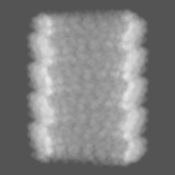
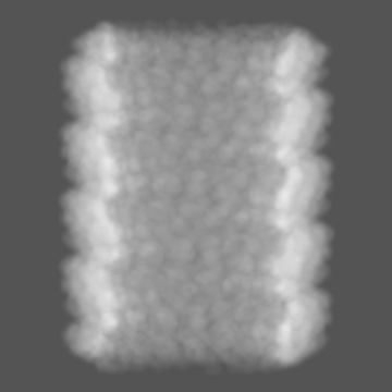
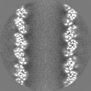
| Projections & slices | Image control
Images are generated by Spider. | ||||||||||||||||||||||||||||||||||||||||||||||||||||||||||||
| Voxel size | X=Y=Z: 1.15 Å | ||||||||||||||||||||||||||||||||||||||||||||||||||||||||||||
| Density |
| ||||||||||||||||||||||||||||||||||||||||||||||||||||||||||||
| Symmetry | Space group: 1 | ||||||||||||||||||||||||||||||||||||||||||||||||||||||||||||
| Details | EMDB XML:
CCP4 map header:
| ||||||||||||||||||||||||||||||||||||||||||||||||||||||||||||
 Z (Sec.)
 Y (Row.)
 X (Col.)


-Supplemental data
-Mask #1
| File | emd_0522_msk_1.map | ||||||||||||
|---|---|---|---|---|---|---|---|---|---|---|---|---|---|
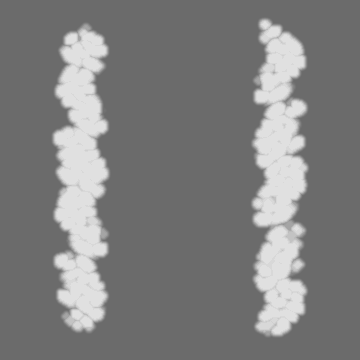
| Projections & Slices |
| ||||||||||||
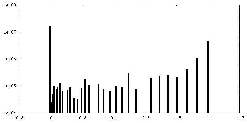
| Density Histograms |


-Half map: Ebola virus nucleoprotein - RNA complex, first half map
| File | emd_0522_half_map_1.map | ||||||||||||
|---|---|---|---|---|---|---|---|---|---|---|---|---|---|
| Annotation | Ebola virus nucleoprotein - RNA complex, first half map | ||||||||||||
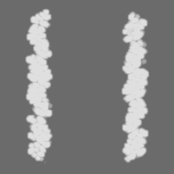
| Projections & Slices |
| ||||||||||||
| Density Histograms |


-Half map: Ebola virus nucleoprotein - RNA complex, second half map
| File | emd_0522_half_map_2.map | ||||||||||||
|---|---|---|---|---|---|---|---|---|---|---|---|---|---|
| Annotation | Ebola virus nucleoprotein - RNA complex, second half map | ||||||||||||
| Projections & Slices |
| ||||||||||||
| Density Histograms |


- Sample components
-Entire : Ebola virus nucleoprotein bound to RNA
| Entire | Name: Ebola virus nucleoprotein bound to RNA |
|---|---|
| Components |
|
-Supramolecule #1: Ebola virus nucleoprotein bound to RNA
| Supramolecule | Name: Ebola virus nucleoprotein bound to RNA / type: complex / ID: 1 / Parent: 0 / Macromolecule list: all |
|---|---|
| Source (natural) | Organism: Ebola virus - Mayinga, Zaire, 1976 |
| Molecular weight | Theoretical: 177 kDa/nm |
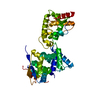
-Macromolecule #1: Nucleoprotein
| Macromolecule | Name: Nucleoprotein / type: protein_or_peptide / ID: 1 / Number of copies: 1 / Enantiomer: LEVO |
|---|---|
| Source (natural) | Organism: Zaire ebolavirus (strain Mayinga-76) / Strain: Mayinga-76 |
| Molecular weight | Theoretical: 50.267098 KDa |
| Recombinant expression | Organism: Homo sapiens (human) |
| Sequence | String: MDSRPQKIWM APSLTESDMD YHKILTAGLS VQQGIVRQRV IPVYQVNNLE EICQLIIQAF EAGVDFQESA DSFLLMLCLH HAYQGDYKL FLESGAVKYL EGHGFRFEVK KRDGVKRLEE LLPAVSSGKN IKRTLAAMPE EETTEANAGQ FLSFASLFLP K LVVGEKAC ...String: MDSRPQKIWM APSLTESDMD YHKILTAGLS VQQGIVRQRV IPVYQVNNLE EICQLIIQAF EAGVDFQESA DSFLLMLCLH HAYQGDYKL FLESGAVKYL EGHGFRFEVK KRDGVKRLEE LLPAVSSGKN IKRTLAAMPE EETTEANAGQ FLSFASLFLP K LVVGEKAC LEKVQRQIQV HAEQGLIQYP TAWQSVGHMM VIFRLMRTNF LIKFLLIHQG MHMVAGHDAN DAVISNSVAQ AR FSGLLIV KTVLDHILQK TERGVRLHPL ARTAKVKNEV NSFKAALSSL AKHGEYAPFA RLLNLSGVNN LEHGLFPQLS AIA LGVATA HGSTLAGVNV GEQYQQLREA ATEAEKQLQQ YAESRELDHL GLDDQEKKIL MNFHQKKNEI SFQQTNAMVT LRKE RLAKL TEAITAASLP KTSGHYDDDD DIPFPGPIND DDNPGHQDDD PTDSQ UniProtKB: Nucleoprotein |
-Macromolecule #2: RNA (5'-R(P*AP*AP*AP*AP*AP*A)-3')
| Macromolecule | Name: RNA (5'-R(P*AP*AP*AP*AP*AP*A)-3') / type: rna / ID: 2 Details: The poly-adenosine sequence was modeled to represent the mixed identity of nucleotide sequences bound to nucleoprotein. Number of copies: 1 |
|---|---|
| Source (natural) | Organism: Homo sapiens (human) |
| Molecular weight | Theoretical: 1.930277 KDa |
| Sequence | String: AAAAAA |
-Experimental details
-Structure determination
| Method | cryo EM |
|---|---|
 Processing Processing | helical reconstruction |
| Aggregation state | filament |
-Sample preparation
| Concentration | 3.8 mg/mL | |||||||||||||||
|---|---|---|---|---|---|---|---|---|---|---|---|---|---|---|---|---|
| Buffer | pH: 7.4 Component:
| |||||||||||||||
| Grid | Model: Quantifoil, UltrAuFoil, R1.2/1.3 / Material: GOLD / Mesh: 300 / Pretreatment - Type: PLASMA CLEANING / Pretreatment - Time: 7 sec. / Pretreatment - Atmosphere: OTHER | |||||||||||||||
| Vitrification | Cryogen name: ETHANE / Chamber humidity: 100 % / Chamber temperature: 277 K / Instrument: FEI VITROBOT MARK IV |
- Electron microscopy
| Microscope | FEI TALOS ARCTICA |
|---|---|
| Image recording | Film or detector model: GATAN K2 SUMMIT (4k x 4k) / Detector mode: COUNTING / Digitization - Dimensions - Width: 3838 pixel / Digitization - Dimensions - Height: 3710 pixel / Digitization - Frames/image: 1-65 / Number grids imaged: 1 / Number real images: 731 / Average exposure time: 13.0 sec. / Average electron dose: 49.7 e/Å2 |
| Electron beam | Acceleration voltage: 200 kV / Electron source:  FIELD EMISSION GUN FIELD EMISSION GUN |
| Electron optics | C2 aperture diameter: 70.0 µm / Calibrated defocus max: 2.0 µm / Calibrated defocus min: 0.6 µm / Calibrated magnification: 47478 / Illumination mode: FLOOD BEAM / Imaging mode: BRIGHT FIELD / Cs: 2.7 mm |
| Sample stage | Specimen holder model: FEI TITAN KRIOS AUTOGRID HOLDER / Cooling holder cryogen: NITROGEN |
| Experimental equipment |  Model: Talos Arctica / Image courtesy: FEI Company |