Movie
Movie Controller


+ Open data

- Basic information
| Entry | Database: PDB / ID: 6nut | |||||||||
|---|---|---|---|---|---|---|---|---|---|---|
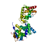
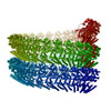
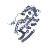
| Title | Ebola virus nucleoprotein - RNA complex | |||||||||
 Components Components |
| |||||||||
 Keywords Keywords | VIRAL PROTEIN/RNA / RNA-binding / nucleoprotein / nucleocapsid / capsid / VIRAL PROTEIN-RNA complex | |||||||||
| Function / homology |  Function and homology information Function and homology informationviral RNA genome packaging / helical viral capsid / viral nucleocapsid / host cell cytoplasm / ribonucleoprotein complex / RNA binding Similarity search - Function | |||||||||
| Biological species |   Zaire ebolavirus Zaire ebolavirus Homo sapiens (human) Homo sapiens (human) | |||||||||
| Method | ELECTRON MICROSCOPY / helical reconstruction / cryo EM / Resolution: 3.1 Å | |||||||||
 Authors Authors | Kirchdoerfer, R.N. / Ward, A.B. | |||||||||
| Funding support |  United States, 2items United States, 2items
| |||||||||
 Citation Citation | Journal: Acta Crystallogr F Struct Biol Commun / Year: 2019 Title: Cryo-EM structure of the Ebola virus nucleoprotein-RNA complex. Authors: Robert N Kirchdoerfer / Erica Ollmann Saphire / Andrew B Ward / Abstract: Ebola virus is an emerging virus that is capable of causing a deadly disease in humans. Replication, transcription and packaging of the viral genome are carried out by the viral nucleocapsid. The ...Ebola virus is an emerging virus that is capable of causing a deadly disease in humans. Replication, transcription and packaging of the viral genome are carried out by the viral nucleocapsid. The nucleocapsid is a complex of the viral nucleoprotein, RNA and several other viral proteins. The nucleoprotein forms large, RNA-bound, helical filaments and acts as a scaffold for additional viral proteins. The 3.1 Å resolution single-particle cryo-electron microscopy structure of the nucleoprotein-RNA helical filament presented here resembles previous structures determined at lower resolution, while providing improved molecular details of protein-protein and protein-RNA interactions. The higher resolution of the structure presented here will facilitate the design and characterization of novel and specific Ebola virus therapeutics targeting the nucleocapsid. | |||||||||
| History |
|
- Structure visualization
| Movie |
Movie viewer |
|---|---|
| Structure viewer | Molecule: MolmilJmol/JSmol |
- Downloads & links
-Download
| PDBx/mmCIF format | 6nut.cif.gz | 92.8 KB | Display | PDBx/mmCIF format |
|---|---|---|---|---|
| PDB format | pdb6nut.ent.gz | 68.1 KB | Display | PDB format |
| PDBx/mmJSON format | 6nut.json.gz | Tree view | PDBx/mmJSON format | |
| Others |  Other downloads Other downloads |
-Validation report
| Arichive directory | https://data.pdbj.org/pub/pdb/validation_reports/nu/6nutftp://data.pdbj.org/pub/pdb/validation_reports/nu/6nut | HTTPS FTP |
|---|
-Related structure data
| Related structure data | 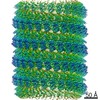 0522MC M: map data used to model this data C: citing same article ( |
|---|---|
| Similar structure data |


-Links
 PDBj
PDBj
- Assembly
| Deposited unit | 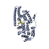
|
|---|---|
| 1 | x 50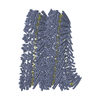
|
| 2 |
|
| Symmetry | Helical symmetry: (Circular symmetry: 1 / Dyad axis: no / N subunits divisor: 1 / Num. of operations: 50 / Rise per n subunits: 2.84 Å / Rotation per n subunits: -14.71 °) |
-Components
| #1: Protein | Mass: 50267.098 Da / Num. of mol.: 1 Source method: isolated from a genetically manipulated source Source: (gene. exp.) Zaire ebolavirus (strain Mayinga-76) / Strain: Mayinga-76 / Gene: NP / Plasmid: pDisplay / Cell line (production host): 293F / Production host: Homo sapiens (human) / References: UniProt: P18272 |
|---|---|
| #2: RNA chain | Mass: 1930.277 Da / Num. of mol.: 1 / Source method: isolated from a natural source Details: The poly-adenosine sequence was modeled to represent the mixed identity of nucleotide sequences bound to nucleoprotein. Source: (natural) Homo sapiens (human) / Cell line: 293F |
-Experimental details
-Experiment
| Experiment | Method: ELECTRON MICROSCOPY |
|---|---|
| EM experiment | Aggregation state: FILAMENT / 3D reconstruction method: helical reconstruction |
- Sample preparation
| Component | Name: Ebola virus nucleoprotein bound to RNA / Type: COMPLEX / Entity ID: all / Source: RECOMBINANT | |||||||||||||||||||||||||
|---|---|---|---|---|---|---|---|---|---|---|---|---|---|---|---|---|---|---|---|---|---|---|---|---|---|---|
| Molecular weight | Value: 177 kDa/nm / Experimental value: NO | |||||||||||||||||||||||||
| Source (natural) | Organism: Ebola virus - Mayinga, Zaire, 1976 | |||||||||||||||||||||||||
| Source (recombinant) | Organism: Homo sapiens (human) / Cell: 293F | |||||||||||||||||||||||||
| Buffer solution | pH: 7.4 | |||||||||||||||||||||||||
| Buffer component |
| |||||||||||||||||||||||||
| Specimen | Conc.: 3.8 mg/ml / Embedding applied: NO / Shadowing applied: NO / Staining applied: NO / Vitrification applied: YES | |||||||||||||||||||||||||
| Specimen support | Grid material: GOLD / Grid mesh size: 300 divisions/in. / Grid type: Quantifoil, UltrAuFoil, R1.2/1.3 | |||||||||||||||||||||||||
| Vitrification | Instrument: FEI VITROBOT MARK IV / Cryogen name: ETHANE / Humidity: 100 % / Chamber temperature: 277 K |
- Electron microscopy imaging
| Experimental equipment |  Model: Talos Arctica / Image courtesy: FEI Company |
|---|---|
| Microscopy | Model: FEI TALOS ARCTICA |
| Electron gun | Electron source:  FIELD EMISSION GUN / Accelerating voltage: 200 kV / Illumination mode: FLOOD BEAM FIELD EMISSION GUN / Accelerating voltage: 200 kV / Illumination mode: FLOOD BEAM |
| Electron lens | Mode: BRIGHT FIELD / Calibrated magnification: 47478 X / Calibrated defocus min: 600 nm / Calibrated defocus max: 2000 nm / Cs: 2.7 mm / C2 aperture diameter: 70 µm |
| Specimen holder | Cryogen: NITROGEN / Specimen holder model: FEI TITAN KRIOS AUTOGRID HOLDER |
| Image recording | Average exposure time: 13 sec. / Electron dose: 49.7 e/Å2 / Detector mode: COUNTING / Film or detector model: GATAN K2 SUMMIT (4k x 4k) / Num. of grids imaged: 1 / Num. of real images: 731 |
| Image scans | Width: 3838 / Height: 3710 / Movie frames/image: 65 / Used frames/image: 1-65 |
- Processing
| EM software |
| ||||||||||||||||||||||||||||||||||||||||
|---|---|---|---|---|---|---|---|---|---|---|---|---|---|---|---|---|---|---|---|---|---|---|---|---|---|---|---|---|---|---|---|---|---|---|---|---|---|---|---|---|---|
| CTF correction | Type: NONE | ||||||||||||||||||||||||||||||||||||||||
| Helical symmerty | Angular rotation/subunit: -14.71 ° / Axial rise/subunit: 2.84 Å / Axial symmetry: C1 | ||||||||||||||||||||||||||||||||||||||||
| Particle selection | Num. of particles selected: 24608 | ||||||||||||||||||||||||||||||||||||||||
| 3D reconstruction | Resolution: 3.1 Å / Resolution method: FSC 0.143 CUT-OFF / Num. of particles: 24609 / Num. of class averages: 1 / Symmetry type: HELICAL | ||||||||||||||||||||||||||||||||||||||||
| Atomic model building | Space: REAL | ||||||||||||||||||||||||||||||||||||||||
| Atomic model building | 3D fitting-ID: 1 / Pdb chain-ID: A / Source name: PDB / Type: experimental model
|