- EMDB-2311: Three-dimensional structure of active, full-length human telomera... -
+
Open data
ID or keywords:
Loading...
-
Basic information
Entry
Database: EMDB / ID: EMD-2311
Title
Three-dimensional structure of active, full-length human telomerase. Independently refined open monomer structure, determined by single-particle electron microscopy in negative stain
Map data
Three-dimensional structure of active, full-length human telomerase. Independently refined open monomer structure, determined by single-particle electron microscopy in negative stain
Sample
Sample: Three-dimensional structure of active, full-length human telomerase dimer, determined by single-particle electron microscopy in negative stain
Protein or peptide: Telomerase reverse transcriptase
Keywords
Telomerase reverse transcriptase / human / telomere length extension
Function / homology
Function and homology information
positive regulation of hair cycle / template-free RNA nucleotidyltransferase / positive regulation of transdifferentiation / TERT-RMRP complex / DNA strand elongation / RNA-directed RNA polymerase complex / positive regulation of protein localization to nucleolus / siRNA transcription / telomerase catalytic core complex / RNA-templated DNA biosynthetic process ...positive regulation of hair cycle / template-free RNA nucleotidyltransferase / positive regulation of transdifferentiation / TERT-RMRP complex / DNA strand elongation / RNA-directed RNA polymerase complex / positive regulation of protein localization to nucleolus / siRNA transcription / telomerase catalytic core complex / RNA-templated DNA biosynthetic process / establishment of protein localization to telomere / Regulation of MITF-M-dependent genes involved in DNA replication, damage repair and senescence / nuclear telomere cap complex / siRNA processing / telomere maintenance via recombination / positive regulation of vascular associated smooth muscle cell migration / telomerase holoenzyme complex / telomerase RNA binding / RNA-templated transcription / DNA biosynthetic process / telomeric DNA binding / positive regulation of stem cell proliferation / mitochondrial nucleoid / negative regulation of cellular senescence / Telomere Extension By Telomerase / replicative senescence / positive regulation of Wnt signaling pathway / negative regulation of extrinsic apoptotic signaling pathway in absence of ligand / positive regulation of G1/S transition of mitotic cell cycle / telomere maintenance via telomerase / RNA-directed DNA polymerase activity / negative regulation of endothelial cell apoptotic process / response to cadmium ion / positive regulation of vascular associated smooth muscle cell proliferation / telomere maintenance / positive regulation of nitric-oxide synthase activity / positive regulation of D-glucose import / mitochondrion organization / Formation of the beta-catenin:TCF transactivating complex / transcription coactivator binding / regulation of protein stability / PML body / positive regulation of miRNA transcription / RNA-directed DNA polymerase / telomerase activity / protein import into nucleus / positive regulation of angiogenesis / positive regulation of protein binding / protein-folding chaperone binding / heart development / cellular response to hypoxia / negative regulation of neuron apoptotic process / tRNA binding / chromosome, telomeric region / nuclear speck / negative regulation of gene expression / RNA-directed RNA polymerase activity / nucleolus / protein homodimerization activity / DNA binding / RNA binding / nucleoplasm / metal ion binding / identical protein binding / nucleus / plasma membrane / cytosol Similarity search - Function
Journal: Nat Struct Mol Biol / Year: 2013 Title: Structure of active dimeric human telomerase. Authors: Anselm Sauerwald / Sara Sandin / Gaël Cristofari / Sjors H W Scheres / Joachim Lingner / Daniela Rhodes / Abstract: Telomerase contains a large RNA subunit, TER, and a protein catalytic subunit, TERT. Whether telomerase functions as a monomer or dimer has been a matter of debate. Here we report biochemical and ...Telomerase contains a large RNA subunit, TER, and a protein catalytic subunit, TERT. Whether telomerase functions as a monomer or dimer has been a matter of debate. Here we report biochemical and labeling data that show that in vivo-assembled human telomerase contains two TERT subunits and binds two telomeric DNA substrates. Notably, catalytic activity requires both TERT active sites to be functional, which demonstrates that human telomerase functions as a dimer. We also present the three-dimensional structure of the active full-length human telomerase dimer, determined by single-particle EM in negative stain. Telomerase has a bilobal architecture with the two monomers linked by a flexible interface. The monomer reconstruction at 23-Å resolution and fitting of the atomic structure of the TERT subunit from beetle Tribolium castaneum into the EM density reveals the spatial relationship between RNA and protein subunits, providing insights into telomerase architecture.
History
Deposition
Feb 7, 2013
-
Header (metadata) release
Mar 13, 2013
-
Map release
Mar 20, 2013
-
Update
Apr 17, 2013
-
Current status
Apr 17, 2013
Processing site: PDBe / Status: Released
-
Structure visualization
Movie
Surface view with section colored by density value
#236 - Aug 2019 Cyclin and Cyclin-dependent Kinase similarity (1)
-
Map
File
Download / File: emd_2311.map.gz / Format: CCP4 / Size: 53.7 KB / Type: IMAGE STORED AS FLOATING POINT NUMBER (4 BYTES)
Annotation
Three-dimensional structure of active, full-length human telomerase. Independently refined open monomer structure, determined by single-particle electron microscopy in negative stain
Entire : Three-dimensional structure of active, full-length human telomera...
Entire
Name: Three-dimensional structure of active, full-length human telomerase dimer, determined by single-particle electron microscopy in negative stain
Components
Sample: Three-dimensional structure of active, full-length human telomerase dimer, determined by single-particle electron microscopy in negative stain
Protein or peptide: Telomerase reverse transcriptase
-
Supramolecule #1000: Three-dimensional structure of active, full-length human telomera...
Supramolecule
Name: Three-dimensional structure of active, full-length human telomerase dimer, determined by single-particle electron microscopy in negative stain type: sample / ID: 1000 Details: Independently refined open monomer structure. The composition analysed by mass spectroscopy.Purified telomerase contains the hTERT subunits and two accessory proteins, Nop10 and dyskerin. ...Details: Independently refined open monomer structure. The composition analysed by mass spectroscopy.Purified telomerase contains the hTERT subunits and two accessory proteins, Nop10 and dyskerin. Telomerase complexes have a molecular weight consistent with that of a dimer consisting of two hTERT (127 kDa) and two hTER (153 kDa) subunits, as well as the two accessory proteins Nop10 (7.7 kDa) and dyskerin (58 kDA). Number unique components: 1
pH: 7.6 / Details: 20 mM Tris, 150 mM KCl, 1 mM MgCl2
Staining
Type: NEGATIVE Details: Continuous carbon-coated grids were freshly prepared and glow-discharged before use. 13 microl of telomerase sample (8-10 nM) were deposited on the grid for 15-30 minutes, blotted with ...Details: Continuous carbon-coated grids were freshly prepared and glow-discharged before use. 13 microl of telomerase sample (8-10 nM) were deposited on the grid for 15-30 minutes, blotted with filter paper and negatively stained with 2 drops of 1-2% (w/v) uranyl acetate solution.
Grid
Details: 200 mesh carbon coated with thin carbon, glow discharged
Vitrification
Cryogen name: NONE / Instrument: OTHER
-
Electron microscopy
Microscope
FEI TECNAI 12
Alignment procedure
Legacy - Astigmatism: Corrected at 100,000 times magnification
Details
The films were developed in Kodak developer at full strength for 12 min.
Date
Jan 1, 2011
Image recording
Category: FILM / Film or detector model: KODAK SO-163 FILM / Digitization - Scanner: ZEISS SCAI / Digitization - Sampling interval: 7 µm / Number real images: 482 / Average electron dose: 10 e/Å2 / Details: The micrographs were compressed x4 / Od range: 1.4
Electron beam
Acceleration voltage: 120 kV / Electron source: TUNGSTEN HAIRPIN
In the structure databanks used in Yorodumi, some data are registered as the other names, "COVID-19 virus" and "2019-nCoV". Here are the details of the virus and the list of structure data.
Jan 31, 2019. EMDB accession codes are about to change! (news from PDBe EMDB page)
EMDB accession codes are about to change! (news from PDBe EMDB page)
The allocation of 4 digits for EMDB accession codes will soon come to an end. Whilst these codes will remain in use, new EMDB accession codes will include an additional digit and will expand incrementally as the available range of codes is exhausted. The current 4-digit format prefixed with “EMD-” (i.e. EMD-XXXX) will advance to a 5-digit format (i.e. EMD-XXXXX), and so on. It is currently estimated that the 4-digit codes will be depleted around Spring 2019, at which point the 5-digit format will come into force.
The EM Navigator/Yorodumi systems omit the EMD- prefix.
Related info.:Q: What is EMD? / ID/Accession-code notation in Yorodumi/EM Navigator
Yorodumi is a browser for structure data from EMDB, PDB, SASBDB, etc.
This page is also the successor to EM Navigator detail page, and also detail information page/front-end page for Omokage search.
The word "yorodu" (or yorozu) is an old Japanese word meaning "ten thousand". "mi" (miru) is to see.
Related info.:EMDB / PDB / SASBDB / Comparison of 3 databanks / Yorodumi Search / Aug 31, 2016. New EM Navigator & Yorodumi / Yorodumi Papers / Jmol/JSmol / Function and homology information / Changes in new EM Navigator and Yorodumi
 Movie
Movie Controller

 Yorodumi
 Open data
 Basic information
 Map data
 Sample
 Keywords
 Function and homology information
 Homo sapiens (human)
 Authors
 Citation
 Structure visualization
 Downloads & links
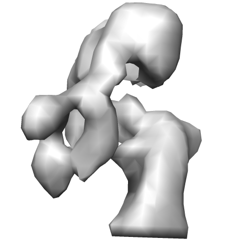 EMD-2311.png
 http://ftp.pdbj.org/pub/emdb/structures/EMD-2311


 Z (Sec.)
 Y (Row.)
 X (Col.)


 Sample components
 Processing
 Electron microscopy
 Chimera
