 Movie
Movie Controller

[English] 日本語
 Yorodumi
- PDB-4ooj: Crystal structure of the N-terminal domain of the Legionella pneu... -
+ Open data

- Basic information
| Entry | Database: PDB / ID: 4ooj | ||||||
|---|---|---|---|---|---|---|---|
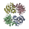
| Title | Crystal structure of the N-terminal domain of the Legionella pneumophila protein SidC at 2.4A resolution | ||||||
 Components Components | SidC, interaptin | ||||||
 Keywords Keywords | UNKNOWN FUNCTION / novel fold / Legionella effector / Legionella containing vacuole / Host-Pathogen Interaction | ||||||
| Function / homology | SidC, N-terminal / : / : / SidC N-terminal domain / SidC, C-terminal domain / SidC, lipid-binding domain / DI(HYDROXYETHYL)ETHER / SidC, interaptin Function and homology information Function and homology information | ||||||
| Biological species |   Legionella pneumophila (bacteria) Legionella pneumophila (bacteria) | ||||||
| Method |  X-RAY DIFFRACTION / SYNCHROTRON / MOLECULAR REPLACEMENT / Resolution: 2.4 Å X-RAY DIFFRACTION / SYNCHROTRON / MOLECULAR REPLACEMENT / Resolution: 2.4 Å | ||||||
 Authors Authors | Gazdag, E.M. / Shoebel, S. / Shkumatov, A.V. / Goody, R.S. / Itzen, A. | ||||||
 Citation Citation | Journal: J.Struct.Biol. / Year: 2014 Title: The structure of the N-terminal domain of the Legionella protein SidC Authors: Gazdag, E.M. / Schobel, S. / Shkumatov, A.V. / Goody, R.S. / Itzen, A. | ||||||
| History |
|
- Structure visualization
| Structure viewer | Molecule: MolmilJmol/JSmol |
|---|
- Downloads & links
-Download
| PDBx/mmCIF format | 4ooj.cif.gz | 947.3 KB | Display | PDBx/mmCIF format |
|---|---|---|---|---|
| PDB format | pdb4ooj.ent.gz | 793.2 KB | Display | PDB format |
| PDBx/mmJSON format | 4ooj.json.gz | Tree view | PDBx/mmJSON format | |
| Others |  Other downloads Other downloads |
-Validation report
| Arichive directory | https://data.pdbj.org/pub/pdb/validation_reports/oo/4oojftp://data.pdbj.org/pub/pdb/validation_reports/oo/4ooj | HTTPS FTP |
|---|
-Related structure data
| Similar structure data |
|---|


-Links
 PDBj
PDBj- Assembly
| Deposited unit | 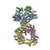
| ||||||||
|---|---|---|---|---|---|---|---|---|---|
| 1 | 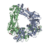
| ||||||||
| 2 | 
| ||||||||
| Unit cell |
|
-Components
| #1: Protein | Mass: 70127.664 Da / Num. of mol.: 4 / Fragment: UNP residues 1-609 Source method: isolated from a genetically manipulated source Source: (gene. exp.) Legionella pneumophila (bacteria) / Strain: Philadelphia 1 / Gene: sidC / Production host: #2: Chemical | ChemComp-MG /   Mass: 24.305 Da / Num. of mol.: 4 / Source method: obtained synthetically / Formula: Mg Mass: 24.305 Da / Num. of mol.: 4 / Source method: obtained synthetically / Formula: Mg#3: Chemical | ChemComp-PEG /   Mass: 106.120 Da / Num. of mol.: 17 / Source method: obtained synthetically / Formula: C4H10O3 Mass: 106.120 Da / Num. of mol.: 17 / Source method: obtained synthetically / Formula: C4H10O3#4: Water | ChemComp-HOH / |  Mass: 18.015 Da / Num. of mol.: 419 / Source method: isolated from a natural source / Formula: H2O Mass: 18.015 Da / Num. of mol.: 419 / Source method: isolated from a natural source / Formula: H2O |
|---|
-Experimental details
-Experiment
| Experiment | Method: X-RAY DIFFRACTION / Number of used crystals: 2 |
|---|
- Sample preparation
| Crystal |
| |||||||||||||||
|---|---|---|---|---|---|---|---|---|---|---|---|---|---|---|---|---|
| Crystal grow |
|
-Data collection
| Diffraction |
| ||||||||||||||||||
|---|---|---|---|---|---|---|---|---|---|---|---|---|---|---|---|---|---|---|---|
| Diffraction source |
| ||||||||||||||||||
| Detector |
| ||||||||||||||||||
| Radiation |
| ||||||||||||||||||
| Radiation wavelength |
| ||||||||||||||||||
| Reflection | Resolution: 2.4→50 Å / Num. obs: 124784 / % possible obs: 99.8 % / Observed criterion σ(I): -3 / Redundancy: 28.65 % / Rmerge(I) obs: 0.101 / Rsym value: 0.054 / Net I/σ(I): 19.03 | ||||||||||||||||||
| Reflection shell | Resolution: 2.4→2.5 Å / Redundancy: 28.32 % / Rmerge(I) obs: 0.579 / Mean I/σ(I) obs: 2.87 / Rsym value: 0.473 / % possible all: 99.8 |

- Processing
| Software |
| |||||||||||||||||||||||||||||||||||||||||||||||||||||||||||||||||||||||||||||||||||||||||||||||||||||||||||||||||||||||||||||
|---|---|---|---|---|---|---|---|---|---|---|---|---|---|---|---|---|---|---|---|---|---|---|---|---|---|---|---|---|---|---|---|---|---|---|---|---|---|---|---|---|---|---|---|---|---|---|---|---|---|---|---|---|---|---|---|---|---|---|---|---|---|---|---|---|---|---|---|---|---|---|---|---|---|---|---|---|---|---|---|---|---|---|---|---|---|---|---|---|---|---|---|---|---|---|---|---|---|---|---|---|---|---|---|---|---|---|---|---|---|---|---|---|---|---|---|---|---|---|---|---|---|---|---|---|---|---|
| Refinement | Method to determine structure: MOLECULAR REPLACEMENT Starting model: partial model after phasing and building in AutoSol Resolution: 2.4→45.9 Å / Cor.coef. Fo:Fc: 0.945 / Cor.coef. Fo:Fc free: 0.909 / SU B: 19.051 / SU ML: 0.201 / Cross valid method: THROUGHOUT / σ(F): 0 / ESU R: 0.352 / ESU R Free: 0.263 / Stereochemistry target values: MAXIMUM LIKELIHOOD / Details: HYDROGENS HAVE BEEN ADDED IN THE RIDING POSITIONS
| |||||||||||||||||||||||||||||||||||||||||||||||||||||||||||||||||||||||||||||||||||||||||||||||||||||||||||||||||||||||||||||
| Solvent computation | Ion probe radii: 0.8 Å / Shrinkage radii: 0.8 Å / VDW probe radii: 1.4 Å / Solvent model: BABINET MODEL WITH MASK | |||||||||||||||||||||||||||||||||||||||||||||||||||||||||||||||||||||||||||||||||||||||||||||||||||||||||||||||||||||||||||||
| Displacement parameters | Biso max: 92.71 Å2 / Biso mean: 50.024 Å2 / Biso min: 16.16 Å2
| |||||||||||||||||||||||||||||||||||||||||||||||||||||||||||||||||||||||||||||||||||||||||||||||||||||||||||||||||||||||||||||
| Refinement step | Cycle: LAST / Resolution: 2.4→45.9 Å
| |||||||||||||||||||||||||||||||||||||||||||||||||||||||||||||||||||||||||||||||||||||||||||||||||||||||||||||||||||||||||||||
| Refine LS restraints |
| |||||||||||||||||||||||||||||||||||||||||||||||||||||||||||||||||||||||||||||||||||||||||||||||||||||||||||||||||||||||||||||
| LS refinement shell | Resolution: 2.4→2.462 Å / Total num. of bins used: 20
| |||||||||||||||||||||||||||||||||||||||||||||||||||||||||||||||||||||||||||||||||||||||||||||||||||||||||||||||||||||||||||||
| Refinement TLS params. | Method: refined / Refine-ID: X-RAY DIFFRACTION
| |||||||||||||||||||||||||||||||||||||||||||||||||||||||||||||||||||||||||||||||||||||||||||||||||||||||||||||||||||||||||||||
| Refinement TLS group |
|
