 Movie
Movie Controller


+ Open data

- Basic information
| Entry | Database: PDB / ID: 4bz5 | ||||||
|---|---|---|---|---|---|---|---|
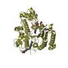
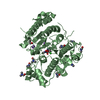
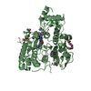
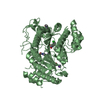
| Title | Crystal structure of Schistosoma mansoni HDAC8 | ||||||
 Components Components | HISTONE DEACETYLASE 8 | ||||||
 Keywords Keywords | HYDROLASE / PLATYHELMINTHS / INHIBITION | ||||||
| Function / homology |  Function and homology information Function and homology informationhistone deacetylase activity, hydrolytic mechanism / histone deacetylase / heterochromatin formation / metal ion binding / nucleus Similarity search - Function | ||||||
| Biological species |  | ||||||
| Method |  X-RAY DIFFRACTION / SYNCHROTRON / MOLECULAR REPLACEMENT / Resolution: 1.785 Å X-RAY DIFFRACTION / SYNCHROTRON / MOLECULAR REPLACEMENT / Resolution: 1.785 Å | ||||||
 Authors Authors | Marek, M. / Romier, C. | ||||||
 Citation Citation | Journal: Plos Pathog. / Year: 2013 Title: Structural Basis for the Inhibition of Histone Deacetylase 8 (Hdac8), a Key Epigenetic Player in the Blood Fluke Schistosoma Mansoni. Authors: Marek, M. / Kannan, S. / Hauser, A. / Moraes Mourao, M. / Caby, S. / Cura, V. / Stolfa, D.A. / Schmidtkunz, K. / Lancelot, J. / Andrade, L. / Renaud, J. / Oliveira, G. / Sippl, W. / Jung, M. ...Authors: Marek, M. / Kannan, S. / Hauser, A. / Moraes Mourao, M. / Caby, S. / Cura, V. / Stolfa, D.A. / Schmidtkunz, K. / Lancelot, J. / Andrade, L. / Renaud, J. / Oliveira, G. / Sippl, W. / Jung, M. / Cavarelli, J. / Pierce, R.J. / Romier, C. | ||||||
| History |
|
- Structure visualization
| Structure viewer | Molecule: MolmilJmol/JSmol |
|---|
- Downloads & links
-Download
| PDBx/mmCIF format | 4bz5.cif.gz | 677.1 KB | Display | PDBx/mmCIF format |
|---|---|---|---|---|
| PDB format | pdb4bz5.ent.gz | 560.6 KB | Display | PDB format |
| PDBx/mmJSON format | 4bz5.json.gz | Tree view | PDBx/mmJSON format | |
| Others |  Other downloads Other downloads |
-Validation report
| Arichive directory | https://data.pdbj.org/pub/pdb/validation_reports/bz/4bz5ftp://data.pdbj.org/pub/pdb/validation_reports/bz/4bz5 | HTTPS FTP |
|---|
-Related structure data
| Related structure data |  4bz6C  4bz7C  4bz8C  4bz9C  1t67S C: citing same article ( S: Starting model for refinement |
|---|---|
| Similar structure data |


-Links
 PDBj
PDBj
- Assembly
| Deposited unit | 
| ||||||||
|---|---|---|---|---|---|---|---|---|---|
| 1 | 
| ||||||||
| 2 | 
| ||||||||
| 3 | 
| ||||||||
| 4 | 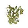
| ||||||||
| Unit cell |
|
-Components
| #1: Protein | Mass: 50444.875 Da / Num. of mol.: 4 Source method: isolated from a genetically manipulated source Source: (gene. exp.)  #2: Chemical | ChemComp-ZN /   Mass: 65.409 Da / Num. of mol.: 4 / Source method: obtained synthetically / Formula: Zn Mass: 65.409 Da / Num. of mol.: 4 / Source method: obtained synthetically / Formula: Zn#3: Chemical | ChemComp-K /   Mass: 39.098 Da / Num. of mol.: 8 / Source method: obtained synthetically / Formula: K Mass: 39.098 Da / Num. of mol.: 8 / Source method: obtained synthetically / Formula: K#4: Chemical | ChemComp-TLA /   Mass: 150.087 Da / Num. of mol.: 4 / Source method: obtained synthetically / Formula: C4H6O6 Mass: 150.087 Da / Num. of mol.: 4 / Source method: obtained synthetically / Formula: C4H6O6#5: Water | ChemComp-HOH / |  Mass: 18.015 Da / Num. of mol.: 1532 / Source method: isolated from a natural source / Formula: H2O Mass: 18.015 Da / Num. of mol.: 1532 / Source method: isolated from a natural source / Formula: H2OSequence details | THE GSLVPR MOTIF AT THE END OF THE SEQUENCE CORRESPONDS TO A BAMHI CLONING SITE (GS) FOLLOWED BY A ...THE GSLVPR MOTIF AT THE END OF THE SEQUENCE CORRESPOND | |
|---|
-Experimental details
-Experiment
| Experiment | Method: X-RAY DIFFRACTION / Number of used crystals: 1 |
|---|
- Sample preparation
| Crystal | Density Matthews: 1.5 Å3/Da / Density % sol: 19 % / Description: NONE |
|---|---|
| Crystal grow | Details: 0.2 M NA,K L-TARTRATE, 21% (W/V) PEG3350 |
-Data collection
| Diffraction | Mean temperature: 100 K |
|---|---|
| Diffraction source | Source: SYNCHROTRON / Site: ESRF  / Beamline: ID23-2 / Wavelength: 0.8726 / Beamline: ID23-2 / Wavelength: 0.8726 |
| Detector | Type: MARMOSAIC 225 mm CCD / Detector: CCD / Date: Jul 19, 2010 |
| Radiation | Protocol: SINGLE WAVELENGTH / Monochromatic (M) / Laue (L): M / Scattering type: x-ray |
| Radiation wavelength | Wavelength: 0.8726 Å / Relative weight: 1 |
| Reflection | Resolution: 1.79→50 Å / Num. obs: 166229 / % possible obs: 97.2 % / Observed criterion σ(I): 2 / Redundancy: 1.9 % / Biso Wilson estimate: 17.94 Å2 / Rmerge(I) obs: 0.03 / Net I/σ(I): 26.5 |
| Reflection shell | Resolution: 1.79→1.82 Å / Redundancy: 1.9 % / Rmerge(I) obs: 0.13 / Mean I/σ(I) obs: 5 / % possible all: 93.4 |
- Processing
| Software |
| |||||||||||||||||||||||||||||||||||||||||||||||||||||||||||||||||||||||||||||||||||||||||||||||||||||||||||||||||||||||||||||||||||||||||||||||||||||||||||||||||||||||||||||||||||||||||||||||||||||||||||||||||||||||||
|---|---|---|---|---|---|---|---|---|---|---|---|---|---|---|---|---|---|---|---|---|---|---|---|---|---|---|---|---|---|---|---|---|---|---|---|---|---|---|---|---|---|---|---|---|---|---|---|---|---|---|---|---|---|---|---|---|---|---|---|---|---|---|---|---|---|---|---|---|---|---|---|---|---|---|---|---|---|---|---|---|---|---|---|---|---|---|---|---|---|---|---|---|---|---|---|---|---|---|---|---|---|---|---|---|---|---|---|---|---|---|---|---|---|---|---|---|---|---|---|---|---|---|---|---|---|---|---|---|---|---|---|---|---|---|---|---|---|---|---|---|---|---|---|---|---|---|---|---|---|---|---|---|---|---|---|---|---|---|---|---|---|---|---|---|---|---|---|---|---|---|---|---|---|---|---|---|---|---|---|---|---|---|---|---|---|---|---|---|---|---|---|---|---|---|---|---|---|---|---|---|---|---|---|---|---|---|---|---|---|---|---|---|---|---|---|---|---|---|
| Refinement | Method to determine structure: MOLECULAR REPLACEMENT Starting model: PDB ENTRY 1T67 Resolution: 1.785→31.37 Å / SU ML: 0.4 / σ(F): 1.98 / Phase error: 18.29 / Stereochemistry target values: ML
| |||||||||||||||||||||||||||||||||||||||||||||||||||||||||||||||||||||||||||||||||||||||||||||||||||||||||||||||||||||||||||||||||||||||||||||||||||||||||||||||||||||||||||||||||||||||||||||||||||||||||||||||||||||||||
| Solvent computation | Shrinkage radii: 0.9 Å / VDW probe radii: 1.11 Å / Solvent model: FLAT BULK SOLVENT MODEL / Bsol: 60.43 Å2 / ksol: 0.4 e/Å3 | |||||||||||||||||||||||||||||||||||||||||||||||||||||||||||||||||||||||||||||||||||||||||||||||||||||||||||||||||||||||||||||||||||||||||||||||||||||||||||||||||||||||||||||||||||||||||||||||||||||||||||||||||||||||||
| Displacement parameters | Biso mean: 23.68 Å2
| |||||||||||||||||||||||||||||||||||||||||||||||||||||||||||||||||||||||||||||||||||||||||||||||||||||||||||||||||||||||||||||||||||||||||||||||||||||||||||||||||||||||||||||||||||||||||||||||||||||||||||||||||||||||||
| Refinement step | Cycle: LAST / Resolution: 1.785→31.37 Å
| |||||||||||||||||||||||||||||||||||||||||||||||||||||||||||||||||||||||||||||||||||||||||||||||||||||||||||||||||||||||||||||||||||||||||||||||||||||||||||||||||||||||||||||||||||||||||||||||||||||||||||||||||||||||||
| Refine LS restraints |
| |||||||||||||||||||||||||||||||||||||||||||||||||||||||||||||||||||||||||||||||||||||||||||||||||||||||||||||||||||||||||||||||||||||||||||||||||||||||||||||||||||||||||||||||||||||||||||||||||||||||||||||||||||||||||
| LS refinement shell |
| |||||||||||||||||||||||||||||||||||||||||||||||||||||||||||||||||||||||||||||||||||||||||||||||||||||||||||||||||||||||||||||||||||||||||||||||||||||||||||||||||||||||||||||||||||||||||||||||||||||||||||||||||||||||||
| Refinement TLS params. | Method: refined / Refine-ID: X-RAY DIFFRACTION
| |||||||||||||||||||||||||||||||||||||||||||||||||||||||||||||||||||||||||||||||||||||||||||||||||||||||||||||||||||||||||||||||||||||||||||||||||||||||||||||||||||||||||||||||||||||||||||||||||||||||||||||||||||||||||
| Refinement TLS group |
|
