 Movie
Movie Controller

[English] 日本語
 Yorodumi
- PDB-5j7i: Crystal structure of a Geobacillus thermoglucosidasius Acetylatin... -
+ Open data

- Basic information
| Entry | Database: PDB / ID: 5j7i | ||||||
|---|---|---|---|---|---|---|---|
| Title | Crystal structure of a Geobacillus thermoglucosidasius Acetylating Aldehyde Dehydrogenase in complex with ADP | ||||||
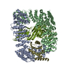
 Components Components | Acetaldehyde dehydrogenase (Acetylating) | ||||||
 Keywords Keywords | OXIDOREDUCTASE / Acetylating aldehyde dehydrogenase / complex | ||||||
| Function / homology |  Function and homology information Function and homology informationacetaldehyde dehydrogenase (acetylating) / acetaldehyde dehydrogenase (acetylating) activity / nucleotide binding Similarity search - Function | ||||||
| Biological species |  Geobacillus thermoglucosidasius (bacteria) Geobacillus thermoglucosidasius (bacteria) | ||||||
| Method |  X-RAY DIFFRACTION / MOLECULAR REPLACEMENT / Resolution: 4 Å X-RAY DIFFRACTION / MOLECULAR REPLACEMENT / Resolution: 4 Å | ||||||
 Authors Authors | Crennell, S.J. / Extance, J.P. / Danson, M.J. | ||||||
 Citation Citation | Journal: Protein Sci. / Year: 2016 Title: Structure of an acetylating aldehyde dehydrogenase from the thermophilic ethanologen Geobacillus thermoglucosidasius. Authors: Extance, J. / Danson, M.J. / Crennell, S.J. | ||||||
| History |
|
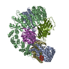
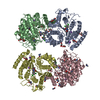
- Structure visualization
| Structure viewer | Molecule: MolmilJmol/JSmol |
|---|
- Downloads & links
-Download
| PDBx/mmCIF format | 5j7i.cif.gz | 960.8 KB | Display | PDBx/mmCIF format |
|---|---|---|---|---|
| PDB format | pdb5j7i.ent.gz | 818.3 KB | Display | PDB format |
| PDBx/mmJSON format | 5j7i.json.gz | Tree view | PDBx/mmJSON format | |
| Others |  Other downloads Other downloads |
-Validation report
| Arichive directory | https://data.pdbj.org/pub/pdb/validation_reports/j7/5j7iftp://data.pdbj.org/pub/pdb/validation_reports/j7/5j7i | HTTPS FTP |
|---|
-Related structure data
| Related structure data |  5j78SC S: Starting model for refinement C: citing same article ( |
|---|---|
| Similar structure data |
-Links
 PDBj
PDBj


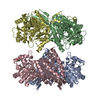
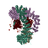
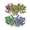
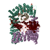
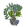
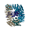
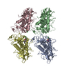
- Assembly
| Deposited unit | 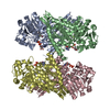
| ||||||||
|---|---|---|---|---|---|---|---|---|---|
| 1 |
| ||||||||
| Unit cell |
|
-Components
| #1: Protein | Mass: 52280.922 Da / Num. of mol.: 4 Source method: isolated from a genetically manipulated source Source: (gene. exp.) Geobacillus thermoglucosidasius (strain C56-YS93) (bacteria)Strain: C56-YS93 / Gene: Geoth_3234 / Plasmid: pET28a / Production host: References: UniProt: A0A0M1QQ83, acetaldehyde dehydrogenase (acetylating) #2: Chemical |   Mass: 427.201 Da / Num. of mol.: 3 / Source method: obtained synthetically / Formula: C10H15N5O10P2 / Comment: ADP, energy-carrying molecule*YM Mass: 427.201 Da / Num. of mol.: 3 / Source method: obtained synthetically / Formula: C10H15N5O10P2 / Comment: ADP, energy-carrying molecule*YM#3: Chemical | ChemComp-APR / |   Mass: 559.316 Da / Num. of mol.: 1 / Source method: obtained synthetically / Formula: C15H23N5O14P2 Mass: 559.316 Da / Num. of mol.: 1 / Source method: obtained synthetically / Formula: C15H23N5O14P2Has protein modification | Y | |
|---|
-Experimental details
-Experiment
| Experiment | Method: X-RAY DIFFRACTION / Number of used crystals: 1 |
|---|
- Sample preparation
| Crystal | Density Matthews: 2.67 Å3/Da / Density % sol: 53.94 % / Description: thin and plate-like |
|---|---|
| Crystal grow | Temperature: 289 K / Method: vapor diffusion, hanging drop / pH: 5 Details: protein solution containing 10.7mg/ml protein, 0.5mM acetyl-CoA and 5mM NAD+ was mixed 1:1 with 0.1M MMT buffer pH5.0, 21 to 23% PEG 1500 |
-Data collection
| Diffraction | Mean temperature: 293 K |
|---|---|
| Diffraction source | Source: ROTATING ANODE / Type: RIGAKU MICROMAX-007 HF / Wavelength: 1.5406 Å |
| Detector | Type: RIGAKU SATURN 944+ / Detector: CCD / Date: Jan 20, 2012 |
| Radiation | Protocol: SINGLE WAVELENGTH / Monochromatic (M) / Laue (L): M / Scattering type: x-ray |
| Radiation wavelength | Wavelength: 1.5406 Å / Relative weight: 1 |
| Reflection | Resolution: 4→50 Å / Num. obs: 14328 / % possible obs: 78 % / Redundancy: 2.5 % / Rmerge(I) obs: 0.267 / Net I/σ(I): 3.87 |
| Reflection shell | Resolution: 4→4.07 Å / Redundancy: 2.26 % / Rmerge(I) obs: 0.466 / Mean I/σ(I) obs: 2.26 / % possible all: 70.5 |
- Processing
| Software |
| ||||||||||||||||||||||||||||||||||||||||||
|---|---|---|---|---|---|---|---|---|---|---|---|---|---|---|---|---|---|---|---|---|---|---|---|---|---|---|---|---|---|---|---|---|---|---|---|---|---|---|---|---|---|---|---|
| Refinement | Method to determine structure: MOLECULAR REPLACEMENT Starting model: 5J78 Resolution: 4→42.668 Å / SU ML: 0.54 / Cross valid method: FREE R-VALUE / σ(F): 0 / Phase error: 25.78 / Stereochemistry target values: ML
| ||||||||||||||||||||||||||||||||||||||||||
| Solvent computation | Shrinkage radii: 0.9 Å / VDW probe radii: 1.11 Å / Solvent model: FLAT BULK SOLVENT MODEL | ||||||||||||||||||||||||||||||||||||||||||
| Refinement step | Cycle: LAST / Resolution: 4→42.668 Å
| ||||||||||||||||||||||||||||||||||||||||||
| Refine LS restraints |
| ||||||||||||||||||||||||||||||||||||||||||
| LS refinement shell |
|
