+ Open data
- Basic information
| Entry | Database: EMDB / ID: EMD-22631 | |||||||||
|---|---|---|---|---|---|---|---|---|---|---|
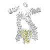
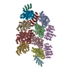
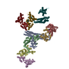
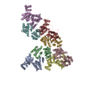
| Title | PIKfyve/Fig4/Vac14 complex centered on Fig4 - map3 | |||||||||
 Map data Map data | PIKfyve complex density map centered on Fig4 | |||||||||
 Sample Sample |
| |||||||||
 Keywords Keywords | Lipid kinase / Lipid phosphatase / protein complex / LIPID BINDING PROTEIN | |||||||||
| Function / homology |  Function and homology information Function and homology informationphosphatidylinositol-3,5-bisphosphate 5-phosphatase activity / myelin assembly / Synthesis of PIPs at the late endosome membrane / Synthesis of PIPs at the early endosome membrane / negative regulation of myelination / phosphatidylinositol-3,4,5-trisphosphate 5-phosphatase / phosphoinositide 5-phosphatase / phosphatidylinositol-3,4,5-trisphosphate 5-phosphatase activity / phosphatidylinositol-4,5-bisphosphate 5-phosphatase activity / Synthesis of PIPs at the Golgi membrane ...phosphatidylinositol-3,5-bisphosphate 5-phosphatase activity / myelin assembly / Synthesis of PIPs at the late endosome membrane / Synthesis of PIPs at the early endosome membrane / negative regulation of myelination / phosphatidylinositol-3,4,5-trisphosphate 5-phosphatase / phosphoinositide 5-phosphatase / phosphatidylinositol-3,4,5-trisphosphate 5-phosphatase activity / phosphatidylinositol-4,5-bisphosphate 5-phosphatase activity / Synthesis of PIPs at the Golgi membrane / phosphatidylinositol dephosphorylation / Hydrolases; Acting on ester bonds; Phosphoric-monoester hydrolases / vacuole organization / phosphatidylinositol biosynthetic process / pigmentation / protein-serine/threonine phosphatase / protein serine/threonine phosphatase activity / neuron development / lipid droplet / locomotory behavior / late endosome membrane / early endosome membrane / endosome membrane / Golgi membrane / intracellular membrane-bounded organelle Similarity search - Function | |||||||||
| Biological species |  Homo sapiens (human) Homo sapiens (human) | |||||||||
| Method | single particle reconstruction / cryo EM / Resolution: 5.1 Å | |||||||||
 Authors Authors | Lees JA / Reinisch KM | |||||||||
| Funding support |  United States, 1 items United States, 1 items
| |||||||||
 Citation Citation |  Journal: Mol Cell / Year: 2020 Journal: Mol Cell / Year: 2020Title: Insights into Lysosomal PI(3,5)P Homeostasis from a Structural-Biochemical Analysis of the PIKfyve Lipid Kinase Complex. Authors: Joshua A Lees / PeiQi Li / Nikit Kumar / Lois S Weisman / Karin M Reinisch /  Abstract: The phosphoinositide PI(3,5)P, generated exclusively by the PIKfyve lipid kinase complex, is key for lysosomal biology. Here, we explore how PI(3,5)P levels within cells are regulated. We find the ...The phosphoinositide PI(3,5)P, generated exclusively by the PIKfyve lipid kinase complex, is key for lysosomal biology. Here, we explore how PI(3,5)P levels within cells are regulated. We find the PIKfyve complex comprises five copies of the scaffolding protein Vac14 and one copy each of the lipid kinase PIKfyve, generating PI(3,5)P from PI3P and the lipid phosphatase Fig4, reversing the reaction. Fig4 is active as a lipid phosphatase in the ternary complex, whereas PIKfyve within the complex cannot access membrane-incorporated phosphoinositides due to steric constraints. We find further that the phosphoinositide-directed activities of both PIKfyve and Fig4 are regulated by protein-directed activities within the complex. PIKfyve autophosphorylation represses its lipid kinase activity and stimulates Fig4 lipid phosphatase activity. Further, Fig4 is also a protein phosphatase acting on PIKfyve to stimulate its lipid kinase activity, explaining why catalytically active Fig4 is required for maximal PI(3,5)P production by PIKfyve in vivo. | |||||||||
| History |
|
- Structure visualization
| Movie |
 Movie viewer Movie viewer |
|---|---|
| Structure viewer | EM map:  SurfView SurfView Molmil Molmil Jmol/JSmol Jmol/JSmol |
| Supplemental images |
- Downloads & links
-EMDB archive
| Map data |  emd_22631.map.gz emd_22631.map.gz | 227.9 MB |  EMDB map data format EMDB map data format | |
|---|---|---|---|---|
| Header (meta data) |  emd-22631-v30.xml emd-22631-v30.xml emd-22631.xml emd-22631.xml | 13.7 KB 13.7 KB | Display Display |  EMDB header EMDB header |
| Images |  emd_22631.png emd_22631.png | 77 KB | ||
| Filedesc metadata |  emd-22631.cif.gz emd-22631.cif.gz | 6 KB | ||
| Archive directory |  http://ftp.pdbj.org/pub/emdb/structures/EMD-22631 http://ftp.pdbj.org/pub/emdb/structures/EMD-22631 ftp://ftp.pdbj.org/pub/emdb/structures/EMD-22631 ftp://ftp.pdbj.org/pub/emdb/structures/EMD-22631 | HTTPS FTP |
-Validation report
| Summary document |  emd_22631_validation.pdf.gz emd_22631_validation.pdf.gz | 565.6 KB | Display |  EMDB validaton report EMDB validaton report |
|---|---|---|---|---|
| Full document |  emd_22631_full_validation.pdf.gz emd_22631_full_validation.pdf.gz | 565.2 KB | Display | |
| Data in XML |  emd_22631_validation.xml.gz emd_22631_validation.xml.gz | 6.7 KB | Display | |
| Data in CIF |  emd_22631_validation.cif.gz emd_22631_validation.cif.gz | 7.7 KB | Display | |
| Arichive directory |  https://ftp.pdbj.org/pub/emdb/validation_reports/EMD-22631 https://ftp.pdbj.org/pub/emdb/validation_reports/EMD-22631 ftp://ftp.pdbj.org/pub/emdb/validation_reports/EMD-22631 ftp://ftp.pdbj.org/pub/emdb/validation_reports/EMD-22631 | HTTPS FTP |
-Related structure data
| Related structure data |  7k1wMC  7k1yC  7k2vC C: citing same article ( M: atomic model generated by this map |
|---|---|
| Similar structure data |
- Links
Links
| EMDB pages |  EMDB (EBI/PDBe) / EMDB (EBI/PDBe) /  EMDataResource EMDataResource |
|---|
- Map
Map
| File |  Download / File: emd_22631.map.gz / Format: CCP4 / Size: 244.1 MB / Type: IMAGE STORED AS FLOATING POINT NUMBER (4 BYTES) Download / File: emd_22631.map.gz / Format: CCP4 / Size: 244.1 MB / Type: IMAGE STORED AS FLOATING POINT NUMBER (4 BYTES) | ||||||||||||||||||||||||||||||||||||||||||||||||||||||||||||||||||||
|---|---|---|---|---|---|---|---|---|---|---|---|---|---|---|---|---|---|---|---|---|---|---|---|---|---|---|---|---|---|---|---|---|---|---|---|---|---|---|---|---|---|---|---|---|---|---|---|---|---|---|---|---|---|---|---|---|---|---|---|---|---|---|---|---|---|---|---|---|---|
| Annotation | PIKfyve complex density map centered on Fig4 | ||||||||||||||||||||||||||||||||||||||||||||||||||||||||||||||||||||
| Projections & slices | Image control
Images are generated by Spider. | ||||||||||||||||||||||||||||||||||||||||||||||||||||||||||||||||||||
| Voxel size | X=Y=Z: 1.05 Å | ||||||||||||||||||||||||||||||||||||||||||||||||||||||||||||||||||||
| Density |
| ||||||||||||||||||||||||||||||||||||||||||||||||||||||||||||||||||||
| Symmetry | Space group: 1 | ||||||||||||||||||||||||||||||||||||||||||||||||||||||||||||||||||||
| Details | EMDB XML:
CCP4 map header:
| ||||||||||||||||||||||||||||||||||||||||||||||||||||||||||||||||||||
-Supplemental data
- Sample components
-Entire : PIKfyve/Fig4/Vac14 complex
| Entire | Name: PIKfyve/Fig4/Vac14 complex |
|---|---|
| Components |
|
-Supramolecule #1: PIKfyve/Fig4/Vac14 complex
| Supramolecule | Name: PIKfyve/Fig4/Vac14 complex / type: complex / ID: 1 / Parent: 0 / Macromolecule list: all |
|---|---|
| Source (natural) | Organism:  Homo sapiens (human) Homo sapiens (human) |
| Molecular weight | Theoretical: 4.28 MDa |
-Macromolecule #1: Fig4 Sac homology model
| Macromolecule | Name: Fig4 Sac homology model / type: protein_or_peptide / ID: 1 / Number of copies: 1 / Enantiomer: LEVO |
|---|---|
| Source (natural) | Organism:  Homo sapiens (human) Homo sapiens (human) |
| Molecular weight | Theoretical: 105.144289 KDa |
| Recombinant expression | Organism:  Homo sapiens (human) Homo sapiens (human) |
| Sequence | String: MHHHHHHHHG SMPTAAAPII SSVQKLVLYE TRARYFLVGS NNAETKYRVL KIDRTEPKDL VIIDDRHVYT QQEVRELLGR LDLGNRTKM GQKGSSGLFR AVSAFGVVGF VRFLEGYYIV LITKRRKMAD IGGHAIYKVE DTNMIYIPND SVRVTHPDEA R YLRIFQNV ...String: MHHHHHHHHG SMPTAAAPII SSVQKLVLYE TRARYFLVGS NNAETKYRVL KIDRTEPKDL VIIDDRHVYT QQEVRELLGR LDLGNRTKM GQKGSSGLFR AVSAFGVVGF VRFLEGYYIV LITKRRKMAD IGGHAIYKVE DTNMIYIPND SVRVTHPDEA R YLRIFQNV DLSSNFYFSY SYDLSHSLQY NLTVLRMPLE MLKSEMTQNR QESFDIFEDE GLITQGGSGV FGICSEPYMK YV WNGELLD IIKSTVHRDW LLYIIHGFCG QSKLLIYGRP VYVTLIARRS SKFAGTRFLK RGANCEGDVA NEVETEQILC DAS VMSFTA GSYSSYVQVR GSVPLYWSQD ISTMMPKPPI TLDQADPFAH VAALHFDQMF QRFGSPIIIL NLVKEREKRK HERI LSEEL VAAVTYLNQF LPPEHTIVYI PWDMAKYTKS KLCNVLDRLN VIAESVVKKT GFFVNRPDSY CSILRPDEKW NELGG CVIP TGRLQTGILR TNCVDCLDRT NTAQFMVGKC ALAYQLYSLG LIDKPNLQFD TDAVRLFEEL YEDHGDTLSL QYGGSQ LVH RVKTYRKIAP WTQHSKDIMQ TLSRYYSNAF SDADRQDSIN LFLGVFHPTE GKPHLWELPT DFYLHHKNTM RLLPTRR SY TYWWTPEVIK HLPLPYDEVI CAVNLKKLIV KKFHKYEEEI DIHNEFFRPY ELSSFDDTFC LAMTSSARDF MPKTVGID P SPFTVRKPDE TGKSVLGNKS NREEAVLQRK TAASAPPPPS EEAVSSSSED DSGTDREEEG SVSQRSTPVK MTDAGDSAK VTENVVQPMK ELYGINLSDG LSEEDFSIYS RFVQLGQSQH KQDKNSQQPC SRCSDGVIKL TPISAFSQDN IYEVQPPRVD RKSTEIFQA HIQASQGIMQ PLGKEDSSMY REYIRNRYL |
-Experimental details
-Structure determination
| Method | cryo EM |
|---|---|
 Processing Processing | single particle reconstruction |
| Aggregation state | particle |
- Sample preparation
| Buffer | pH: 7.8 |
|---|---|
| Vitrification | Cryogen name: ETHANE |
- Electron microscopy
| Microscope | FEI TITAN KRIOS |
|---|---|
| Image recording | Film or detector model: GATAN K2 SUMMIT (4k x 4k) / Average electron dose: 58.4 e/Å2 |
| Electron beam | Acceleration voltage: 300 kV / Electron source:  FIELD EMISSION GUN FIELD EMISSION GUN |
| Electron optics | Illumination mode: FLOOD BEAM / Imaging mode: BRIGHT FIELD / Cs: 2.7 mm |
| Experimental equipment |  Model: Titan Krios / Image courtesy: FEI Company |
 Movie
Movie Controller


 Z (Sec.)
 Y (Row.)
 X (Col.)


