[English] 日本語
Yorodumi
- EMDB-22631: PIKfyve/Fig4/Vac14 complex centered on Fig4 - map3 -

+
Open data


ID or keywords:

Loading...

-
Basic information

Entry
Database: EMDB / ID: EMD-22631
TitlePIKfyve/Fig4/Vac14 complex centered on Fig4 - map3
Map dataPIKfyve complex density map centered on Fig4
Sample
  • Complex: PIKfyve/Fig4/Vac14 complex
    • Protein or peptide: Fig4 Sac homology model
KeywordsLipid kinase / Lipid phosphatase / protein complex / LIPID BINDING PROTEIN
Function / homology
Function and homology information


phosphatidylinositol-3,5-bisphosphate 5-phosphatase activity / Synthesis of PIPs at the late endosome membrane / Synthesis of PIPs at the early endosome membrane / Synthesis of PIPs at the Golgi membrane / Hydrolases; Acting on ester bonds; Phosphoric-monoester hydrolases / phosphatidylinositol dephosphorylation / phosphatidylinositol biosynthetic process / lipid droplet / late endosome membrane / early endosome membrane ...phosphatidylinositol-3,5-bisphosphate 5-phosphatase activity / Synthesis of PIPs at the late endosome membrane / Synthesis of PIPs at the early endosome membrane / Synthesis of PIPs at the Golgi membrane / Hydrolases; Acting on ester bonds; Phosphoric-monoester hydrolases / phosphatidylinositol dephosphorylation / phosphatidylinositol biosynthetic process / lipid droplet / late endosome membrane / early endosome membrane / endosome membrane / Golgi membrane / intracellular membrane-bounded organelle
Similarity search - Function
Polyphosphoinositide phosphatase Fig4-like / SAC domain / SacI homology domain / Sac phosphatase domain profile.
Similarity search - Domain/homology
Polyphosphoinositide phosphatase
Similarity search - Component
Biological speciesHomo sapiens (human)
Methodsingle particle reconstruction / cryo EM / Resolution: 5.1 Å
AuthorsLees JA / Reinisch KM
Funding support United States, 1 items
OrganizationGrant numberCountry
National Institutes of Health/National Institute of General Medical Sciences (NIH/NIGMS)GM131715 United States
Citation
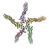
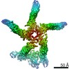
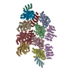
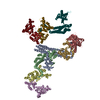
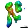
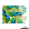
Journal: Mol Cell / Year: 2020
Title: Insights into Lysosomal PI(3,5)P Homeostasis from a Structural-Biochemical Analysis of the PIKfyve Lipid Kinase Complex.
Authors: Joshua A Lees / PeiQi Li / Nikit Kumar / Lois S Weisman / Karin M Reinisch /
Abstract: The phosphoinositide PI(3,5)P, generated exclusively by the PIKfyve lipid kinase complex, is key for lysosomal biology. Here, we explore how PI(3,5)P levels within cells are regulated. We find the ...The phosphoinositide PI(3,5)P, generated exclusively by the PIKfyve lipid kinase complex, is key for lysosomal biology. Here, we explore how PI(3,5)P levels within cells are regulated. We find the PIKfyve complex comprises five copies of the scaffolding protein Vac14 and one copy each of the lipid kinase PIKfyve, generating PI(3,5)P from PI3P and the lipid phosphatase Fig4, reversing the reaction. Fig4 is active as a lipid phosphatase in the ternary complex, whereas PIKfyve within the complex cannot access membrane-incorporated phosphoinositides due to steric constraints. We find further that the phosphoinositide-directed activities of both PIKfyve and Fig4 are regulated by protein-directed activities within the complex. PIKfyve autophosphorylation represses its lipid kinase activity and stimulates Fig4 lipid phosphatase activity. Further, Fig4 is also a protein phosphatase acting on PIKfyve to stimulate its lipid kinase activity, explaining why catalytically active Fig4 is required for maximal PI(3,5)P production by PIKfyve in vivo.
History
DepositionSep 8, 2020-
Header (metadata) releaseOct 21, 2020-
Map releaseOct 21, 2020-
UpdateMar 6, 2024-
Current statusMar 6, 2024Processing site: RCSB / Status: Released

-
Structure visualization

Movie
  • Surface view with section colored by density value
  • Surface level: 0.01
  • Imaged by UCSF Chimera
  • Download
  • Surface view colored by radius
  • Surface level: 0.01
  • Imaged by UCSF Chimera
  • Download
  • Surface view with fitted model
  • Atomic models: PDB-7k1w
  • Surface level: 0.01
  • Imaged by UCSF Chimera
  • Download
  • Simplified surface model + fitted atomic model
  • Atomic modelsPDB-7k1w
  • Imaged by Jmol
  • Download
Movie viewer
Structure viewerEM map:
SurfViewMolmilJmol/JSmol
Supplemental images

Downloads & links

-
Map

FileDownload / File: emd_22631.map.gz / Format: CCP4 / Size: 244.1 MB / Type: IMAGE STORED AS FLOATING POINT NUMBER (4 BYTES)
AnnotationPIKfyve complex density map centered on Fig4
Voxel sizeX=Y=Z: 1.05 Å
Density
Contour LevelBy AUTHOR: 0.01 / Movie #1: 0.01
Minimum - Maximum-0.022210082 - 0.046168517
Average (Standard dev.)-0.00006561446 (±0.0013303686)
SymmetrySpace group: 1
Details

EMDB XML:

Map geometry
Axis orderXYZ
Origin000
Dimensions400400400
Spacing400400400
CellA=B=C: 419.99997 Å
α=β=γ: 90.0 °

CCP4 map header:

modeImage stored as Reals
Å/pix. X/Y/Z1.051.051.05
M x/y/z400400400
origin x/y/z0.0000.0000.000
length x/y/z420.000420.000420.000
α/β/γ90.00090.00090.000
start NX/NY/NZ000
NX/NY/NZ360360360
MAP C/R/S123
start NC/NR/NS000
NC/NR/NS400400400
D min/max/mean-0.0220.046-0.000

-
Supplemental data

-
Sample components

-
Entire : PIKfyve/Fig4/Vac14 complex

EntireName: PIKfyve/Fig4/Vac14 complex
Components
  • Complex: PIKfyve/Fig4/Vac14 complex
    • Protein or peptide: Fig4 Sac homology model

-
Supramolecule #1: PIKfyve/Fig4/Vac14 complex

SupramoleculeName: PIKfyve/Fig4/Vac14 complex / type: complex / ID: 1 / Parent: 0 / Macromolecule list: all
Source (natural)Organism: Homo sapiens (human)
Molecular weightTheoretical: 4.28 MDa

-
Macromolecule #1: Fig4 Sac homology model

MacromoleculeName: Fig4 Sac homology model / type: protein_or_peptide / ID: 1 / Number of copies: 1 / Enantiomer: LEVO
Source (natural)Organism: Homo sapiens (human)
Molecular weightTheoretical: 105.144289 KDa
Recombinant expressionOrganism: Homo sapiens (human)
SequenceString: MHHHHHHHHG SMPTAAAPII SSVQKLVLYE TRARYFLVGS NNAETKYRVL KIDRTEPKDL VIIDDRHVYT QQEVRELLGR LDLGNRTKM GQKGSSGLFR AVSAFGVVGF VRFLEGYYIV LITKRRKMAD IGGHAIYKVE DTNMIYIPND SVRVTHPDEA R YLRIFQNV ...String:
MHHHHHHHHG SMPTAAAPII SSVQKLVLYE TRARYFLVGS NNAETKYRVL KIDRTEPKDL VIIDDRHVYT QQEVRELLGR LDLGNRTKM GQKGSSGLFR AVSAFGVVGF VRFLEGYYIV LITKRRKMAD IGGHAIYKVE DTNMIYIPND SVRVTHPDEA R YLRIFQNV DLSSNFYFSY SYDLSHSLQY NLTVLRMPLE MLKSEMTQNR QESFDIFEDE GLITQGGSGV FGICSEPYMK YV WNGELLD IIKSTVHRDW LLYIIHGFCG QSKLLIYGRP VYVTLIARRS SKFAGTRFLK RGANCEGDVA NEVETEQILC DAS VMSFTA GSYSSYVQVR GSVPLYWSQD ISTMMPKPPI TLDQADPFAH VAALHFDQMF QRFGSPIIIL NLVKEREKRK HERI LSEEL VAAVTYLNQF LPPEHTIVYI PWDMAKYTKS KLCNVLDRLN VIAESVVKKT GFFVNRPDSY CSILRPDEKW NELGG CVIP TGRLQTGILR TNCVDCLDRT NTAQFMVGKC ALAYQLYSLG LIDKPNLQFD TDAVRLFEEL YEDHGDTLSL QYGGSQ LVH RVKTYRKIAP WTQHSKDIMQ TLSRYYSNAF SDADRQDSIN LFLGVFHPTE GKPHLWELPT DFYLHHKNTM RLLPTRR SY TYWWTPEVIK HLPLPYDEVI CAVNLKKLIV KKFHKYEEEI DIHNEFFRPY ELSSFDDTFC LAMTSSARDF MPKTVGID P SPFTVRKPDE TGKSVLGNKS NREEAVLQRK TAASAPPPPS EEAVSSSSED DSGTDREEEG SVSQRSTPVK MTDAGDSAK VTENVVQPMK ELYGINLSDG LSEEDFSIYS RFVQLGQSQH KQDKNSQQPC SRCSDGVIKL TPISAFSQDN IYEVQPPRVD RKSTEIFQA HIQASQGIMQ PLGKEDSSMY REYIRNRYL

-
Experimental details

-
Structure determination

Methodcryo EM
Processingsingle particle reconstruction
Aggregation stateparticle

-
Sample preparation

BufferpH: 7.8
VitrificationCryogen name: ETHANE

-
Electron microscopy

MicroscopeFEI TITAN KRIOS
Image recordingFilm or detector model: GATAN K2 SUMMIT (4k x 4k) / Average electron dose: 58.4 e/Å2
Electron beamAcceleration voltage: 300 kV / Electron source: FIELD EMISSION GUN
Electron opticsIllumination mode: FLOOD BEAM / Imaging mode: BRIGHT FIELD / Cs: 2.7 mm
Experimental equipment
Model: Titan Krios / Image courtesy: FEI Company

-
Image processing

Startup modelType of model: NONE
Final reconstructionResolution.type: BY AUTHOR / Resolution: 5.1 Å / Resolution method: FSC 0.143 CUT-OFF / Number images used: 19998
Initial angle assignmentType: MAXIMUM LIKELIHOOD
Final angle assignmentType: MAXIMUM LIKELIHOOD

+
About Yorodumi

-
News

-
Feb 9, 2022. New format data for meta-information of EMDB entries

New format data for meta-information of EMDB entries

  • Version 3 of the EMDB header file is now the official format.
  • The previous official version 1.9 will be removed from the archive.

Related info.:EMDB header

External links:wwPDB to switch to version 3 of the EMDB data model

-
Aug 12, 2020. Covid-19 info

Covid-19 info

URL: https://pdbjlvh1.pdbj.org/emnavi/covid19.php

New page: Covid-19 featured information page in EM Navigator.

Related info.:Covid-19 info / Mar 5, 2020. Novel coronavirus structure data

+
Mar 5, 2020. Novel coronavirus structure data

Novel coronavirus structure data

Related info.:Yorodumi Speices / Aug 12, 2020. Covid-19 info

External links:COVID-19 featured content - PDBj / Molecule of the Month (242):Coronavirus Proteases

+
Jan 31, 2019. EMDB accession codes are about to change! (news from PDBe EMDB page)

EMDB accession codes are about to change! (news from PDBe EMDB page)

  • The allocation of 4 digits for EMDB accession codes will soon come to an end. Whilst these codes will remain in use, new EMDB accession codes will include an additional digit and will expand incrementally as the available range of codes is exhausted. The current 4-digit format prefixed with “EMD-” (i.e. EMD-XXXX) will advance to a 5-digit format (i.e. EMD-XXXXX), and so on. It is currently estimated that the 4-digit codes will be depleted around Spring 2019, at which point the 5-digit format will come into force.
  • The EM Navigator/Yorodumi systems omit the EMD- prefix.

Related info.:Q: What is EMD? / ID/Accession-code notation in Yorodumi/EM Navigator

External links:EMDB Accession Codes are Changing Soon! / Contact to PDBj

+
Jul 12, 2017. Major update of PDB

Major update of PDB

  • wwPDB released updated PDB data conforming to the new PDBx/mmCIF dictionary.
  • This is a major update changing the version number from 4 to 5, and with Remediation, in which all the entries are updated.
  • In this update, many items about electron microscopy experimental information are reorganized (e.g. em_software).
  • Now, EM Navigator and Yorodumi are based on the updated data.

External links:wwPDB Remediation / Enriched Model Files Conforming to OneDep Data Standards Now Available in the PDB FTP Archive

-
Yorodumi

Thousand views of thousand structures

  • Yorodumi is a browser for structure data from EMDB, PDB, SASBDB, etc.
  • This page is also the successor to EM Navigator detail page, and also detail information page/front-end page for Omokage search.
  • The word "yorodu" (or yorozu) is an old Japanese word meaning "ten thousand". "mi" (miru) is to see.

Related info.:EMDB / PDB / SASBDB / Comparison of 3 databanks / Yorodumi Search / Aug 31, 2016. New EM Navigator & Yorodumi / Yorodumi Papers / Jmol/JSmol / Function and homology information / Changes in new EM Navigator and Yorodumi

Read more