+ データを開く
- 基本情報
基本情報
| 登録情報 | データベース: EMDB / ID: EMD-2221 | |||||||||
|---|---|---|---|---|---|---|---|---|---|---|
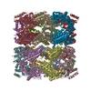
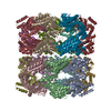
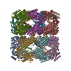
| タイトル | GroEL at sub-nanometer resolution by Constrained Single Particle Tomography | |||||||||
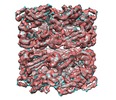
 マップデータ マップデータ | Reconstruction of GroEL | |||||||||
 試料 試料 |
| |||||||||
| 機能・相同性 |  機能・相同性情報 機能・相同性情報chaperonin ATPase / isomerase activity / ATP-dependent protein folding chaperone / unfolded protein binding / protein refolding / ATP binding / cytoplasm 類似検索 - 分子機能 | |||||||||
| 生物種 |  | |||||||||
| 手法 | サブトモグラム平均法 / クライオ電子顕微鏡法 / 解像度: 8.4 Å | |||||||||
 データ登録者 データ登録者 | Bartesaghi A / Lecumberry F / Sapiro G / Subramaniam S | |||||||||
 引用 引用 |  ジャーナル: Structure / 年: 2012 ジャーナル: Structure / 年: 2012タイトル: Protein secondary structure determination by constrained single-particle cryo-electron tomography. 著者: Alberto Bartesaghi / Federico Lecumberry / Guillermo Sapiro / Sriram Subramaniam /  要旨: Cryo-electron microscopy (cryo-EM) is a powerful technique for 3D structure determination of protein complexes by averaging information from individual molecular images. The resolutions that can be ...Cryo-electron microscopy (cryo-EM) is a powerful technique for 3D structure determination of protein complexes by averaging information from individual molecular images. The resolutions that can be achieved with single-particle cryo-EM are frequently limited by inaccuracies in assigning molecular orientations based solely on 2D projection images. Tomographic data collection schemes, however, provide powerful constraints that can be used to more accurately determine molecular orientations necessary for 3D reconstruction. Here, we propose "constrained single-particle tomography" as a general strategy for 3D structure determination in cryo-EM. A key component of our approach is the effective use of images recorded in tilt series to extract high-resolution information and correct for the contrast transfer function. By incorporating geometric constraints into the refinement to improve orientational accuracy of images, we reduce model bias and overrefinement artifacts and demonstrate that protein structures can be determined at resolutions of ∼8 Å starting from low-dose tomographic tilt series. | |||||||||
| 履歴 |
|
- 構造の表示
構造の表示
| ムービー |
 ムービービューア ムービービューア |
|---|---|
| 構造ビューア | EMマップ:  SurfView SurfView Molmil Molmil Jmol/JSmol Jmol/JSmol |
| 添付画像 |
- ダウンロードとリンク
-EMDBアーカイブ
| マップデータ |  emd_2221.map.gz emd_2221.map.gz | 19.9 MB |  EMDBマップデータ形式 EMDBマップデータ形式 | |
|---|---|---|---|---|
| ヘッダ (付随情報) |  emd-2221-v30.xml emd-2221-v30.xml emd-2221.xml emd-2221.xml | 11.9 KB 11.9 KB | 表示 表示 |  EMDBヘッダ EMDBヘッダ |
| 画像 |  emd_2221.png emd_2221.png | 985.6 KB | ||
| アーカイブディレクトリ |  http://ftp.pdbj.org/pub/emdb/structures/EMD-2221 http://ftp.pdbj.org/pub/emdb/structures/EMD-2221 ftp://ftp.pdbj.org/pub/emdb/structures/EMD-2221 ftp://ftp.pdbj.org/pub/emdb/structures/EMD-2221 | HTTPS FTP |
-検証レポート
| 文書・要旨 |  emd_2221_validation.pdf.gz emd_2221_validation.pdf.gz | 270.1 KB | 表示 |  EMDB検証レポート EMDB検証レポート |
|---|---|---|---|---|
| 文書・詳細版 |  emd_2221_full_validation.pdf.gz emd_2221_full_validation.pdf.gz | 269.2 KB | 表示 | |
| XML形式データ |  emd_2221_validation.xml.gz emd_2221_validation.xml.gz | 5.7 KB | 表示 | |
| アーカイブディレクトリ |  https://ftp.pdbj.org/pub/emdb/validation_reports/EMD-2221 https://ftp.pdbj.org/pub/emdb/validation_reports/EMD-2221 ftp://ftp.pdbj.org/pub/emdb/validation_reports/EMD-2221 ftp://ftp.pdbj.org/pub/emdb/validation_reports/EMD-2221 | HTTPS FTP |
-関連構造データ
- リンク
リンク
| EMDBのページ |  EMDB (EBI/PDBe) / EMDB (EBI/PDBe) /  EMDataResource EMDataResource |
|---|---|
| 「今月の分子」の関連する項目 |
- マップ
マップ
| ファイル |  ダウンロード / ファイル: emd_2221.map.gz / 形式: CCP4 / 大きさ: 21.7 MB / タイプ: IMAGE STORED AS FLOATING POINT NUMBER (4 BYTES) ダウンロード / ファイル: emd_2221.map.gz / 形式: CCP4 / 大きさ: 21.7 MB / タイプ: IMAGE STORED AS FLOATING POINT NUMBER (4 BYTES) | ||||||||||||||||||||||||||||||||||||||||||||||||||||||||||||||||||||
|---|---|---|---|---|---|---|---|---|---|---|---|---|---|---|---|---|---|---|---|---|---|---|---|---|---|---|---|---|---|---|---|---|---|---|---|---|---|---|---|---|---|---|---|---|---|---|---|---|---|---|---|---|---|---|---|---|---|---|---|---|---|---|---|---|---|---|---|---|---|
| 注釈 | Reconstruction of GroEL | ||||||||||||||||||||||||||||||||||||||||||||||||||||||||||||||||||||
| 投影像・断面図 | 画像のコントロール
画像は Spider により作成 | ||||||||||||||||||||||||||||||||||||||||||||||||||||||||||||||||||||
| ボクセルのサイズ | X=Y=Z: 1.74 Å | ||||||||||||||||||||||||||||||||||||||||||||||||||||||||||||||||||||
| 密度 |
| ||||||||||||||||||||||||||||||||||||||||||||||||||||||||||||||||||||
| 対称性 | 空間群: 1 | ||||||||||||||||||||||||||||||||||||||||||||||||||||||||||||||||||||
| 詳細 | EMDB XML:
CCP4マップ ヘッダ情報:
| ||||||||||||||||||||||||||||||||||||||||||||||||||||||||||||||||||||
-添付データ
- 試料の構成要素
-全体 : GroEL
| 全体 | 名称: GroEL |
|---|---|
| 要素 |
|
-超分子 #1000: GroEL
| 超分子 | 名称: GroEL / タイプ: sample / ID: 1000 / 集合状態: 14-mer / Number unique components: 1 |
|---|---|
| 分子量 | 理論値: 800 KDa |
-分子 #1: GroEL
| 分子 | 名称: GroEL / タイプ: protein_or_peptide / ID: 1 / コピー数: 1 / 集合状態: 14-mer / 組換発現: Yes |
|---|---|
| 由来(天然) | 生物種:  |
| 分子量 | 理論値: 800 KDa |
| 組換発現 | 生物種:  |
-実験情報
-構造解析
| 手法 | クライオ電子顕微鏡法 |
|---|---|
 解析 解析 | サブトモグラム平均法 |
| 試料の集合状態 | particle |
- 試料調製
試料調製
| 濃度 | 3 mg/mL |
|---|---|
| 緩衝液 | pH: 7.5 詳細: 100 mM Hepes, pH 7.5, 10 mM Mg(OAc)2, 10 mM KOAc, 2 mM DTT |
| グリッド | 詳細: 400 mesh C-flat, 2um hole size (CF/2/2 grids), plasma cleaned for 6s with a Gatan Solarus plasma cleaner. |
| 凍結 | 凍結剤: ETHANE / チャンバー内湿度: 100 % / チャンバー内温度: 93 K / 装置: FEI VITROBOT MARK IV / 手法: Blot for 4 seconds before plunging |
- 電子顕微鏡法
| 顕微鏡 | FEI TITAN KRIOS |
|---|---|
| 温度 | 最低: 80 K / 最高: 93 K / 平均: 80 K |
| アライメント法 | Legacy - 非点収差: Astigmatism corrected at 76000x |
| 詳細 | The total dose of 25 (electrons/square Angstrom) was fractionated evenly across 11 tilted projections taken between 0 and -20 degrees tilt (every 2 degrees). |
| 日付 | 2011年10月7日 |
| 撮影 | カテゴリ: CCD / フィルム・検出器のモデル: GENERIC CCD / 実像数: 1595 / 平均電子線量: 25 e/Å2 詳細: The 1595 micrographs corresponded to 145 tilt-series each containing 11 projections with tilt-angles between 0 and -20 degrees (every 2-degrees). ビット/ピクセル: 16 |
| 電子線 | 加速電圧: 80 kV / 電子線源:  FIELD EMISSION GUN FIELD EMISSION GUN |
| 電子光学系 | 倍率(補正後): 47000 / 照射モード: FLOOD BEAM / 撮影モード: BRIGHT FIELD / Cs: 2.7 mm / 最大 デフォーカス(公称値): 3.0 µm / 最小 デフォーカス(公称値): 2.0 µm / 倍率(公称値): 47000 |
| 試料ステージ | 試料ホルダー: Nitrogen cooled 試料ホルダーモデル: FEI TITAN KRIOS AUTOGRID HOLDER Tilt series - Axis1 - Min angle: -20 ° / Tilt series - Axis1 - Max angle: 0 ° |
| 実験機器 |  モデル: Titan Krios / 画像提供: FEI Company |
- 画像解析
画像解析
| 詳細 | Particles were manually selected in 3D from reconstructed tomograms and their corresponding raw 2D projections extracted for further processing using Constrained Single Particle Tomography. Average number of projections used in the 3D reconstructions: 10000. |
|---|---|
| 最終 再構成 | アルゴリズム: OTHER / 解像度のタイプ: BY AUTHOR / 解像度: 8.4 Å / 解像度の算出法: FSC 0.5 CUT-OFF / ソフトウェア - 名称: FREALIGN 詳細: Final map was amplitude corrected with BSOFT's command 'bampweight' using a density map generated from the PDB ID 3e76 coordinates as a reference. |
| CTF補正 | 詳細: Defocus values were assigned to each particle projection based on the defocus at the untilted plane of each tilt-series and a correction according to the relative height of each particle. to this plane |
| 最終 角度割当 | 詳細: FREALIGN |
-原子モデル構築 1
| 初期モデル | PDB ID: Chain - Chain ID: H |
|---|---|
| ソフトウェア | 名称:  Chimera Chimera |
| 詳細 | Protocol: Rigid body. Coordinates of chain H from 3e76 were fit using D7-symmetric fitting operation in Chimera. |
| 精密化 | 空間: REAL / プロトコル: RIGID BODY FIT / 当てはまり具合の基準: Correlation |
| 得られたモデル |  PDB-2ynj: |
 ムービー
ムービー コントローラー








 Z (Sec.)
 Y (Row.)
 X (Col.)


