 Movie
Movie Controller


+ Open data

- Basic information
| Entry |  | |||||||||
|---|---|---|---|---|---|---|---|---|---|---|
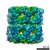
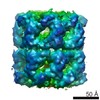
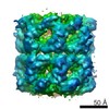
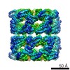
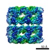
| Title | Symmetry-free cryo-EM map of GroEL-actin | |||||||||
 Map data Map data | ||||||||||
 Sample Sample |
| |||||||||
| Biological species |  | |||||||||
| Method | single particle reconstruction / cryo EM / Resolution: 10.3 Å | |||||||||
 Authors Authors | Milicic G / Balchin D / Hayer-Hartl M / Hartl FU | |||||||||
 Citation Citation | Journal: Cell / Year: 2018 Title: Pathway of Actin Folding Directed by the Eukaryotic Chaperonin TRiC. Authors: David Balchin / Goran Miličić / Mike Strauss / Manajit Hayer-Hartl / F Ulrich Hartl /  Abstract: The hetero-oligomeric chaperonin of eukarya, TRiC, is required to fold the cytoskeletal protein actin. The simpler bacterial chaperonin system, GroEL/GroES, is unable to mediate actin folding. Here, ...The hetero-oligomeric chaperonin of eukarya, TRiC, is required to fold the cytoskeletal protein actin. The simpler bacterial chaperonin system, GroEL/GroES, is unable to mediate actin folding. Here, we use spectroscopic and structural techniques to determine how TRiC promotes the conformational progression of actin to the native state. We find that actin fails to fold spontaneously even in the absence of aggregation but populates a kinetically trapped, conformationally dynamic state. Binding of this frustrated intermediate to TRiC specifies an extended topology of actin with native-like secondary structure. In contrast, GroEL stabilizes bound actin in an unfolded state. ATP binding to TRiC effects an asymmetric conformational change in the chaperonin ring. This step induces the partial release of actin, priming it for folding upon complete release into the chaperonin cavity, mediated by ATP hydrolysis. Our results reveal how the unique features of TRiC direct the folding pathway of an obligate eukaryotic substrate. | |||||||||
| History |
|
- Structure visualization
| Structure viewer | EM map:  SurfViewMolmilJmol/JSmol SurfViewMolmilJmol/JSmol |
|---|---|
| Supplemental images |

- Downloads & links
-EMDB archive
| Map data | emd_0015.map.gz | 5 MB | EMDB map data format | |
|---|---|---|---|---|
| Header (meta data) | emd-0015-v30.xmlemd-0015.xml | 7.5 KB 7.5 KB | Display Display | EMDB header |
| FSC (resolution estimation) | emd_0015_fsc.xml | 8.9 KB | Display | FSC data file |
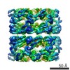
| Images | 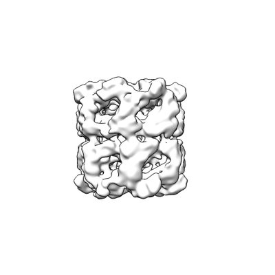 emd_0015.png emd_0015.png | 22 KB | ||
| Archive directory |  http://ftp.pdbj.org/pub/emdb/structures/EMD-0015ftp://ftp.pdbj.org/pub/emdb/structures/EMD-0015 http://ftp.pdbj.org/pub/emdb/structures/EMD-0015ftp://ftp.pdbj.org/pub/emdb/structures/EMD-0015 | HTTPS FTP |
-Related structure data


-Links
| EMDB pages | EMDB (EBI/PDBe) / EMDataResource |
|---|
-Map
| File | Download / File: emd_0015.map.gz / Format: CCP4 / Size: 64 MB / Type: IMAGE STORED AS FLOATING POINT NUMBER (4 BYTES) | ||||||||||||||||||||||||||||||||||||
|---|---|---|---|---|---|---|---|---|---|---|---|---|---|---|---|---|---|---|---|---|---|---|---|---|---|---|---|---|---|---|---|---|---|---|---|---|---|
| Projections & slices | Image control
Images are generated by Spider. | ||||||||||||||||||||||||||||||||||||
| Voxel size | X=Y=Z: 1.85 Å | ||||||||||||||||||||||||||||||||||||
| Density |
| ||||||||||||||||||||||||||||||||||||
| Symmetry | Space group: 1 | ||||||||||||||||||||||||||||||||||||
| Details | EMDB XML:
|
 Z (Sec.)
 Y (Row.)
 X (Col.)


-Supplemental data
- Sample components
-Entire : GroEL in complex with actin
| Entire | Name: GroEL in complex with actin |
|---|---|
| Components |
|
-Supramolecule #1: GroEL in complex with actin
| Supramolecule | Name: GroEL in complex with actin / type: complex / ID: 1 / Parent: 0 / Macromolecule list: #1 |
|---|---|
| Source (natural) | Organism: |
| Recombinant expression | Organism: |
| Molecular weight | Theoretical: 842 KDa |
-Experimental details
-Structure determination
| Method | cryo EM |
|---|---|
 Processing Processing | single particle reconstruction |
| Aggregation state | particle |
-Sample preparation
| Buffer | pH: 7.4 |
|---|---|
| Vitrification | Cryogen name: ETHANE |
- Electron microscopy
| Microscope | FEI TITAN |
|---|---|
| Image recording | Film or detector model: FEI FALCON II (4k x 4k) / Average electron dose: 40.0 e/Å2 |
| Electron beam | Acceleration voltage: 300 kV / Electron source:  FIELD EMISSION GUN FIELD EMISSION GUN |
| Electron optics | Illumination mode: FLOOD BEAM / Imaging mode: BRIGHT FIELD |
-Image processing
| Final reconstruction | Applied symmetry - Point group: C1 (asymmetric) / Resolution.type: BY AUTHOR / Resolution: 10.3 Å / Resolution method: FSC 0.143 CUT-OFF / Number images used: 10424 |
|---|---|
| Initial angle assignment | Type: ANGULAR RECONSTITUTION |
| Final angle assignment | Type: ANGULAR RECONSTITUTION |
| FSC plot (resolution estimation) |  |
