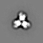
 Movie
Movie Controller

[English] 日本語
 Yorodumi
- EMDB-22112: HIV-1 Envelope Glycoprotein BG505 SOSIP.664, expressed in HEK293S... -
+ Open data

- Basic information
| Entry | Database: EMDB / ID: EMD-22112 | ||||||||||||
|---|---|---|---|---|---|---|---|---|---|---|---|---|---|
| Title | HIV-1 Envelope Glycoprotein BG505 SOSIP.664, expressed in HEK293S cells and deglycosylated by endoglycosidase H, in complex with RM20A3 Fab | ||||||||||||
 Map data Map data | |||||||||||||
 Sample Sample |
| ||||||||||||
 Keywords Keywords | HIV-1 / Envelope / glycoprotein / spike / VIRAL PROTEIN-IMMUNE SYSTEM complex | ||||||||||||
| Function / homology |  Function and homology information Function and homology informationsymbiont-mediated perturbation of host defense response / positive regulation of plasma membrane raft polarization / positive regulation of receptor clustering / host cell endosome membrane / clathrin-dependent endocytosis of virus by host cell / viral protein processing / fusion of virus membrane with host plasma membrane / fusion of virus membrane with host endosome membrane / viral envelope / virion attachment to host cell ...symbiont-mediated perturbation of host defense response / positive regulation of plasma membrane raft polarization / positive regulation of receptor clustering / host cell endosome membrane / clathrin-dependent endocytosis of virus by host cell / viral protein processing / fusion of virus membrane with host plasma membrane / fusion of virus membrane with host endosome membrane / viral envelope / virion attachment to host cell / host cell plasma membrane / virion membrane / structural molecule activity / membrane / identical protein binding Similarity search - Function | ||||||||||||
| Biological species |   Human immunodeficiency virus 1 / Human immunodeficiency virus 1 /  | ||||||||||||
| Method | single particle reconstruction / cryo EM / Resolution: 3.5 Å | ||||||||||||
 Authors Authors | Berndsen ZT / Ward AB | ||||||||||||
| Funding support |  United States, 3 items United States, 3 items
| ||||||||||||
 Citation Citation | Journal: Proc Natl Acad Sci U S A / Year: 2020 Title: Visualization of the HIV-1 Env glycan shield across scales. Authors: Zachary T Berndsen / Srirupa Chakraborty / Xiaoning Wang / Christopher A Cottrell / Jonathan L Torres / Jolene K Diedrich / Cesar A López / John R Yates / Marit J van Gils / James C Paulson ...Authors: Zachary T Berndsen / Srirupa Chakraborty / Xiaoning Wang / Christopher A Cottrell / Jonathan L Torres / Jolene K Diedrich / Cesar A López / John R Yates / Marit J van Gils / James C Paulson / Sandrasegaram Gnanakaran / Andrew B Ward /  Abstract: The dense array of N-linked glycans on the HIV-1 envelope glycoprotein (Env), known as the "glycan shield," is a key determinant of immunogenicity, yet intrinsic heterogeneity confounds typical ...The dense array of N-linked glycans on the HIV-1 envelope glycoprotein (Env), known as the "glycan shield," is a key determinant of immunogenicity, yet intrinsic heterogeneity confounds typical structure-function analysis. Here, we present an integrated approach of single-particle electron cryomicroscopy (cryo-EM), computational modeling, and site-specific mass spectrometry (MS) to probe glycan shield structure and behavior at multiple levels. We found that dynamics lead to an extensive network of interglycan interactions that drive the formation of higher-order structure within the glycan shield. This structure defines diffuse boundaries between buried and exposed protein surface and creates a mapping of potentially immunogenic sites on Env. Analysis of Env expressed in different cell lines revealed how cryo-EM can detect subtle changes in glycan occupancy, composition, and dynamics that impact glycan shield structure and epitope accessibility. Importantly, this identified unforeseen changes in the glycan shield of Env obtained from expression in the same cell line used for vaccine production. Finally, by capturing the enzymatic deglycosylation of Env in a time-resolved manner, we found that highly connected glycan clusters are resistant to digestion and help stabilize the prefusion trimer, suggesting the glycan shield may function beyond immune evasion. | ||||||||||||
| History |
|
- Structure visualization
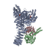
| Movie |
Movie viewer |
|---|---|
| Structure viewer | EM map: SurfViewMolmilJmol/JSmol |
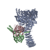
| Supplemental images |

- Downloads & links
-EMDB archive
| Map data | emd_22112.map.gz | 78.4 MB | EMDB map data format | |
|---|---|---|---|---|
| Header (meta data) | emd-22112-v30.xmlemd-22112.xml | 25.8 KB 25.8 KB | Display Display | EMDB header |
| FSC (resolution estimation) | emd_22112_fsc.xml | 10 KB | Display | FSC data file |
| Images | 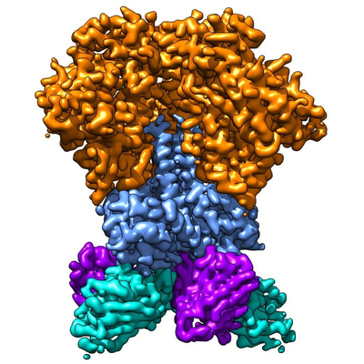 emd_22112.png emd_22112.png | 206 KB | ||
| Filedesc metadata | emd-22112.cif.gz | 6.7 KB | ||
| Others | emd_22112_additional_1.map.gzemd_22112_additional_2.map.gzemd_22112_half_map_1.map.gzemd_22112_half_map_2.map.gz | 5.1 MB 5.2 MB 65.4 MB 65.6 MB | ||
| Archive directory |  http://ftp.pdbj.org/pub/emdb/structures/EMD-22112ftp://ftp.pdbj.org/pub/emdb/structures/EMD-22112 http://ftp.pdbj.org/pub/emdb/structures/EMD-22112ftp://ftp.pdbj.org/pub/emdb/structures/EMD-22112 | HTTPS FTP |
-Related structure data
| Related structure data |  6x9vMC  6x9rC  6x9sC  6x9tC  6x9uC C: citing same article ( M: atomic model generated by this map |
|---|---|
| Similar structure data |


-Links
| EMDB pages | EMDB (EBI/PDBe) / EMDataResource |
|---|---|
| Related items in Molecule of the Month |


-Map
| File | Download / File: emd_22112.map.gz / Format: CCP4 / Size: 83.7 MB / Type: IMAGE STORED AS FLOATING POINT NUMBER (4 BYTES) | ||||||||||||||||||||||||||||||||||||||||||||||||||||||||||||
|---|---|---|---|---|---|---|---|---|---|---|---|---|---|---|---|---|---|---|---|---|---|---|---|---|---|---|---|---|---|---|---|---|---|---|---|---|---|---|---|---|---|---|---|---|---|---|---|---|---|---|---|---|---|---|---|---|---|---|---|---|---|
| Projections & slices | Image control
Images are generated by Spider. | ||||||||||||||||||||||||||||||||||||||||||||||||||||||||||||
| Voxel size | X=Y=Z: 1.15 Å | ||||||||||||||||||||||||||||||||||||||||||||||||||||||||||||
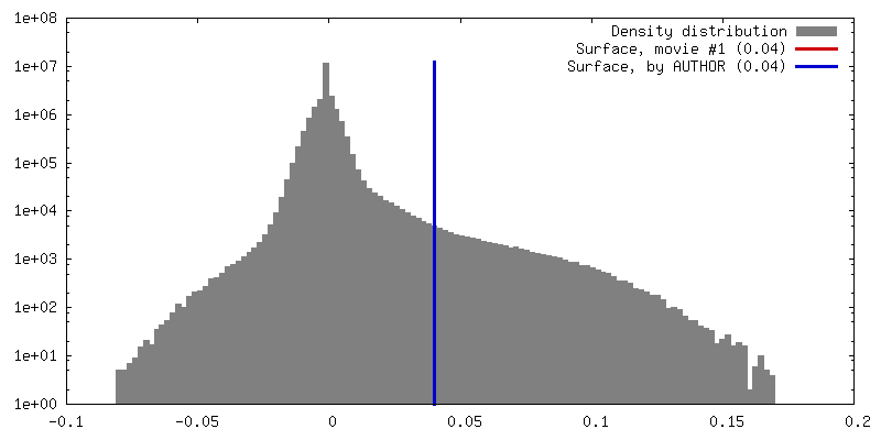
| Density |
| ||||||||||||||||||||||||||||||||||||||||||||||||||||||||||||
| Symmetry | Space group: 1 | ||||||||||||||||||||||||||||||||||||||||||||||||||||||||||||
| Details | EMDB XML:
CCP4 map header:
| ||||||||||||||||||||||||||||||||||||||||||||||||||||||||||||
 Z (Sec.)
 Y (Row.)
 X (Col.)


-Supplemental data
-Additional map: SPARX 3D Variability
| File | emd_22112_additional_1.map | ||||||||||||
|---|---|---|---|---|---|---|---|---|---|---|---|---|---|
| Annotation | SPARX 3D Variability | ||||||||||||
| Projections & Slices |
| ||||||||||||
| Density Histograms |


-Additional map: #1
| File | emd_22112_additional_2.map | ||||||||||||
|---|---|---|---|---|---|---|---|---|---|---|---|---|---|
| Projections & Slices |
| ||||||||||||
| Density Histograms |


-Half map: #1
| File | emd_22112_half_map_1.map | ||||||||||||
|---|---|---|---|---|---|---|---|---|---|---|---|---|---|
| Projections & Slices |
| ||||||||||||
| Density Histograms |


-Half map: #2
| File | emd_22112_half_map_2.map | ||||||||||||
|---|---|---|---|---|---|---|---|---|---|---|---|---|---|
| Projections & Slices |
| ||||||||||||
| Density Histograms |


- Sample components
-Entire : HIV-1 Envelope Glycoprotein BG505 SOSIP.664, expressed in HEK293S...
| Entire | Name: HIV-1 Envelope Glycoprotein BG505 SOSIP.664, expressed in HEK293S cells and deglycosylated by endoglycosidase H, in complex with RM20A3 Fab |
|---|---|
| Components |
|
-Supramolecule #1: HIV-1 Envelope Glycoprotein BG505 SOSIP.664, expressed in HEK293S...
| Supramolecule | Name: HIV-1 Envelope Glycoprotein BG505 SOSIP.664, expressed in HEK293S cells and deglycosylated by endoglycosidase H, in complex with RM20A3 Fab type: complex / ID: 1 / Parent: 0 / Macromolecule list: #1-#4 |
|---|
-Supramolecule #2: HIV-1 Envelope Glycoprotein BG505 SOSIP.664
| Supramolecule | Name: HIV-1 Envelope Glycoprotein BG505 SOSIP.664 / type: complex / ID: 2 / Parent: 1 / Macromolecule list: #1-#2 / Details: deglycosylated by endoglycosidase H |
|---|---|
| Source (natural) | Organism: Human immunodeficiency virus 1 |
-Supramolecule #3: RM20A3 Fab
| Supramolecule | Name: RM20A3 Fab / type: complex / ID: 3 / Parent: 1 / Macromolecule list: #3-#4 |
|---|---|
| Source (natural) | Organism: |
-Macromolecule #1: HIV-1 Envelope Glycoprotein BG505 SOSIP.664 gp120
| Macromolecule | Name: HIV-1 Envelope Glycoprotein BG505 SOSIP.664 gp120 / type: protein_or_peptide / ID: 1 / Number of copies: 1 / Enantiomer: LEVO |
|---|---|
| Source (natural) | Organism: Human immunodeficiency virus 1 |
| Molecular weight | Theoretical: 57.945977 KDa |
| Recombinant expression | Organism:  Homo sapiens (human) Homo sapiens (human) |
| Sequence | String: MDAMKRGLCC VLLLCGAVFV SPSQEIHARF RRGARAENLW VTVYYGVPVW KDAETTLFCA SDAKAYETEK HNVWATHACV PTDPNPQEI HLENVTEEFN MWKNNMVEQM HTDIISLWDQ SLKPCVKLTP LCVTLQCTNV TNNITDDMRG ELKNCSFNMT T ELRDKKQK ...String: MDAMKRGLCC VLLLCGAVFV SPSQEIHARF RRGARAENLW VTVYYGVPVW KDAETTLFCA SDAKAYETEK HNVWATHACV PTDPNPQEI HLENVTEEFN MWKNNMVEQM HTDIISLWDQ SLKPCVKLTP LCVTLQCTNV TNNITDDMRG ELKNCSFNMT T ELRDKKQK VYSLFYRLDV VQINENQGNR SNNSNKEYRL INCNTSAITQ ACPKVSFEPI PIHYCAPAGF AILKCKDKKF NG TGPCPSV STVQCTHGIK PVVSTQLLLN GSLAEEEVMI RSENITNNAK NILVQFNTPV QINCTRPNNN TRKSIRIGPG QAF YATGDI IGDIRQAHCN VSKATWNETL GKVVKQLRKH FGNNTIIRFA NSSGGDLEVT THSFNCGGEF FYCNTSGLFN STWI SNTSV QGSNSTGSND SITLPCRIKQ IINMWQRIGQ AMYAPPIQGV IRCVSNITGL ILTRDGGSTN STTETFRPGG GDMRD NWRS ELYKYKVVKI EPLGVAPTRC KRRVVGRRRR RR UniProtKB: Envelope glycoprotein gp160 |
-Macromolecule #2: HIV-1 Envelope Glycoprotein BG505 SOSIP.664 gp41
| Macromolecule | Name: HIV-1 Envelope Glycoprotein BG505 SOSIP.664 gp41 / type: protein_or_peptide / ID: 2 / Number of copies: 1 / Enantiomer: LEVO |
|---|---|
| Source (natural) | Organism: Human immunodeficiency virus 1 |
| Molecular weight | Theoretical: 17.146482 KDa |
| Recombinant expression | Organism: Homo sapiens (human) |
| Sequence | String: AVGIGAVFLG FLGAAGSTMG AASMTLTVQA RNLLSGIVQQ QSNLLRAPEA QQHLLKLTVW GIKQLQARVL AVERYLRDQQ LLGIWGCSG KLICCTNVPW NSSWSNRNLS EIWDNMTWLQ WDKEISNYTQ IIYGLLEESQ NQQEKNEQDL LALD UniProtKB: Envelope glycoprotein gp160 |
-Macromolecule #3: RM20A3 Fab Heavy Chain
| Macromolecule | Name: RM20A3 Fab Heavy Chain / type: protein_or_peptide / ID: 3 / Number of copies: 1 / Enantiomer: LEVO |
|---|---|
| Source (natural) | Organism: |
| Molecular weight | Theoretical: 13.511111 KDa |
| Recombinant expression | Organism: Homo sapiens (human) |
| Sequence | String: EVQLVETGGG LVQPGGSLKL SCRASGYTFS SFAMSWVRQA PGKGLEWVSL INDRGGLTFY VDSVKGRFTI SRDNSKNTLS LQMHSLRDG DTAVYYCATG GMSSALQSSK YYFDFWGQGA LVTVSS |
-Macromolecule #4: RM20A3 Fab Light Chain
| Macromolecule | Name: RM20A3 Fab Light Chain / type: protein_or_peptide / ID: 4 / Number of copies: 1 / Enantiomer: LEVO |
|---|---|
| Source (natural) | Organism: |
| Molecular weight | Theoretical: 13.5088 KDa |
| Recombinant expression | Organism: Homo sapiens (human) |
| Sequence | String: ALTQPPSVSG SPGQSVTISC TGTSSDIGSY NYVSWYQQHP GKAPKLMIYD VTQRPSGVSD RFSGSKSGNT ASLTISGLQA DDEADYYCS AYAGRQTFYI FGGGTRLTVL GQPKASPTVT LFPPSSEEL |
-Macromolecule #7: 2-acetamido-2-deoxy-beta-D-glucopyranose
| Macromolecule | Name: 2-acetamido-2-deoxy-beta-D-glucopyranose / type: ligand / ID: 7 / Number of copies: 17 / Formula: NAG |
|---|---|
| Molecular weight | Theoretical: 221.208 Da |
| Chemical component information |  ChemComp-NAG: |
-Experimental details
-Structure determination
| Method | cryo EM |
|---|---|
 Processing Processing | single particle reconstruction |
| Aggregation state | particle |
-Sample preparation
| Concentration | 7 mg/mL |
|---|---|
| Buffer | pH: 7.6 / Component - Name: TBS |
| Grid | Details: unspecified |
| Vitrification | Cryogen name: ETHANE / Chamber humidity: 100 % / Chamber temperature: 277.15 K / Instrument: FEI VITROBOT MARK IV |
- Electron microscopy
| Microscope | FEI TITAN KRIOS |
|---|---|
| Image recording | Film or detector model: GATAN K2 SUMMIT (4k x 4k) / Detector mode: COUNTING / Average electron dose: 50.0 e/Å2 |
| Electron beam | Acceleration voltage: 300 kV / Electron source:  FIELD EMISSION GUN FIELD EMISSION GUN |
| Electron optics | Illumination mode: FLOOD BEAM / Imaging mode: DARK FIELD |
| Sample stage | Specimen holder model: FEI TITAN KRIOS AUTOGRID HOLDER / Cooling holder cryogen: NITROGEN |
| Experimental equipment |  Model: Titan Krios / Image courtesy: FEI Company |
+Image processing

-Atomic model buiding 1
| Refinement | Space: REAL / Protocol: FLEXIBLE FIT |
|---|---|
| Output model | PDB-6x9v: |
