 ムービー
ムービー コントローラー


+ データを開く

- 基本情報
基本情報
| 登録情報 | データベース: EMDB / ID: EMD-21969 | |||||||||
|---|---|---|---|---|---|---|---|---|---|---|
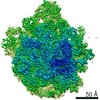
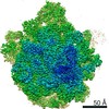
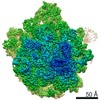
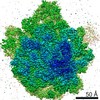
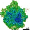
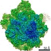
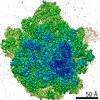
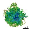
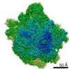
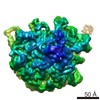
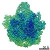
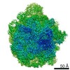
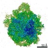
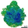
| タイトル | E. coli 50S ribosome bound to compounds 47 and VS1 | |||||||||
 マップデータ マップデータ | ||||||||||
 試料 試料 |
| |||||||||
 キーワード キーワード | E. coli ribosome / streptogramin A analog / antibiotics / RIBOSOME | |||||||||
| 機能・相同性 |  機能・相同性情報 機能・相同性情報transcriptional attenuation / endoribonuclease inhibitor activity / RNA-binding transcription regulator activity / negative regulation of cytoplasmic translation / DnaA-L2 complex / translation repressor activity / negative regulation of DNA-templated DNA replication initiation / ribosome assembly / DNA-templated transcription termination / mRNA 5'-UTR binding ...transcriptional attenuation / endoribonuclease inhibitor activity / RNA-binding transcription regulator activity / negative regulation of cytoplasmic translation / DnaA-L2 complex / translation repressor activity / negative regulation of DNA-templated DNA replication initiation / ribosome assembly / DNA-templated transcription termination / mRNA 5'-UTR binding / large ribosomal subunit / transferase activity / ribosomal large subunit assembly / large ribosomal subunit rRNA binding / cytosolic large ribosomal subunit / cytoplasmic translation / negative regulation of translation / rRNA binding / ribosome / structural constituent of ribosome / translation / response to antibiotic / negative regulation of DNA-templated transcription / mRNA binding / DNA binding / RNA binding / zinc ion binding / cytosol / cytoplasm 類似検索 - 分子機能 | |||||||||
| 生物種 |  Streptomyces virginiae (バクテリア) Streptomyces virginiae (バクテリア) | |||||||||
| 手法 | 単粒子再構成法 / クライオ電子顕微鏡法 / 解像度: 2.75 Å | |||||||||
 データ登録者 データ登録者 | Pellegrino J / Lee DJ | |||||||||
| 資金援助 |  米国, 2件 米国, 2件
| |||||||||
 引用 引用 | ジャーナル: Nature / 年: 2020 タイトル: Synthetic group A streptogramin antibiotics that overcome Vat resistance. 著者: Qi Li / Jenna Pellegrino / D John Lee / Arthur A Tran / Hector A Chaires / Ruoxi Wang / Jesslyn E Park / Kaijie Ji / David Chow / Na Zhang / Axel F Brilot / Justin T Biel / Gydo van Zundert / ...著者: Qi Li / Jenna Pellegrino / D John Lee / Arthur A Tran / Hector A Chaires / Ruoxi Wang / Jesslyn E Park / Kaijie Ji / David Chow / Na Zhang / Axel F Brilot / Justin T Biel / Gydo van Zundert / Kenneth Borrelli / Dean Shinabarger / Cindy Wolfe / Beverly Murray / Matthew P Jacobson / Estelle Mühle / Olivier Chesneau / James S Fraser / Ian B Seiple /   要旨: Natural products serve as chemical blueprints for most antibiotics in clinical use. The evolutionary process by which these molecules arise is inherently accompanied by the co-evolution of resistance ...Natural products serve as chemical blueprints for most antibiotics in clinical use. The evolutionary process by which these molecules arise is inherently accompanied by the co-evolution of resistance mechanisms that shorten the clinical lifetime of any given class of antibiotics. Virginiamycin acetyltransferase (Vat) enzymes are resistance proteins that provide protection against streptogramins, potent antibiotics against Gram-positive bacteria that inhibit the bacterial ribosome. Owing to the challenge of selectively modifying the chemically complex, 23-membered macrocyclic scaffold of group A streptogramins, analogues that overcome the resistance conferred by Vat enzymes have not been previously developed. Here we report the design, synthesis, and antibacterial evaluation of group A streptogramin antibiotics with extensive structural variability. Using cryo-electron microscopy and forcefield-based refinement, we characterize the binding of eight analogues to the bacterial ribosome at high resolution, revealing binding interactions that extend into the peptidyl tRNA-binding site and towards synergistic binders that occupy the nascent peptide exit tunnel. One of these analogues has excellent activity against several streptogramin-resistant strains of Staphylococcus aureus, exhibits decreased rates of acetylation in vitro, and is effective at lowering bacterial load in a mouse model of infection. Our results demonstrate that the combination of rational design and modular chemical synthesis can revitalize classes of antibiotics that are limited by naturally arising resistance mechanisms. | |||||||||
| 履歴 |
|
- 構造の表示
構造の表示
| ムービー |
ムービービューア |
|---|---|
| 構造ビューア | EMマップ: SurfViewMolmilJmol/JSmol |
| 添付画像 |

- ダウンロードとリンク
-EMDBアーカイブ
| マップデータ | emd_21969.map.gz | 763.4 MB | EMDBマップデータ形式 | |
|---|---|---|---|---|
| ヘッダ (付随情報) | emd-21969-v30.xmlemd-21969.xml | 19.9 KB 19.9 KB | 表示 表示 | EMDBヘッダ |
| 画像 |  emd_21969.png emd_21969.png | 175 KB | ||
| Filedesc metadata | emd-21969.cif.gz | 7.8 KB | ||
| アーカイブディレクトリ |  http://ftp.pdbj.org/pub/emdb/structures/EMD-21969ftp://ftp.pdbj.org/pub/emdb/structures/EMD-21969 http://ftp.pdbj.org/pub/emdb/structures/EMD-21969ftp://ftp.pdbj.org/pub/emdb/structures/EMD-21969 | HTTPS FTP |
-検証レポート
| 文書・要旨 | emd_21969_validation.pdf.gz | 708.6 KB | 表示 | EMDB検証レポート |
|---|---|---|---|---|
| 文書・詳細版 | emd_21969_full_validation.pdf.gz | 708.2 KB | 表示 | |
| XML形式データ | emd_21969_validation.xml.gz | 8.8 KB | 表示 | |
| CIF形式データ | emd_21969_validation.cif.gz | 10.3 KB | 表示 | |
| アーカイブディレクトリ | https://ftp.pdbj.org/pub/emdb/validation_reports/EMD-21969ftp://ftp.pdbj.org/pub/emdb/validation_reports/EMD-21969 | HTTPS FTP |
-関連構造データ
| 関連構造データ |  6wyvMC 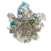 6pc5C 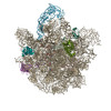 6pc6C  6pc7C  6pc8C  6pchC 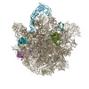 6pcqC  6pcrC 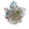 6pcsC  6pctC  6x3cC 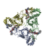 6x3jC M: このマップから作成された原子モデル C: 同じ文献を引用 ( |
|---|---|
| 類似構造データ | |
| 電子顕微鏡画像生データ | EMPIAR-10528 (タイトル: E. coli 50S ribosome bound to compounds 47 and VS1 Data size: 127.5 Data #1: Unaligned movies of 50S ribosome complex bound to compound 47 and VS1 [micrographs - multiframe]) |


-リンク
| EMDBのページ | EMDB (EBI/PDBe) / EMDataResource |
|---|---|
| 「今月の分子」の関連する項目 |


-マップ
| ファイル | ダウンロード / ファイル: emd_21969.map.gz / 形式: CCP4 / 大きさ: 824 MB / タイプ: IMAGE STORED AS FLOATING POINT NUMBER (4 BYTES) | ||||||||||||||||||||||||||||||||||||||||||||||||||||||||||||||||||||
|---|---|---|---|---|---|---|---|---|---|---|---|---|---|---|---|---|---|---|---|---|---|---|---|---|---|---|---|---|---|---|---|---|---|---|---|---|---|---|---|---|---|---|---|---|---|---|---|---|---|---|---|---|---|---|---|---|---|---|---|---|---|---|---|---|---|---|---|---|---|
| 投影像・断面図 | 画像のコントロール
画像は Spider により作成 | ||||||||||||||||||||||||||||||||||||||||||||||||||||||||||||||||||||
| ボクセルのサイズ | X=Y=Z: 0.8261 Å | ||||||||||||||||||||||||||||||||||||||||||||||||||||||||||||||||||||
| 密度 |
| ||||||||||||||||||||||||||||||||||||||||||||||||||||||||||||||||||||
| 対称性 | 空間群: 1 | ||||||||||||||||||||||||||||||||||||||||||||||||||||||||||||||||||||
| 詳細 | EMDB XML:
CCP4マップ ヘッダ情報:
| ||||||||||||||||||||||||||||||||||||||||||||||||||||||||||||||||||||
 Z (Sec.)
 Y (Row.)
 X (Col.)


-添付データ
- 試料の構成要素
+全体 : 50S E. coli ribosome
+超分子 #1: 50S E. coli ribosome
+分子 #1: 23S ribosomal RNA
+分子 #2: 5S ribosomal RNA
+分子 #3: 50S ribosomal protein L2
+分子 #4: 50S ribosomal protein L15
+分子 #5: 50S ribosomal protein L4
+分子 #6: 50S ribosomal protein L3
+分子 #7: 50S ribosomal protein L13
+分子 #8: VIRGINIAMYCIN S1
+分子 #9: (3R,4R,5E,10E,12E,14S,16R,26aR)-16-fluoro-14-hydroxy-12-methyl-3-...

-実験情報
-構造解析
| 手法 | クライオ電子顕微鏡法 |
|---|---|
 解析 解析 | 単粒子再構成法 |
| 試料の集合状態 | particle |
-試料調製
| 緩衝液 | pH: 7.5 |
|---|---|
| 凍結 | 凍結剤: ETHANE |
- 電子顕微鏡法
| 顕微鏡 | FEI TITAN KRIOS |
|---|---|
| 撮影 | フィルム・検出器のモデル: GATAN K3 BIOQUANTUM (6k x 4k) 平均電子線量: 79.3 e/Å2 |
| 電子線 | 加速電圧: 300 kV / 電子線源:  FIELD EMISSION GUN FIELD EMISSION GUN |
| 電子光学系 | 照射モード: FLOOD BEAM / 撮影モード: BRIGHT FIELD |
| 実験機器 |  モデル: Titan Krios / 画像提供: FEI Company |
-画像解析
| 初期モデル | モデルのタイプ: NONE |
|---|---|
| 最終 再構成 | 解像度のタイプ: BY AUTHOR / 解像度: 2.75 Å / 解像度の算出法: FSC 0.143 CUT-OFF / 使用した粒子像数: 19484 |
| 初期 角度割当 | タイプ: PROJECTION MATCHING |
| 最終 角度割当 | タイプ: PROJECTION MATCHING |
