 Movie
Movie Controller


+ Open data

- Basic information
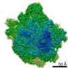
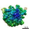
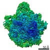
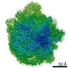
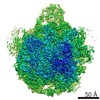
| Entry | Database: EMDB / ID: EMD-20353 | ||||||||||||
|---|---|---|---|---|---|---|---|---|---|---|---|---|---|
| Title | High resolution cryo-EM structure of E.coli 50S | ||||||||||||
 Map data Map data | |||||||||||||
 Sample Sample |
| ||||||||||||
 Keywords Keywords | RIBOSOME | ||||||||||||
| Function / homology |  Function and homology information Function and homology informationlarge ribosomal subunit / transferase activity / 5S rRNA binding / ribosomal large subunit assembly / cytosolic large ribosomal subunit / cytoplasmic translation / tRNA binding / rRNA binding / structural constituent of ribosome / ribosome ...large ribosomal subunit / transferase activity / 5S rRNA binding / ribosomal large subunit assembly / cytosolic large ribosomal subunit / cytoplasmic translation / tRNA binding / rRNA binding / structural constituent of ribosome / ribosome / translation / ribonucleoprotein complex / metal ion binding / cytosol / cytoplasm Similarity search - Function | ||||||||||||
| Biological species |  | ||||||||||||
| Method | single particle reconstruction / cryo EM / Resolution: 2.2 Å | ||||||||||||
 Authors Authors | Stojkovic V / Myasnikov A | ||||||||||||
| Funding support |  United States, 3 items United States, 3 items
| ||||||||||||
 Citation Citation | Journal: Nucleic Acids Res / Year: 2020 Title: Assessment of the nucleotide modifications in the high-resolution cryo-electron microscopy structure of the Escherichia coli 50S subunit. Authors: Vanja Stojković / Alexander G Myasnikov / Iris D Young / Adam Frost / James S Fraser / Danica Galonić Fujimori / Abstract: Post-transcriptional ribosomal RNA (rRNA) modifications are present in all organisms, but their exact functional roles and positions are yet to be fully characterized. Modified nucleotides have been ...Post-transcriptional ribosomal RNA (rRNA) modifications are present in all organisms, but their exact functional roles and positions are yet to be fully characterized. Modified nucleotides have been implicated in the stabilization of RNA structure and regulation of ribosome biogenesis and protein synthesis. In some instances, rRNA modifications can confer antibiotic resistance. High-resolution ribosome structures are thus necessary for precise determination of modified nucleotides' positions, a task that has previously been accomplished by X-ray crystallography. Here, we present a cryo-electron microscopy (cryo-EM) structure of the Escherichia coli 50S subunit at an average resolution of 2.2 Å as an additional approach for mapping modification sites. Our structure confirms known modifications present in 23S rRNA and additionally allows for localization of Mg2+ ions and their coordinated water molecules. Using our cryo-EM structure as a testbed, we developed a program for assessment of cryo-EM map quality. This program can be easily used on any RNA-containing cryo-EM structure, and an associated Coot plugin allows for visualization of validated modifications, making it highly accessible. | ||||||||||||
| History |
|
- Structure visualization
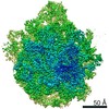
| Movie |
Movie viewer |
|---|---|
| Structure viewer | EM map: SurfViewMolmilJmol/JSmol |
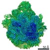
| Supplemental images |

- Downloads & links
-EMDB archive
| Map data | emd_20353.map.gz | 475.8 MB | EMDB map data format | |
|---|---|---|---|---|
| Header (meta data) | emd-20353-v30.xmlemd-20353.xml | 54.2 KB 54.2 KB | Display Display | EMDB header |
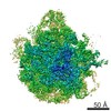
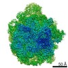
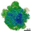
| Images | 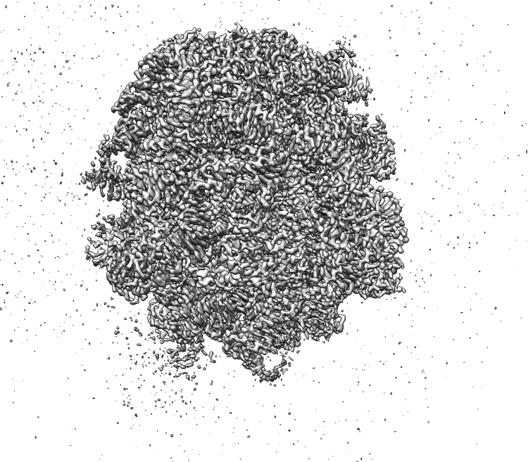 emd_20353.png emd_20353.png | 398.3 KB | ||
| Filedesc metadata | emd-20353.cif.gz | 12.1 KB | ||
| Archive directory |  http://ftp.pdbj.org/pub/emdb/structures/EMD-20353ftp://ftp.pdbj.org/pub/emdb/structures/EMD-20353 http://ftp.pdbj.org/pub/emdb/structures/EMD-20353ftp://ftp.pdbj.org/pub/emdb/structures/EMD-20353 | HTTPS FTP |
-Related structure data
| Related structure data | 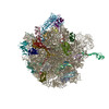 6pj6MC M: atomic model generated by this map C: citing same article ( |
|---|---|
| Similar structure data |
-Links
| EMDB pages | EMDB (EBI/PDBe) / EMDataResource |
|---|---|
| Related items in Molecule of the Month |


-Map
| File | Download / File: emd_20353.map.gz / Format: CCP4 / Size: 512 MB / Type: IMAGE STORED AS FLOATING POINT NUMBER (4 BYTES) | ||||||||||||||||||||||||||||||||||||||||||||||||||||||||||||
|---|---|---|---|---|---|---|---|---|---|---|---|---|---|---|---|---|---|---|---|---|---|---|---|---|---|---|---|---|---|---|---|---|---|---|---|---|---|---|---|---|---|---|---|---|---|---|---|---|---|---|---|---|---|---|---|---|---|---|---|---|---|
| Projections & slices | Image control
Images are generated by Spider. | ||||||||||||||||||||||||||||||||||||||||||||||||||||||||||||
| Voxel size | X=Y=Z: 0.822 Å | ||||||||||||||||||||||||||||||||||||||||||||||||||||||||||||
| Density |
| ||||||||||||||||||||||||||||||||||||||||||||||||||||||||||||
| Symmetry | Space group: 1 | ||||||||||||||||||||||||||||||||||||||||||||||||||||||||||||
| Details | EMDB XML:
CCP4 map header:
| ||||||||||||||||||||||||||||||||||||||||||||||||||||||||||||
 Z (Sec.)
 Y (Row.)
 X (Col.)


-Supplemental data
- Sample components
+Entire : Escherichia coli 50S subunit
+Supramolecule #1: Escherichia coli 50S subunit
+Macromolecule #1: 23S rRNA
+Macromolecule #2: 5S rRNA
+Macromolecule #3: 50S ribosomal protein L2
+Macromolecule #4: 50S ribosomal protein L3
+Macromolecule #5: 50S ribosomal protein L4
+Macromolecule #6: 50S ribosomal protein L5
+Macromolecule #7: 50S ribosomal protein L6
+Macromolecule #8: 50S ribosomal protein L9
+Macromolecule #9: 50S ribosomal protein L11
+Macromolecule #10: 50S ribosomal protein L13
+Macromolecule #11: 50S ribosomal protein L14
+Macromolecule #12: 50S ribosomal protein L15
+Macromolecule #13: 50S ribosomal protein L16
+Macromolecule #14: 50S ribosomal protein L17
+Macromolecule #15: 50S ribosomal protein L18
+Macromolecule #16: 50S ribosomal protein L19
+Macromolecule #17: 50S ribosomal protein L20
+Macromolecule #18: 50S ribosomal protein L21
+Macromolecule #19: 50S ribosomal protein L22
+Macromolecule #20: 50S ribosomal protein L23
+Macromolecule #21: 50S ribosomal protein L24
+Macromolecule #22: 50S ribosomal protein L25
+Macromolecule #23: 50S ribosomal protein L27
+Macromolecule #24: 50S ribosomal protein L28
+Macromolecule #25: 50S ribosomal protein L29
+Macromolecule #26: 50S ribosomal protein L30
+Macromolecule #27: 50S ribosomal protein L32
+Macromolecule #28: 50S ribosomal protein L33
+Macromolecule #29: 50S ribosomal protein L34
+Macromolecule #30: 50S ribosomal protein L35
+Macromolecule #31: 50S ribosomal protein L36
+Macromolecule #32: MAGNESIUM ION
+Macromolecule #33: SODIUM ION
+Macromolecule #34: ZINC ION
+Macromolecule #35: water

-Experimental details
-Structure determination
| Method | cryo EM |
|---|---|
 Processing Processing | single particle reconstruction |
| Aggregation state | particle |
-Sample preparation
| Concentration | 1 mg/mL |
|---|---|
| Buffer | pH: 7.5 / Component - Concentration: 10.0 mM / Component - Formula: TRIS / Component - Name: TRIS / Details: Buffer A |
| Grid | Model: Quantifoil R1.2/1.3 / Material: COPPER / Mesh: 300 / Support film - Material: CARBON / Support film - topology: HOLEY / Support film - Film thickness: 2 / Pretreatment - Type: GLOW DISCHARGE / Pretreatment - Time: 30 sec. / Pretreatment - Atmosphere: AIR / Details: easyGlow settings |
| Vitrification | Cryogen name: ETHANE / Chamber humidity: 95 % / Chamber temperature: 10 K / Instrument: FEI VITROBOT MARK IV Details: Blot Force 5 Blot Time 10sec Hum 95% Temperature 10C Waiting time 1 min. |
| Details | 50S ribosomal subunit was purified from E. coli MRE600 using modified version of previously published protocol (Mehta et al. 2012) |
- Electron microscopy
| Microscope | FEI TITAN KRIOS |
|---|---|
| Temperature | Min: 86.0 K / Max: 130.0 K |
| Alignment procedure | Coma free - Residual tilt: 10.0 mrad |
| Image recording | Film or detector model: GATAN K2 SUMMIT (4k x 4k) / Detector mode: SUPER-RESOLUTION / Digitization - Dimensions - Width: 3838 pixel / Digitization - Dimensions - Height: 3710 pixel / Digitization - Frames/image: 0-80 / Number grids imaged: 1 / Number real images: 1889 / Average exposure time: 8.0 sec. / Average electron dose: 80.0 e/Å2 |
| Electron beam | Acceleration voltage: 300 kV / Electron source:  FIELD EMISSION GUN FIELD EMISSION GUN |
| Electron optics | C2 aperture diameter: 70.0 µm / Calibrated defocus max: 1.5 µm / Calibrated defocus min: 0.5 µm / Calibrated magnification: 60827 / Illumination mode: OTHER / Imaging mode: BRIGHT FIELD / Cs: 2.7 mm / Nominal defocus max: 1.2 µm / Nominal defocus min: 0.3 µm / Nominal magnification: 59000 |
| Sample stage | Specimen holder model: FEI TITAN KRIOS AUTOGRID HOLDER / Cooling holder cryogen: NITROGEN |
| Experimental equipment |  Model: Titan Krios / Image courtesy: FEI Company |
