 Movie
Movie Controller

[English] 日本語
 Yorodumi
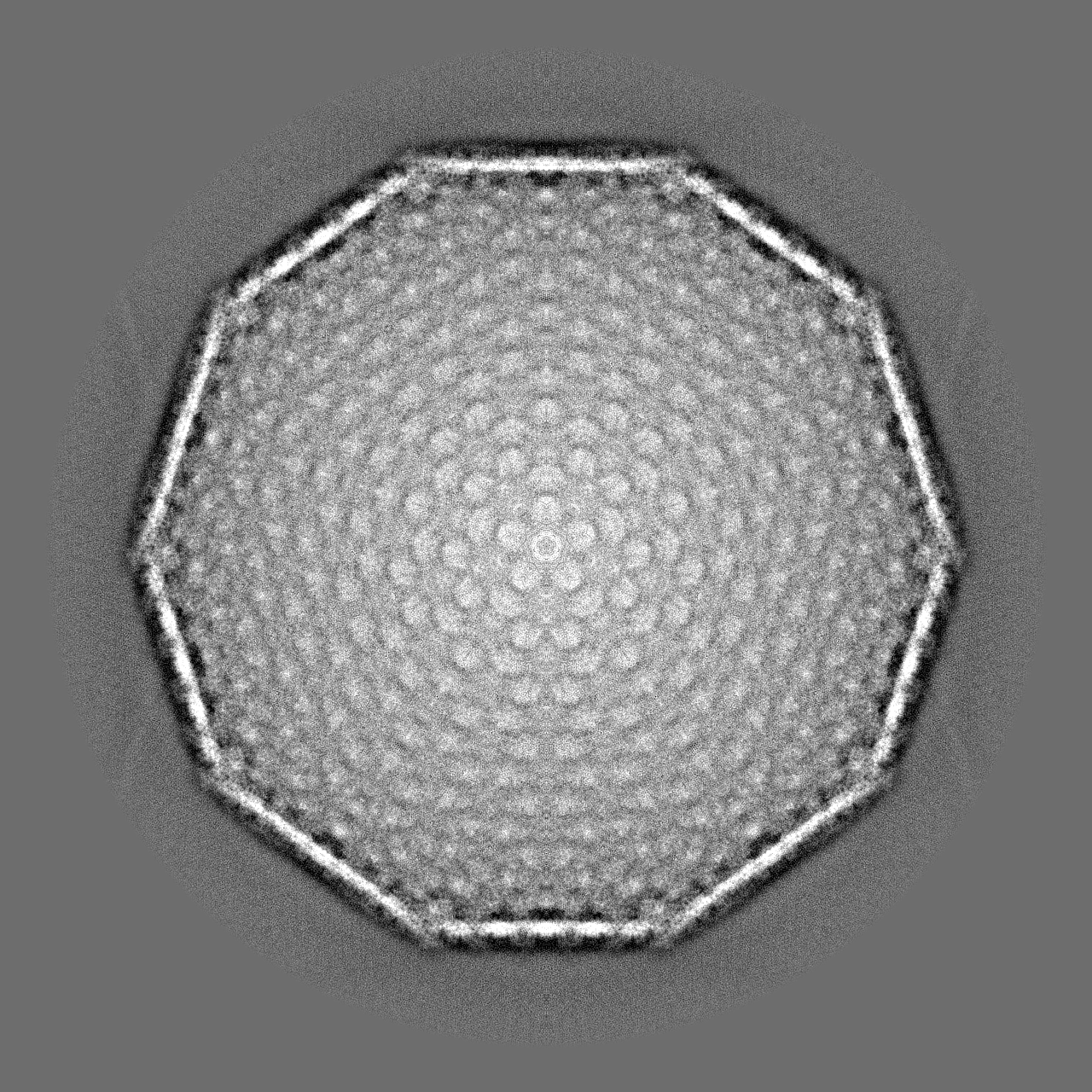
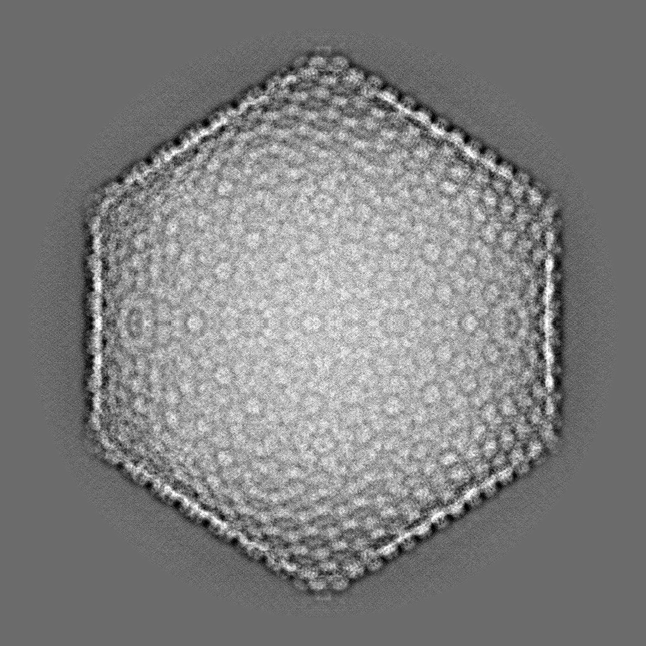
- EMDB-21695: Full phage G capsid cryoEM structure at 6.1 Angstrom resolution -
+ Open data

- Basic information
| Entry | Database: EMDB / ID: EMD-21695 | |||||||||
|---|---|---|---|---|---|---|---|---|---|---|
| Title | Full phage G capsid cryoEM structure at 6.1 Angstrom resolution | |||||||||
 Map data Map data | ||||||||||
 Sample Sample |
| |||||||||
| Function / homology | : / Phage capsid / Phage capsid family / Gp26 / Gp27 Function and homology information Function and homology information | |||||||||
| Biological species |  Bacillus virus G Bacillus virus G | |||||||||
| Method | single particle reconstruction / cryo EM / Resolution: 6.1 Å | |||||||||
 Authors Authors | Gonzalez B / Monroe L / Kunpeng L / Yan R / Wright E / Walter T / Kihara D / Weintraub SE / Julie AT / Philip S / Jiang W | |||||||||
 Citation Citation | Journal: J Mol Biol / Year: 2020 Title: Phage G Structure at 6.1 Å Resolution, Condensed DNA, and Host Identity Revision to a Lysinibacillus. Authors: Brenda González / Lyman Monroe / Kunpeng Li / Rui Yan / Elena Wright / Thomas Walter / Daisuke Kihara / Susan T Weintraub / Julie A Thomas / Philip Serwer / Wen Jiang /  Abstract: Phage G has the largest capsid and genome of any known propagated phage. Many aspects of its structure, assembly, and replication have not been elucidated. Herein, we present the dsDNA-packed and ...Phage G has the largest capsid and genome of any known propagated phage. Many aspects of its structure, assembly, and replication have not been elucidated. Herein, we present the dsDNA-packed and empty phage G capsid at 6.1 and 9 Å resolution, respectively, using cryo-EM for structure determination and mass spectrometry for protein identification. The major capsid protein, gp27, is identified and found to share the HK97-fold universally conserved in all previously solved dsDNA phages. Trimers of the decoration protein, gp26, sit on the 3-fold axes and are thought to enhance the interactions of the hexameric capsomeres of gp27, for other phages encoding decoration proteins. Phage G's decoration protein is longer than what has been reported in other phages, and we suspect the extra interaction surface area helps stabilize the capsid. We identified several additional capsid proteins, including a candidate for the prohead protease responsible for processing gp27. Furthermore, cryo-EM reveals a range of partially full, condensed DNA densities that appear to have no contact with capsid shell. Three analyses confirm that the phage G host is a Lysinibacillus, and not Bacillus megaterium: identity of host proteins in our mass spectrometry analyses, genome sequence of the phage G host, and host range of phage G. | |||||||||
| History |
|
- Structure visualization
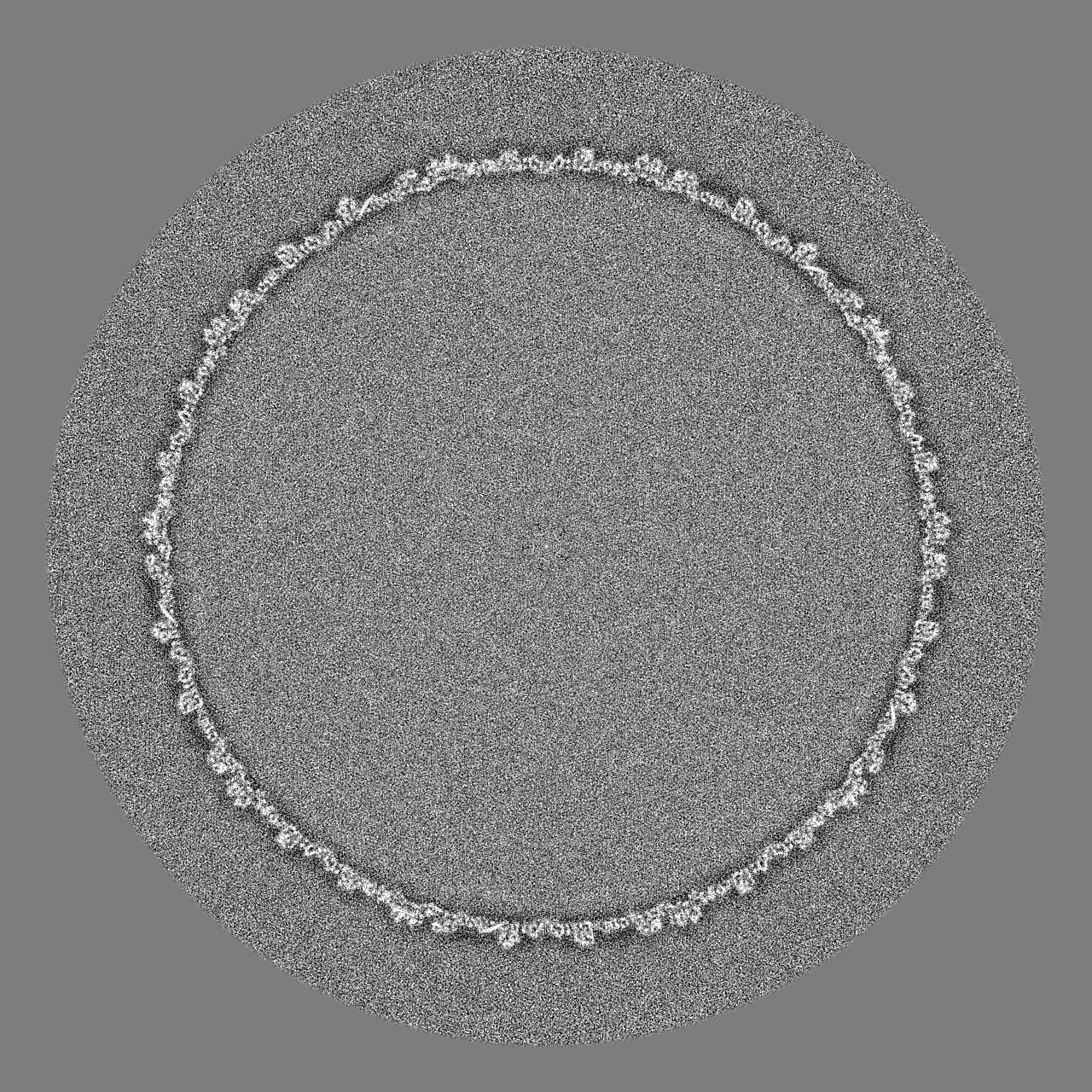
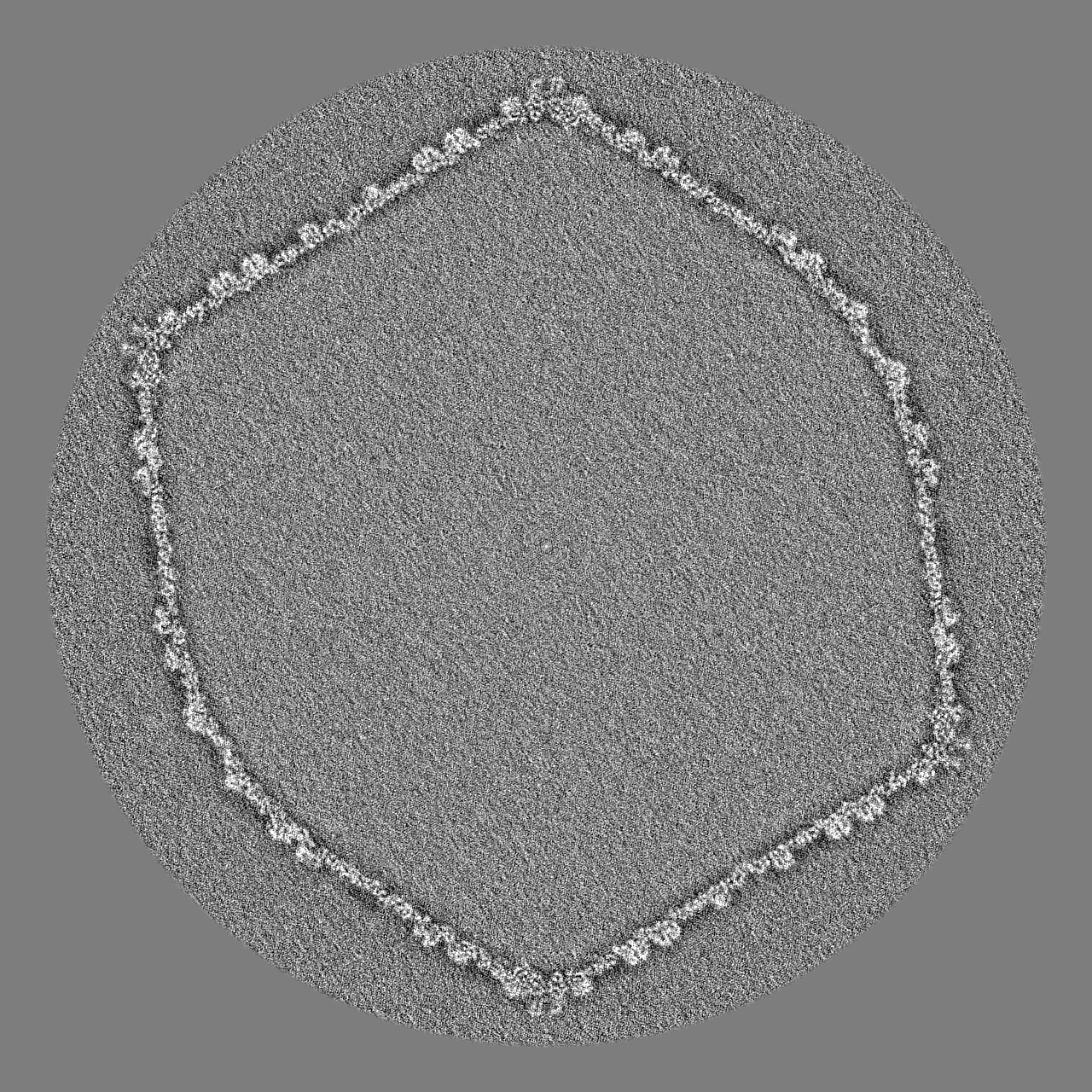
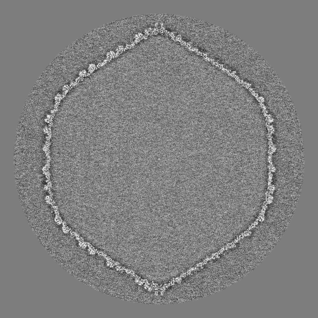
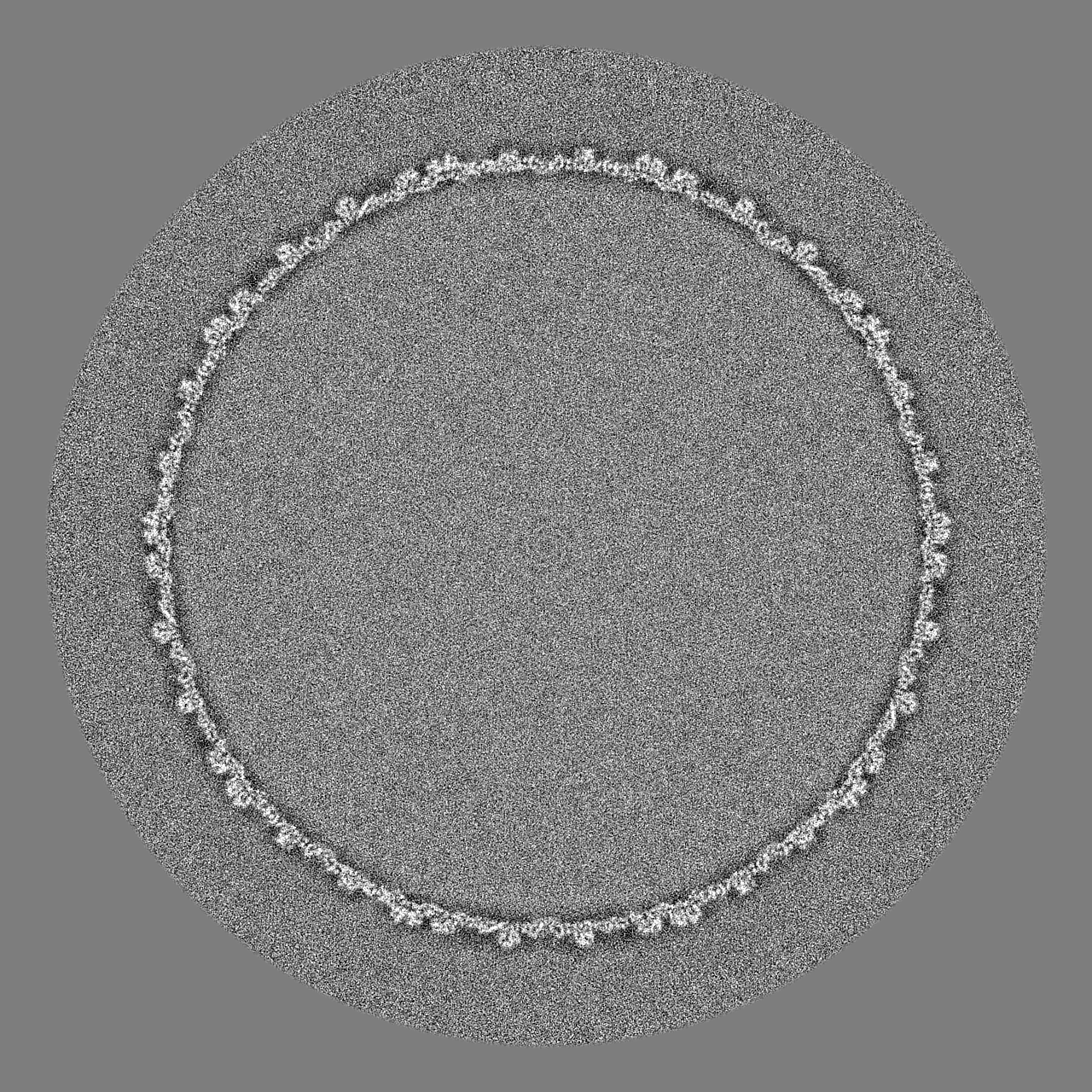
| Movie |
Movie viewer |
|---|---|
| Structure viewer | EM map: SurfViewMolmilJmol/JSmol |
| Supplemental images |

- Downloads & links
-EMDB archive
| Map data | emd_21695.map.gz | 7.3 GB | EMDB map data format | |
|---|---|---|---|---|
| Header (meta data) | emd-21695-v30.xmlemd-21695.xml | 14.8 KB 14.8 KB | Display Display | EMDB header |
| FSC (resolution estimation) | emd_21695_fsc.xml | 45.1 KB | Display | FSC data file |
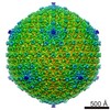
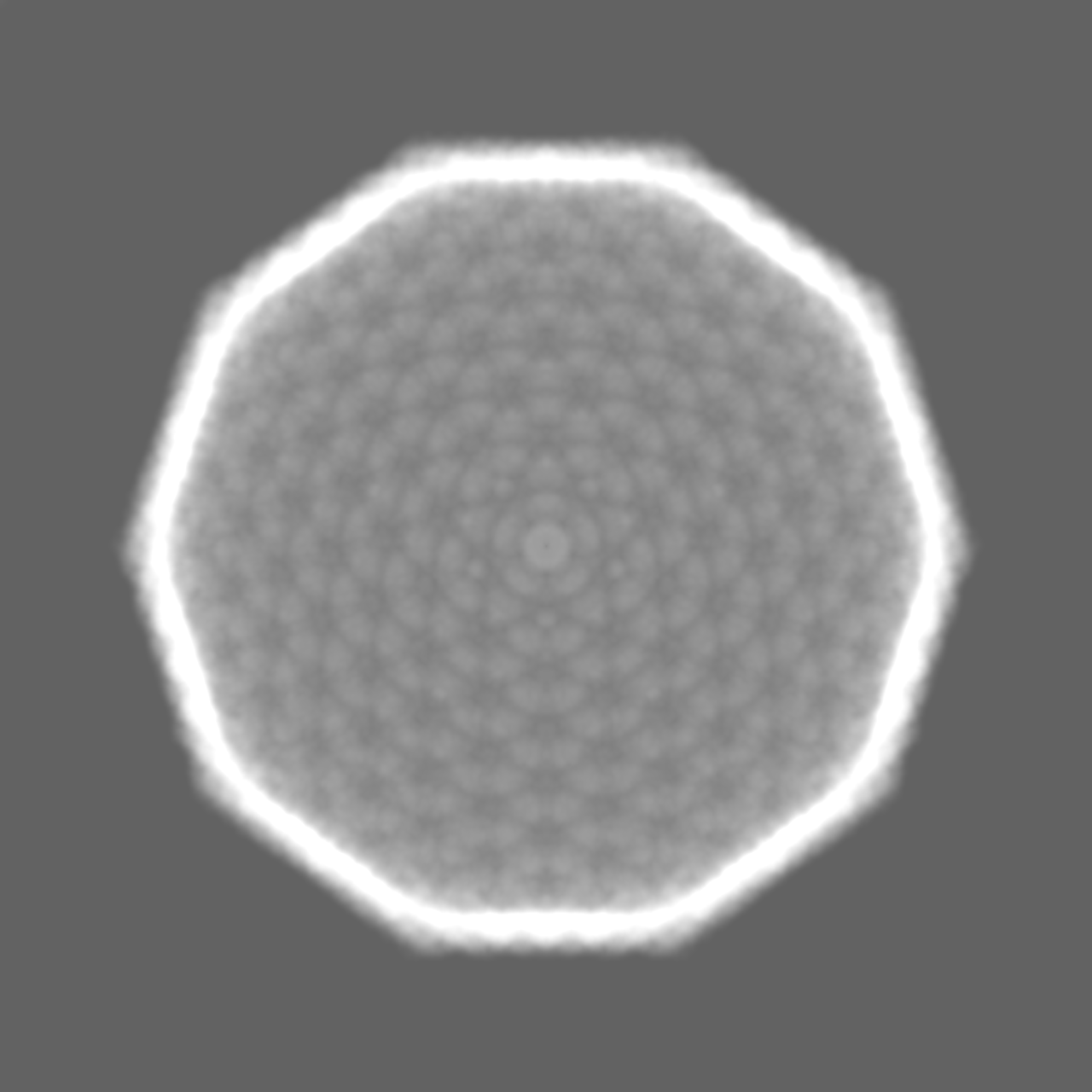
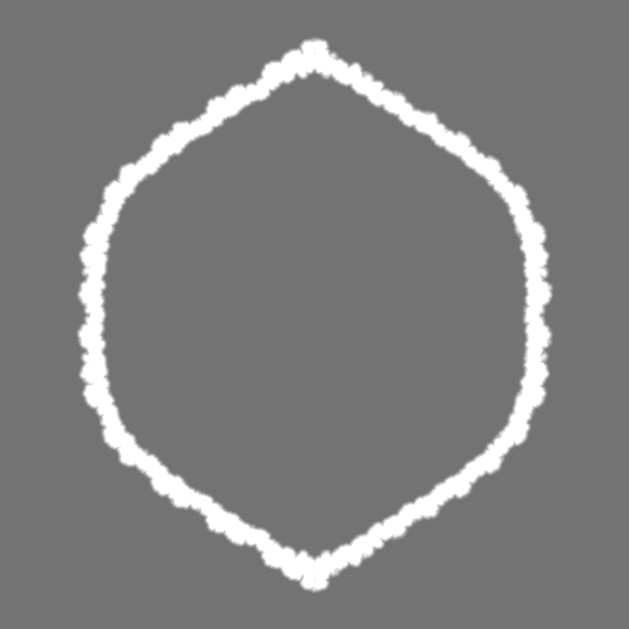
| Images | 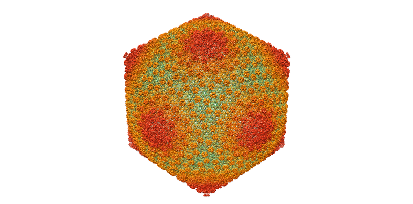 emd_21695.png emd_21695.png | 232.2 KB | ||
| Masks | emd_21695_msk_1.mapemd_21695_msk_2.map | 7.8 GB 7.8 GB | Mask map | |
| Others | emd_21695_half_map_1.map.gzemd_21695_half_map_2.map.gz | 3 GB 3 GB | ||
| Archive directory |  http://ftp.pdbj.org/pub/emdb/structures/EMD-21695ftp://ftp.pdbj.org/pub/emdb/structures/EMD-21695 http://ftp.pdbj.org/pub/emdb/structures/EMD-21695ftp://ftp.pdbj.org/pub/emdb/structures/EMD-21695 | HTTPS FTP |
-Related structure data
| Related structure data |  6wkkMC M: atomic model generated by this map C: citing same article ( |
|---|---|
| Similar structure data |


-Links
| EMDB pages | EMDB (EBI/PDBe) / EMDataResource |
|---|---|
| Related items in Molecule of the Month |


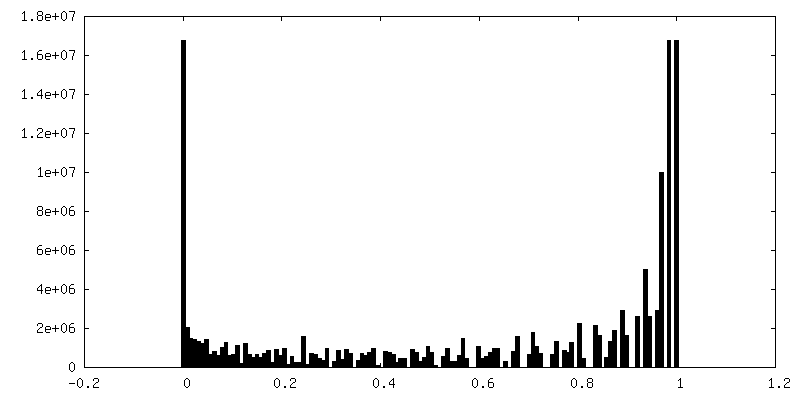
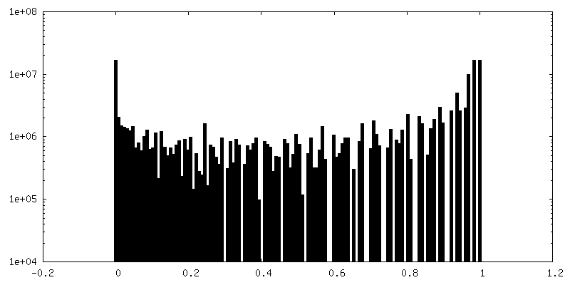
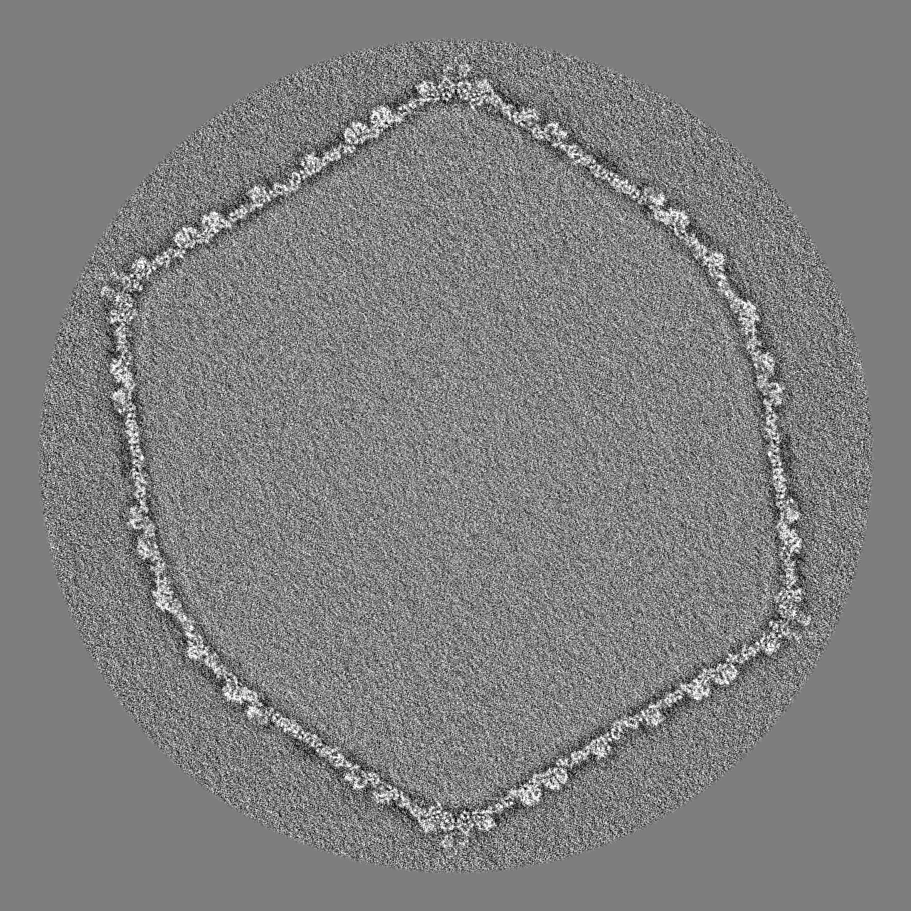
-Map
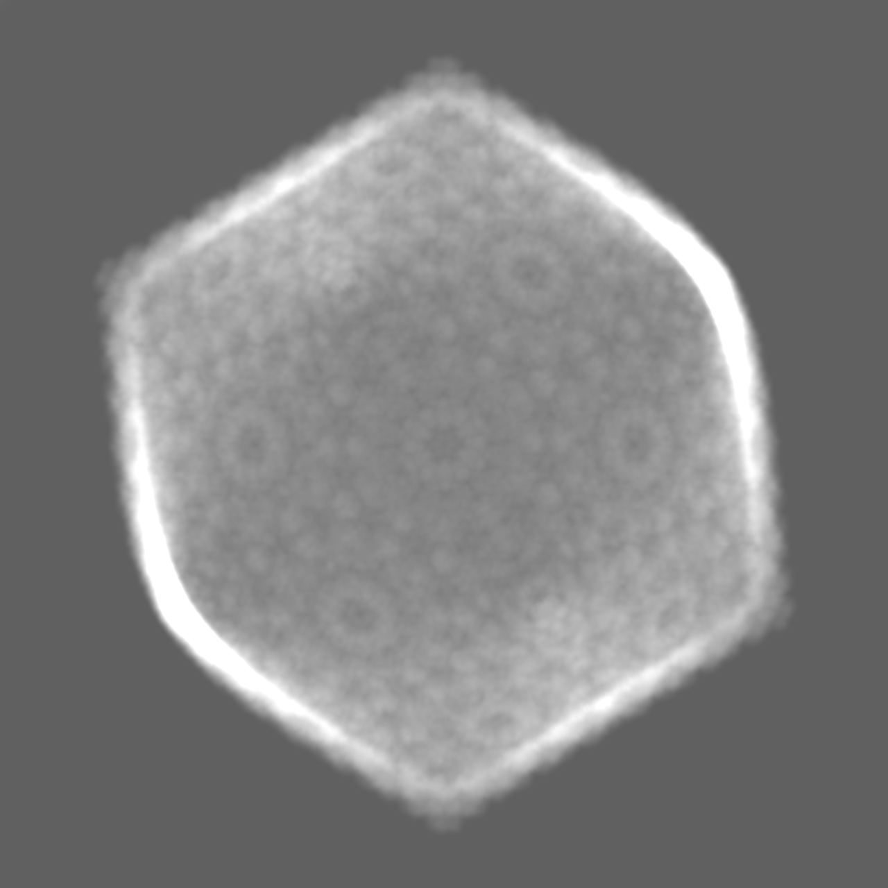
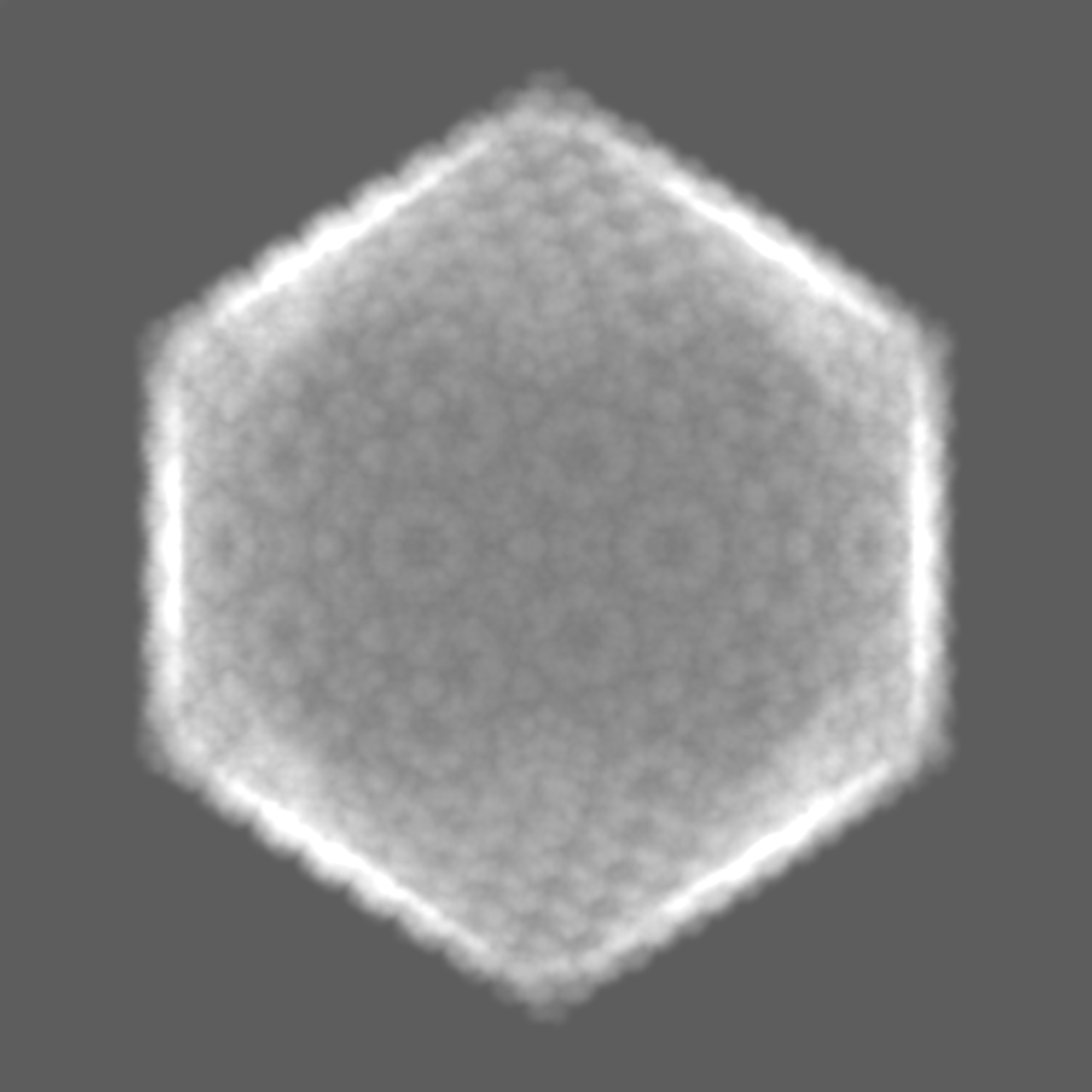
| File | Download / File: emd_21695.map.gz / Format: CCP4 / Size: 7.8 GB / Type: IMAGE STORED AS FLOATING POINT NUMBER (4 BYTES) | ||||||||||||||||||||||||||||||||||||||||||||||||||||||||||||
|---|---|---|---|---|---|---|---|---|---|---|---|---|---|---|---|---|---|---|---|---|---|---|---|---|---|---|---|---|---|---|---|---|---|---|---|---|---|---|---|---|---|---|---|---|---|---|---|---|---|---|---|---|---|---|---|---|---|---|---|---|---|
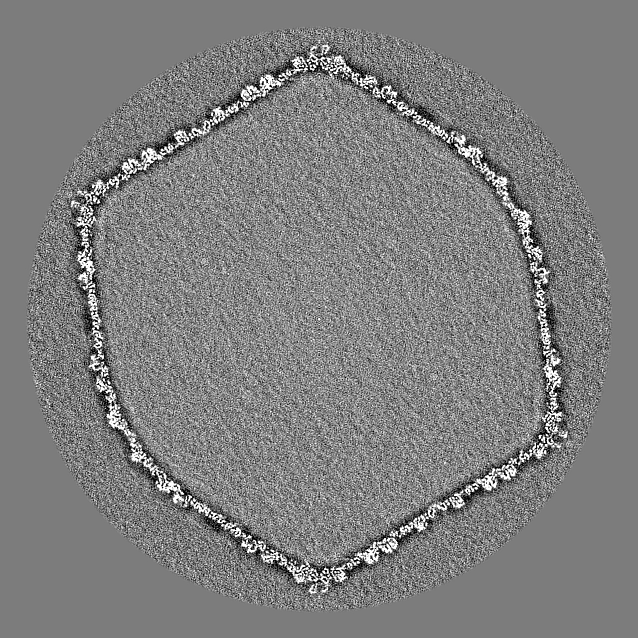
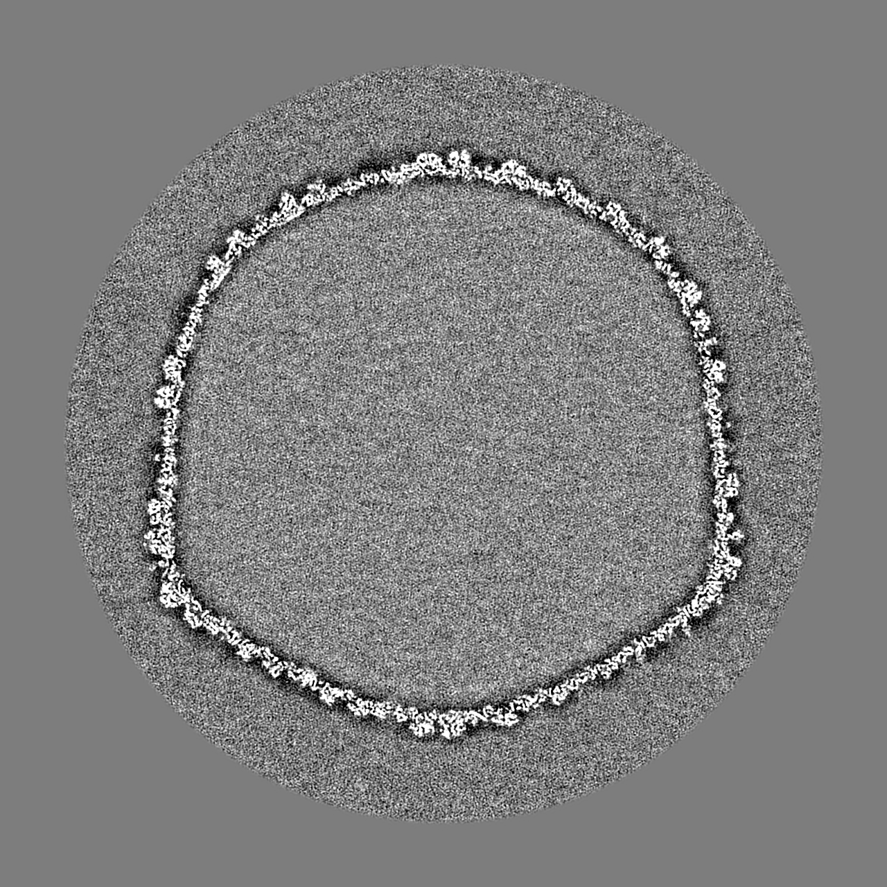
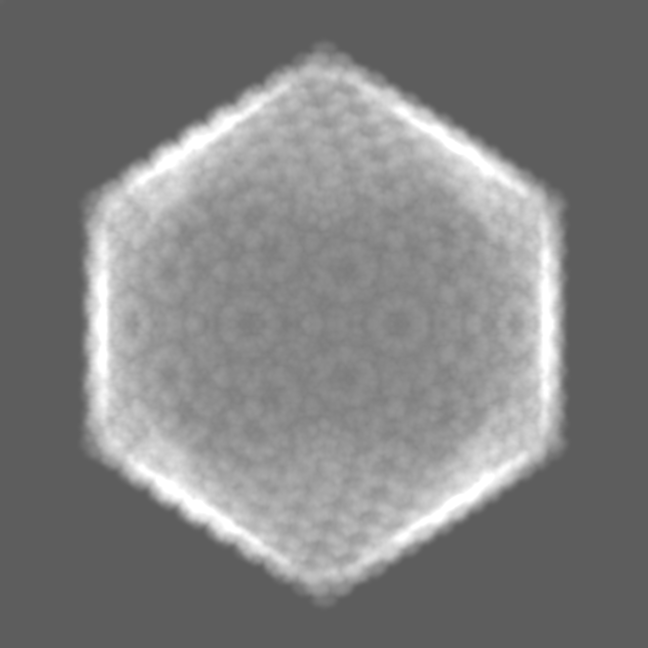
| Projections & slices | Image control
Images are generated by Spider. | ||||||||||||||||||||||||||||||||||||||||||||||||||||||||||||
| Voxel size | X=Y=Z: 1.742 Å | ||||||||||||||||||||||||||||||||||||||||||||||||||||||||||||
| Density |
| ||||||||||||||||||||||||||||||||||||||||||||||||||||||||||||
| Symmetry | Space group: 1 | ||||||||||||||||||||||||||||||||||||||||||||||||||||||||||||
| Details | EMDB XML:
CCP4 map header:
| ||||||||||||||||||||||||||||||||||||||||||||||||||||||||||||
 Z (Sec.)
 Y (Row.)
 X (Col.)


-Supplemental data
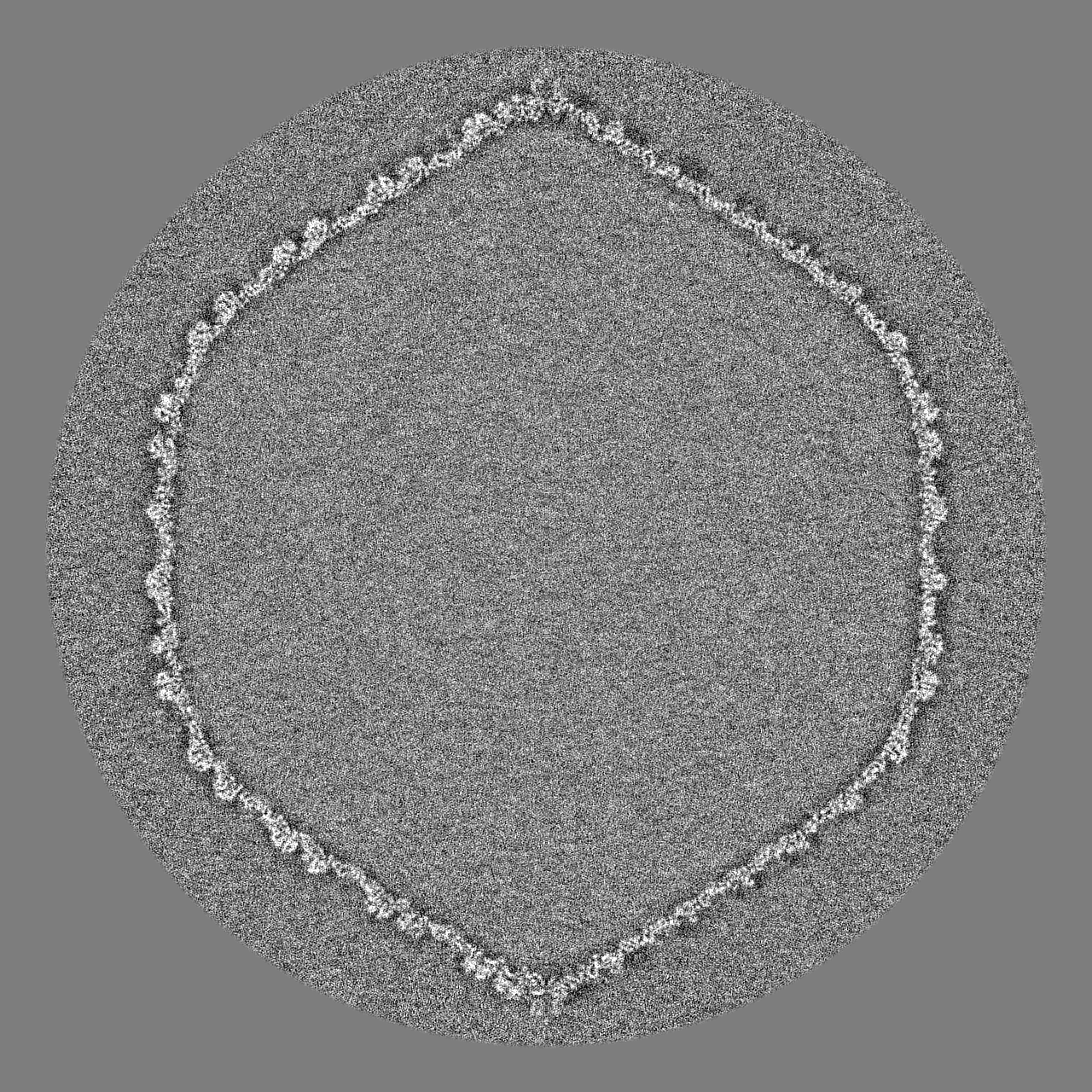
-Mask #1
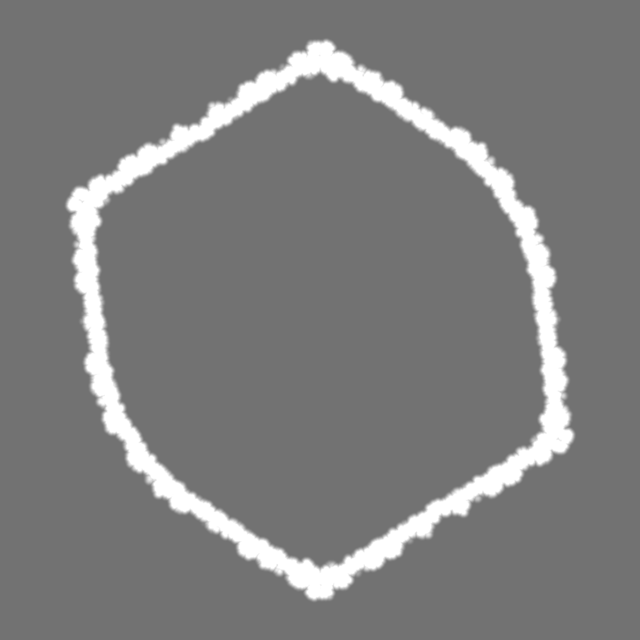
| File | emd_21695_msk_1.map | ||||||||||||
|---|---|---|---|---|---|---|---|---|---|---|---|---|---|
| Projections & Slices |
| ||||||||||||
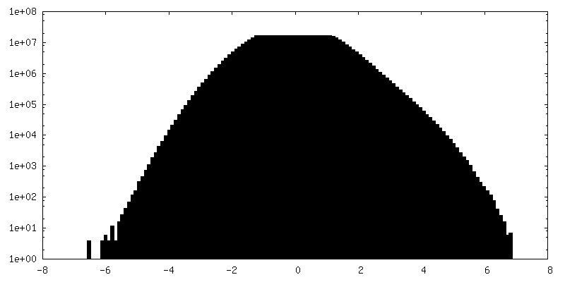
| Density Histograms |


-Mask #2
| File | emd_21695_msk_2.map | ||||||||||||
|---|---|---|---|---|---|---|---|---|---|---|---|---|---|
| Projections & Slices |
| ||||||||||||
| Density Histograms |


-Half map: #1
| File | emd_21695_half_map_1.map | ||||||||||||
|---|---|---|---|---|---|---|---|---|---|---|---|---|---|
| Projections & Slices |
| ||||||||||||
| Density Histograms |


-Half map: #2
| File | emd_21695_half_map_2.map | ||||||||||||
|---|---|---|---|---|---|---|---|---|---|---|---|---|---|
| Projections & Slices |
| ||||||||||||
| Density Histograms |


- Sample components
-Entire : Bacillus virus G
| Entire | Name: Bacillus virus G |
|---|---|
| Components |
|
-Supramolecule #1: Bacillus virus G
| Supramolecule | Name: Bacillus virus G / type: virus / ID: 1 / Parent: 0 / NCBI-ID: 1084719 / Sci species name: Bacillus virus G / Virus type: VIRION / Virus isolate: SPECIES / Virus enveloped: No / Virus empty: No |
|---|---|
| Virus shell | Shell ID: 1 / Diameter: 1600.0 Å / T number (triangulation number): 52 |
-Experimental details
-Structure determination
| Method | cryo EM |
|---|---|
 Processing Processing | single particle reconstruction |
| Aggregation state | particle |
-Sample preparation
| Buffer | pH: 7.4 Details: 0.01 M Tris-Cl (pH 7.4), 0.01 M MgSO4, 6% polyethylene glycol MW 3350 |
|---|---|
| Vitrification | Cryogen name: ETHANE / Chamber humidity: 65 % / Chamber temperature: 298 K / Instrument: GATAN CRYOPLUNGE 3 Details: Three microliters of phage G sample was deposited on a 400 mesh Ted Pella ultrathin lacey carbon grid and incubated for 30 minutes in a humid chamber on ice. The grid was then washed with 10 ...Details: Three microliters of phage G sample was deposited on a 400 mesh Ted Pella ultrathin lacey carbon grid and incubated for 30 minutes in a humid chamber on ice. The grid was then washed with 10 microliters of 0.2 TM buffer. Using a Gatan CP3 plunger, the grid was then blotted for 9 seconds with Whatman 1 filter paper at 65% humidity, then plunge-frozen in liquid ethane. The plunge-frozen, phage G grid was then imaged using a Titan Krios equipped with a K2 detector in super-resolution mode at the Purdue Cryo-EM Facility with a nominal magnification of 8,700 resulting in 1.742 Angstroms per pixel. A total of 375 movies was collected.. |
- Electron microscopy
| Microscope | FEI TITAN KRIOS |
|---|---|
| Image recording | Film or detector model: GATAN K2 QUANTUM (4k x 4k) / Detector mode: SUPER-RESOLUTION / Number real images: 375 / Average electron dose: 14.5 e/Å2 |
| Electron beam | Acceleration voltage: 300 kV / Electron source:  FIELD EMISSION GUN FIELD EMISSION GUN |
| Electron optics | Illumination mode: FLOOD BEAM / Imaging mode: BRIGHT FIELD / Cs: 2.7 mm / Nominal defocus max: 5.0 µm / Nominal defocus min: 1.0 µm / Nominal magnification: 8700 |
| Sample stage | Specimen holder model: FEI TITAN KRIOS AUTOGRID HOLDER / Cooling holder cryogen: NITROGEN |
| Experimental equipment |  Model: Titan Krios / Image courtesy: FEI Company |

