 Movie
Movie Controller


+ Open data

- Basic information
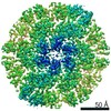
| Entry | Database: PDB / ID: 6wkk | ||||||
|---|---|---|---|---|---|---|---|
| Title | Phage G gp27 major capsid proteins and gp26 decoration proteins | ||||||
 Components Components |
| ||||||
 Keywords Keywords | VIRUS / phage G / major capsid protein / decoration protein / capsid / icosahedral / gp26 / gp27 | ||||||
| Function / homology | : / Phage capsid / Phage capsid family / Gp26 / Gp27 Function and homology information Function and homology information | ||||||
| Biological species |  Bacillus virus G Bacillus virus G | ||||||
| Method | ELECTRON MICROSCOPY / single particle reconstruction / cryo EM / Resolution: 6.1 Å | ||||||
 Authors Authors | Monroe, L. / Gonzalez, B. / Jiang, W. / Kihara, D. | ||||||
 Citation Citation | Journal: J Mol Biol / Year: 2020 Title: Phage G Structure at 6.1 Å Resolution, Condensed DNA, and Host Identity Revision to a Lysinibacillus. Authors: Brenda González / Lyman Monroe / Kunpeng Li / Rui Yan / Elena Wright / Thomas Walter / Daisuke Kihara / Susan T Weintraub / Julie A Thomas / Philip Serwer / Wen Jiang /  Abstract: Phage G has the largest capsid and genome of any known propagated phage. Many aspects of its structure, assembly, and replication have not been elucidated. Herein, we present the dsDNA-packed and ...Phage G has the largest capsid and genome of any known propagated phage. Many aspects of its structure, assembly, and replication have not been elucidated. Herein, we present the dsDNA-packed and empty phage G capsid at 6.1 and 9 Å resolution, respectively, using cryo-EM for structure determination and mass spectrometry for protein identification. The major capsid protein, gp27, is identified and found to share the HK97-fold universally conserved in all previously solved dsDNA phages. Trimers of the decoration protein, gp26, sit on the 3-fold axes and are thought to enhance the interactions of the hexameric capsomeres of gp27, for other phages encoding decoration proteins. Phage G's decoration protein is longer than what has been reported in other phages, and we suspect the extra interaction surface area helps stabilize the capsid. We identified several additional capsid proteins, including a candidate for the prohead protease responsible for processing gp27. Furthermore, cryo-EM reveals a range of partially full, condensed DNA densities that appear to have no contact with capsid shell. Three analyses confirm that the phage G host is a Lysinibacillus, and not Bacillus megaterium: identity of host proteins in our mass spectrometry analyses, genome sequence of the phage G host, and host range of phage G. | ||||||
| History |
|
- Structure visualization
| Movie |
Movie viewer |
|---|---|
| Structure viewer | Molecule: MolmilJmol/JSmol |
- Downloads & links
-Download
| PDBx/mmCIF format | 6wkk.cif.gz | 704.3 KB | Display | PDBx/mmCIF format |
|---|---|---|---|---|
| PDB format | pdb6wkk.ent.gz | 560.5 KB | Display | PDB format |
| PDBx/mmJSON format | 6wkk.json.gz | Tree view | PDBx/mmJSON format | |
| Others |  Other downloads Other downloads |
-Validation report
| Arichive directory | https://data.pdbj.org/pub/pdb/validation_reports/wk/6wkkftp://data.pdbj.org/pub/pdb/validation_reports/wk/6wkk | HTTPS FTP |
|---|
-Related structure data
| Related structure data |  21695MC M: map data used to model this data C: citing same article ( |
|---|---|
| Similar structure data |


-Links
 PDBj
PDBj

- Assembly
| Deposited unit | 
|
|---|---|
| 1 |
|
-Components
| #1: Protein | Mass: 30981.451 Da / Num. of mol.: 6 / Source method: isolated from a natural source / Source: (natural) Bacillus virus G / References: UniProt: G3MB97#2: Protein | Mass: 15763.595 Da / Num. of mol.: 18 / Source method: isolated from a natural source / Source: (natural) Bacillus virus G / References: UniProt: G3MB96 |
|---|
-Experimental details
-Experiment
| Experiment | Method: ELECTRON MICROSCOPY |
|---|---|
| EM experiment | Aggregation state: PARTICLE / 3D reconstruction method: single particle reconstruction |
- Sample preparation
| Component | Name: Bacillus virus G / Type: VIRUS / Entity ID: all / Source: NATURAL |
|---|---|
| Source (natural) | Organism: Bacillus virus G |
| Details of virus | Empty: NO / Enveloped: NO / Isolate: SPECIES / Type: VIRION |
| Virus shell | Diameter: 1600 nm / Triangulation number (T number): 52 |
| Buffer solution | pH: 7.4 |
| Specimen | Embedding applied: NO / Shadowing applied: NO / Staining applied: NO / Vitrification applied: YES Details: In brief, phage G was grown in agarose overlays, concentrated by centrifugal pelleting and subjected to rate zonal centrifugation in a sucrose gradient. After recovery from the gradient, ...Details: In brief, phage G was grown in agarose overlays, concentrated by centrifugal pelleting and subjected to rate zonal centrifugation in a sucrose gradient. After recovery from the gradient, phage G was stored in 0.01 M Tris-Cl (pH 7.4), 0.01 M MgSO4, 6% polyethylene glycol MW 3350. |
| Vitrification | Instrument: GATAN CRYOPLUNGE 3 / Cryogen name: ETHANE / Humidity: 65 % / Chamber temperature: 298 K |
- Electron microscopy imaging
| Experimental equipment |  Model: Titan Krios / Image courtesy: FEI Company |
|---|---|
| Microscopy | Model: FEI TITAN KRIOS |
| Electron gun | Electron source:  FIELD EMISSION GUN / Accelerating voltage: 300 kV / Illumination mode: FLOOD BEAM FIELD EMISSION GUN / Accelerating voltage: 300 kV / Illumination mode: FLOOD BEAM |
| Electron lens | Mode: BRIGHT FIELD |
| Image recording | Electron dose: 14.5 e/Å2 / Film or detector model: GATAN K2 SUMMIT (4k x 4k) / Num. of real images: 375 |
- Processing
| EM software |
| ||||||||||||||||||||||||||||||||
|---|---|---|---|---|---|---|---|---|---|---|---|---|---|---|---|---|---|---|---|---|---|---|---|---|---|---|---|---|---|---|---|---|---|
| CTF correction | Type: PHASE FLIPPING AND AMPLITUDE CORRECTION | ||||||||||||||||||||||||||||||||
| Particle selection | Num. of particles selected: 2564 | ||||||||||||||||||||||||||||||||
| 3D reconstruction | Resolution: 6.1 Å / Resolution method: FSC 0.143 CUT-OFF / Num. of particles: 2564 Details: Phage G's capsid was reconstructed with icosahedral symmetry Symmetry type: POINT |
