National Institutes of Health/National Institute Of Allergy and Infectious Diseases (NIH/NIAID)
R01AI081059
United States
National Institutes of Health/National Institute of General Medical Sciences (NIH/NIGMS)
F32GM108258
United States
National Institutes of Health/National Institute of General Medical Sciences (NIH/NIGMS)
F31GM116210
United States
Howard Hughes Medical Institute (HHMI)
United States
Citation
Journal: Nature / Year: 2020 Title: Structure of a nascent membrane protein as it folds on the BAM complex. Authors: David Tomasek / Shaun Rawson / James Lee / Joseph S Wzorek / Stephen C Harrison / Zongli Li / Daniel Kahne / Abstract: Mitochondria, chloroplasts and Gram-negative bacteria are encased in a double layer of membranes. The outer membrane contains proteins with a β-barrel structure. β-Barrels are sheets of β-strands ...Mitochondria, chloroplasts and Gram-negative bacteria are encased in a double layer of membranes. The outer membrane contains proteins with a β-barrel structure. β-Barrels are sheets of β-strands wrapped into a cylinder, in which the first strand is hydrogen-bonded to the final strand. Conserved multi-subunit molecular machines fold and insert these proteins into the outer membrane. One subunit of the machines is itself a β-barrel protein that has a central role in folding other β-barrels. In Gram-negative bacteria, the β-barrel assembly machine (BAM) consists of the β-barrel protein BamA, and four lipoproteins. To understand how the BAM complex accelerates folding without using exogenous energy (for example, ATP), we trapped folding intermediates on this machine. Here we report the structure of the BAM complex of Escherichia coli folding BamA itself. The BamA catalyst forms an asymmetric hybrid β-barrel with the BamA substrate. The N-terminal edge of the BamA catalyst has an antiparallel hydrogen-bonded interface with the C-terminal edge of the BamA substrate, consistent with previous crosslinking studies; the other edges of the BamA catalyst and substrate are close to each other, but curl inward and do not pair. Six hydrogen bonds in a membrane environment make the interface between the two proteins very stable. This stability allows folding, but creates a high kinetic barrier to substrate release after folding has finished. Features at each end of the substrate overcome this barrier and promote release by stepwise exchange of hydrogen bonds. This mechanism of substrate-assisted product release explains how the BAM complex can stably associate with the substrate during folding and then turn over rapidly when folding is complete.
History
Deposition
Feb 3, 2020
-
Header (metadata) release
Mar 25, 2020
-
Map release
Jun 10, 2020
-
Update
Jul 29, 2020
-
Current status
Jul 29, 2020
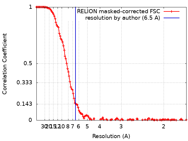
Processing site: RCSB / Status: Released
-
Structure visualization
Movie
Surface view with section colored by density value
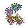
Entire : BamABCDE bound to substrate BamA with loop 1 deleted
Entire
Name: BamABCDE bound to substrate BamA with loop 1 deleted
Components
Complex: BamABCDE bound to substrate BamA with loop 1 deleted
-
Supramolecule #1: BamABCDE bound to substrate BamA with loop 1 deleted
Supramolecule
Name: BamABCDE bound to substrate BamA with loop 1 deleted / type: complex / ID: 1 / Parent: 0 / Macromolecule list: #1 Details: contains a cysteine crosslink between S439C in BamA and E800C in the substrate
Source (natural)
Organism: Escherichia coli K-12 (bacteria)
Recombinant expression
Organism: Escherichia coli BL21(DE3) (bacteria)
-
Experimental details
-
Structure determination
Method
cryo EM
Processing
single particle reconstruction
Aggregation state
particle
-
Sample preparation
Concentration
5 mg/mL
Buffer
pH: 8 Component:
Concentration
Formula
Name
20.0 mM
Tris-HCl
tris(hydroxymethyl)aminomethane hydrochloride
150.0 mM
NaCl
sodium chloride
0.02 %
GDN
glyco-diosgenin
Grid
Details: unspecified
Vitrification
Cryogen name: ETHANE / Chamber humidity: 100 % / Chamber temperature: 298 K / Instrument: FEI VITROBOT MARK IV
Details
contains a cysteine crosslink between S439C in BamA and E800C in the substrate
-
Electron microscopy
Microscope
FEI TITAN KRIOS
Specialist optics
Energy filter - Name: GIF Bioquantum / Energy filter - Slit width: 25 eV
Image recording
Film or detector model: GATAN K3 BIOQUANTUM (6k x 4k) / Average electron dose: 55.0 e/Å2
Electron beam
Acceleration voltage: 300 kV / Electron source: FIELD EMISSION GUN
In the structure databanks used in Yorodumi, some data are registered as the other names, "COVID-19 virus" and "2019-nCoV". Here are the details of the virus and the list of structure data.
Jan 31, 2019. EMDB accession codes are about to change! (news from PDBe EMDB page)
EMDB accession codes are about to change! (news from PDBe EMDB page)
The allocation of 4 digits for EMDB accession codes will soon come to an end. Whilst these codes will remain in use, new EMDB accession codes will include an additional digit and will expand incrementally as the available range of codes is exhausted. The current 4-digit format prefixed with “EMD-” (i.e. EMD-XXXX) will advance to a 5-digit format (i.e. EMD-XXXXX), and so on. It is currently estimated that the 4-digit codes will be depleted around Spring 2019, at which point the 5-digit format will come into force.
The EM Navigator/Yorodumi systems omit the EMD- prefix.
Related info.:Q: What is EMD? / ID/Accession-code notation in Yorodumi/EM Navigator
Yorodumi is a browser for structure data from EMDB, PDB, SASBDB, etc.
This page is also the successor to EM Navigator detail page, and also detail information page/front-end page for Omokage search.
The word "yorodu" (or yorozu) is an old Japanese word meaning "ten thousand". "mi" (miru) is to see.
Related info.:EMDB / PDB / SASBDB / Comparison of 3 databanks / Yorodumi Search / Aug 31, 2016. New EM Navigator & Yorodumi / Yorodumi Papers / Jmol/JSmol / Function and homology information / Changes in new EM Navigator and Yorodumi
 Movie
Movie Controller

 Yorodumi
 Open data
 Basic information
 Map data
 Sample
 Authors
 United States, 4 items
  Citation
 Structure visualization
 Movie viewer
 Downloads & links
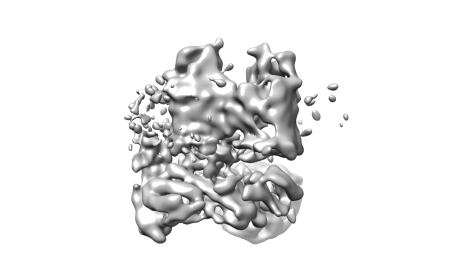 emd_21313.png
 http://ftp.pdbj.org/pub/emdb/structures/EMD-21313


 Z (Sec.)
 Y (Row.)
 X (Col.)


 Sample components
 Processing
 Electron microscopy
 FIELD EMISSION GUN