 Movie
Movie Controller


+ Open data

- Basic information
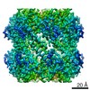
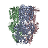
| Entry | Database: EMDB / ID: EMD-21198 | |||||||||
|---|---|---|---|---|---|---|---|---|---|---|
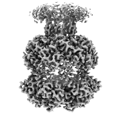
| Title | ClpP1P2 complex from M. tuberculosis bound to ADEP | |||||||||
 Map data Map data | ClpP1P2 complex from M. tuberculosis with ADEP | |||||||||
 Sample Sample |
| |||||||||
 Keywords Keywords | Complex / protease / ClpP / tuberculosis / HYDROLASE / HYDROLASE-ANTIBIOTIC complex | |||||||||
| Function / homology |  Function and homology information Function and homology informationendopeptidase Clp / endopeptidase Clp complex / ATP-dependent peptidase activity / protein quality control for misfolded or incompletely synthesized proteins / peptidoglycan-based cell wall / ATPase binding / serine-type endopeptidase activity / plasma membrane / cytoplasm / cytosol Similarity search - Function | |||||||||
| Biological species |   Mycobacterium tuberculosis (bacteria) / synthetic construct (others) Mycobacterium tuberculosis (bacteria) / synthetic construct (others) | |||||||||
| Method | single particle reconstruction / cryo EM / Resolution: 3.1 Å | |||||||||
 Authors Authors | Ripstein ZA / Vahidi S | |||||||||
| Funding support |  Canada, 2 items Canada, 2 items
| |||||||||
 Citation Citation | Journal: Proc Natl Acad Sci U S A / Year: 2020 Title: An allosteric switch regulates ClpP1P2 protease function as established by cryo-EM and methyl-TROSY NMR. Authors: Siavash Vahidi / Zev A Ripstein / Jordan B Juravsky / Enrico Rennella / Alfred L Goldberg / Anthony K Mittermaier / John L Rubinstein / Lewis E Kay /  Abstract: The 300-kDa ClpP1P2 protease from collaborates with the AAA+ (ATPases associated with a variety of cellular activities) unfoldases, ClpC1 and ClpX, to degrade substrate proteins. Unlike in other ...The 300-kDa ClpP1P2 protease from collaborates with the AAA+ (ATPases associated with a variety of cellular activities) unfoldases, ClpC1 and ClpX, to degrade substrate proteins. Unlike in other bacteria, all of the components of the Clp system are essential for growth and virulence of mycobacteria, and their inhibitors show promise as antibiotics. MtClpP1P2 is unique in that it contains a pair of distinct ClpP1 and ClpP2 rings and also requires the presence of activator peptides, such as benzoyl-leucyl-leucine (Bz-LL), for function. Understanding the structural basis for this requirement has been elusive but is critical for the rational design and improvement of antituberculosis (anti-TB) therapeutics that target the Clp system. Here, we present a combined biophysical and biochemical study to explore the structure-dynamics-function relationship in MtClpP1P2. Electron cryomicroscopy (cryo-EM) structures of apo and acyldepsipeptide-bound MtClpP1P2 explain their lack of activity by showing loss of a key β-sheet in a sequence known as the handle region that is critical for the proper formation of the catalytic triad. Methyl transverse relaxation-optimized spectroscopy (TROSY)-based NMR, cryo-EM, and biochemical assays show that, on binding Bz-LL or covalent inhibitors, MtClpP1P2 undergoes a conformational change from an inactive compact state to an active extended structure that can be explained by a modified Monod-Wyman-Changeux model. Our study establishes a critical role for the handle region as an on/off switch for function and shows extensive allosteric interactions involving both intra- and interring communication that regulate MtClpP1P2 activity and that can potentially be exploited by small molecules to target . | |||||||||
| History |
|
- Structure visualization
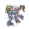
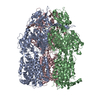
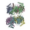
| Movie |
Movie viewer |
|---|---|
| Structure viewer | EM map: SurfViewMolmilJmol/JSmol |
| Supplemental images |
- Downloads & links
-EMDB archive
| Map data | emd_21198.map.gz | 22.4 MB | EMDB map data format | |
|---|---|---|---|---|
| Header (meta data) | emd-21198-v30.xmlemd-21198.xml | 21.2 KB 21.2 KB | Display Display | EMDB header |
| Images |  emd_21198.png emd_21198.png | 76.3 KB | ||
| Filedesc metadata | emd-21198.cif.gz | 7.4 KB | ||
| Archive directory |  http://ftp.pdbj.org/pub/emdb/structures/EMD-21198ftp://ftp.pdbj.org/pub/emdb/structures/EMD-21198 http://ftp.pdbj.org/pub/emdb/structures/EMD-21198ftp://ftp.pdbj.org/pub/emdb/structures/EMD-21198 | HTTPS FTP |
-Related structure data
| Related structure data |  6vgnMC  6vgkC  6vgqC C: citing same article ( M: atomic model generated by this map |
|---|---|
| Similar structure data |

-Links
| EMDB pages | EMDB (EBI/PDBe) / EMDataResource |
|---|
-Map
| File | Download / File: emd_21198.map.gz / Format: CCP4 / Size: 23.8 MB / Type: IMAGE STORED AS FLOATING POINT NUMBER (4 BYTES) | ||||||||||||||||||||||||||||||||||||||||||||||||||||||||||||
|---|---|---|---|---|---|---|---|---|---|---|---|---|---|---|---|---|---|---|---|---|---|---|---|---|---|---|---|---|---|---|---|---|---|---|---|---|---|---|---|---|---|---|---|---|---|---|---|---|---|---|---|---|---|---|---|---|---|---|---|---|---|
| Annotation | ClpP1P2 complex from M. tuberculosis with ADEP | ||||||||||||||||||||||||||||||||||||||||||||||||||||||||||||
| Projections & slices | Image control
Images are generated by Spider. | ||||||||||||||||||||||||||||||||||||||||||||||||||||||||||||
| Voxel size | X=Y=Z: 1.06 Å | ||||||||||||||||||||||||||||||||||||||||||||||||||||||||||||
| Density |
| ||||||||||||||||||||||||||||||||||||||||||||||||||||||||||||
| Symmetry | Space group: 1 | ||||||||||||||||||||||||||||||||||||||||||||||||||||||||||||
| Details | EMDB XML:
CCP4 map header:
| ||||||||||||||||||||||||||||||||||||||||||||||||||||||||||||
 Z (Sec.)
 Y (Row.)
 X (Col.)


-Supplemental data
- Sample components
-Entire : ClpP1P2 complex with ADEP
| Entire | Name: ClpP1P2 complex with ADEP |
|---|---|
| Components |
|
-Supramolecule #1: ClpP1P2 complex with ADEP
| Supramolecule | Name: ClpP1P2 complex with ADEP / type: complex / ID: 1 / Parent: 0 / Macromolecule list: #1-#2 Details: Complex formed between P1 and P2 heptameric rings with bound ADEP |
|---|---|
| Source (natural) | Organism: Mycobacterium tuberculosis (bacteria) |
| Molecular weight | Theoretical: 300 KDa |
-Macromolecule #1: ATP-dependent Clp protease proteolytic subunit
| Macromolecule | Name: ATP-dependent Clp protease proteolytic subunit / type: protein_or_peptide / ID: 1 / Number of copies: 7 / Enantiomer: LEVO / EC number: endopeptidase Clp |
|---|---|
| Source (natural) | Organism: Mycobacterium tuberculosis (bacteria) |
| Molecular weight | Theoretical: 21.914957 KDa |
| Recombinant expression | Organism: |
| Sequence | String: ILPSFIEHSS FGVKESNPYN KLFEERIIFL GVQVDDASAN DIMAQLLVLE SLDPDRDITM YINSPGGGFT SLMAIYDTMQ YVRADIQTV CLGQAASAAA VLLAAGTPGK RMALPNARVL IHQPSLSGVI QGQFSDLEIQ AAEIERMRTL METTLARHTG K DAGVIRKD ...String: ILPSFIEHSS FGVKESNPYN KLFEERIIFL GVQVDDASAN DIMAQLLVLE SLDPDRDITM YINSPGGGFT SLMAIYDTMQ YVRADIQTV CLGQAASAAA VLLAAGTPGK RMALPNARVL IHQPSLSGVI QGQFSDLEIQ AAEIERMRTL METTLARHTG K DAGVIRKD TDRDKILTAE EAKDYGIIDT VLEYRKLSAQ TA UniProtKB: ATP-dependent Clp protease proteolytic subunit |
-Macromolecule #2: ATP-dependent Clp protease proteolytic subunit 1
| Macromolecule | Name: ATP-dependent Clp protease proteolytic subunit 1 / type: protein_or_peptide / ID: 2 / Number of copies: 7 / Enantiomer: LEVO / EC number: endopeptidase Clp |
|---|---|
| Source (natural) | Organism: Mycobacterium tuberculosis (bacteria) |
| Molecular weight | Theoretical: 21.065934 KDa |
| Recombinant expression | Organism: |
| Sequence | String: MRSNSQGLSL TDSVYERLLS ERIIFLGSEV NDEIANRLCA QILLLAAEDA SKDISLYINS PGGSISAGMA IYDTMVLAPC DIATYAMGM AASMGEFLLA AGTKGKRYAL PHARILMHQP LGGVTGSAAD IAIQAEQFAV IKKEMFRLNA EFTGQPIERI E ADSDRDRW ...String: MRSNSQGLSL TDSVYERLLS ERIIFLGSEV NDEIANRLCA QILLLAAEDA SKDISLYINS PGGSISAGMA IYDTMVLAPC DIATYAMGM AASMGEFLLA AGTKGKRYAL PHARILMHQP LGGVTGSAAD IAIQAEQFAV IKKEMFRLNA EFTGQPIERI E ADSDRDRW FTAAEALEYG FVDHIITRAH VNGEAQ UniProtKB: ATP-dependent Clp protease proteolytic subunit 1 |
-Macromolecule #3: R0M-WFP-ALO-PRO-YCP-ALA-MP8
| Macromolecule | Name: R0M-WFP-ALO-PRO-YCP-ALA-MP8 / type: protein_or_peptide / ID: 3 / Number of copies: 7 / Enantiomer: LEVO |
|---|---|
| Source (natural) | Organism: synthetic construct (others) |
| Molecular weight | Theoretical: 800.888 Da |
| Sequence | String: (R0M)(WFP)(ALO)P(YCP)A(MP8) |
-Experimental details
-Structure determination
| Method | cryo EM |
|---|---|
 Processing Processing | single particle reconstruction |
| Aggregation state | particle |
-Sample preparation
| Concentration | 20 mg/mL | ||||||||||
|---|---|---|---|---|---|---|---|---|---|---|---|
| Buffer | pH: 7 Component:
Details: IGEPAL-CA630 was added shortly prior to vitrification | ||||||||||
| Grid | Support film - topology: HOLEY / Support film - Film thickness: 30 / Details: unspecified | ||||||||||
| Vitrification | Cryogen name: ETHANE-PROPANE / Chamber humidity: 100 % / Chamber temperature: 277 K / Instrument: FEI VITROBOT MARK III / Details: Blotted for 4.5 seconds at an offset of -5 mm. | ||||||||||
| Details | Mono-disperse complexes |
- Electron microscopy
| Microscope | TFS KRIOS |
|---|---|
| Temperature | Min: 70.0 K / Max: 77.0 K |
| Image recording | Film or detector model: FEI FALCON III (4k x 4k) / Detector mode: COUNTING / Digitization - Dimensions - Width: 4096 pixel / Digitization - Dimensions - Height: 4096 pixel / Number grids imaged: 1 / Number real images: 725 / Average exposure time: 60.0 sec. / Average electron dose: 43.0 e/Å2 |
| Electron beam | Acceleration voltage: 300 kV / Electron source:  FIELD EMISSION GUN FIELD EMISSION GUN |
| Electron optics | C2 aperture diameter: 50.0 µm / Illumination mode: FLOOD BEAM / Imaging mode: BRIGHT FIELD / Cs: 2.7 mm / Nominal defocus max: 2.0 µm / Nominal defocus min: 0.7000000000000001 µm / Nominal magnification: 75000 |
| Sample stage | Specimen holder model: FEI TITAN KRIOS AUTOGRID HOLDER / Cooling holder cryogen: NITROGEN |
| Experimental equipment |  Model: Titan Krios / Image courtesy: FEI Company |

