National Institutes of Health/National Institute of General Medical Sciences (NIH/NIGMS)
R35 GM131747
United States
Citation
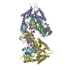
Journal: Elife / Year: 2020 Title: Limited dishevelled/Axin oligomerization determines efficiency of Wnt/β-catenin signal transduction. Authors: Wei Kan / Michael D Enos / Elgin Korkmazhan / Stefan Muennich / Dong-Hua Chen / Melissa V Gammons / Mansi Vasishtha / Mariann Bienz / Alexander R Dunn / Georgios Skiniotis / William I Weis / Abstract: In Wnt/β-catenin signaling, the transcriptional coactivator β-catenin is regulated by its phosphorylation in a complex that includes the scaffold protein Axin and associated kinases. Wnt binding to ...In Wnt/β-catenin signaling, the transcriptional coactivator β-catenin is regulated by its phosphorylation in a complex that includes the scaffold protein Axin and associated kinases. Wnt binding to its coreceptors activates the cytosolic effector Dishevelled (Dvl), leading to the recruitment of Axin and the inhibition of β-catenin phosphorylation. This process requires interaction of homologous DIX domains present in Dvl and Axin, but is mechanistically undefined. We show that Dvl DIX forms antiparallel, double-stranded oligomers in vitro, and that Dvl in cells forms oligomers typically <10 molecules at endogenous expression levels. Axin DIX (DAX) forms small single-stranded oligomers, but its self-association is stronger than that of DIX. DAX caps the ends of DIX oligomers, such that a DIX oligomer has at most four DAX binding sites. The relative affinities and stoichiometry of the DIX-DAX interaction provide a mechanism for efficient inhibition of β-catenin phosphorylation upon Axin recruitment to the Wnt receptor complex.
History
Deposition
Dec 20, 2019
-
Header (metadata) release
Jan 29, 2020
-
Map release
Apr 29, 2020
-
Update
Mar 6, 2024
-
Current status
Mar 6, 2024
Processing site: RCSB / Status: Released
-
Structure visualization
Movie
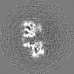
Surface view with section colored by density value
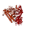
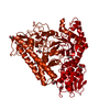
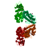
Entire : Segment polarity protein dishevelled homolog DVL-2
Entire
Name: Segment polarity protein dishevelled homolog DVL-2
Components
Complex: Segment polarity protein dishevelled homolog DVL-2
Protein or peptide: Segment polarity protein dishevelled homolog DVL-2
-
Supramolecule #1: Segment polarity protein dishevelled homolog DVL-2
Supramolecule
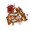
Name: Segment polarity protein dishevelled homolog DVL-2 / type: complex / ID: 1 / Parent: 0 / Macromolecule list: all Details: Protein spontaneously formed a double-stranded, antiparallel helical filament upon removal of the MBP tag with TEV protease.
Source (natural)
Organism: Mus musculus (house mouse)
-
Macromolecule #1: Segment polarity protein dishevelled homolog DVL-2
Macromolecule
Name: Segment polarity protein dishevelled homolog DVL-2 / type: protein_or_peptide / ID: 1 / Number of copies: 12 / Enantiomer: LEVO
UniProtKB: Segment polarity protein dishevelled homolog DVL-2
-
Experimental details
-
Structure determination
Method
cryo EM
Processing
helical reconstruction
Aggregation state
helical array
-
Sample preparation
Concentration
0.12 mg/mL
Buffer
pH: 8 Component:
Concentration
Formula
Name
20.0 mM
C8H18N2O4S
HEPES
150.0 mM
NaCl
sodium chloride
4.0 mM
C4H10O2S2
dithiothreitol
1.0 mM
C10H16N2O8
EDTA
Details: Dithiothreitol was added fresh the day of use.
Grid
Material: COPPER / Mesh: 200 / Support film - Material: CARBON / Support film - topology: LACEY / Pretreatment - Type: GLOW DISCHARGE / Pretreatment - Time: 25 sec. / Pretreatment - Atmosphere: AIR / Pretreatment - Pressure: 0.04 kPa
Vitrification
Cryogen name: ETHANE / Chamber humidity: 95 % / Chamber temperature: 295 K / Instrument: LEICA EM GP Details: The grid was blotted for 2.0 s using Whatman #1 filter paper (GE Healthcare)..
Details
Sample was taken from a single fraction of a gel filtration run performed immediately before grid preparation.
-
Electron microscopy
Microscope
FEI TITAN KRIOS
Image recording
Film or detector model: GATAN K2 SUMMIT (4k x 4k) / Detector mode: COUNTING / Digitization - Dimensions - Width: 3838 pixel / Digitization - Dimensions - Height: 3710 pixel / Digitization - Frames/image: 4-50 / Number real images: 540 / Average exposure time: 10.0 sec. / Average electron dose: 98.0 e/Å2
Electron beam
Acceleration voltage: 300 kV / Electron source: FIELD EMISSION GUN
Type of model: INSILICO MODEL In silico model: A starting model was generated using the stochastic gradient descent option from the "Initial model" jobtype in RELION 3.0.8.
Final angle assignment
Type: NOT APPLICABLE / Software - Name: RELION (ver. 3.0.8)
Chain - Chain ID: A / Chain - Residue range: 12-92 / Chain - Source name: PDB / Chain - Initial model type: experimental model
Details
The two mutations in the source (Y27W and C80S) were reverted to Trp and Ser, respectively, before fitting. Model refinement was carried out through alternating rounds of real-space refinement in PHENIX and manual building in Coot.
Refinement
Space: REAL / Protocol: RIGID BODY FIT
Output model
PDB-6vcc: Cryo-EM structure of the Dvl2 DIX filament
+
About Yorodumi
-
News
-
Feb 9, 2022. New format data for meta-information of EMDB entries
New format data for meta-information of EMDB entries
Version 3 of the EMDB header file is now the official format.
The previous official version 1.9 will be removed from the archive.
In the structure databanks used in Yorodumi, some data are registered as the other names, "COVID-19 virus" and "2019-nCoV". Here are the details of the virus and the list of structure data.
Jan 31, 2019. EMDB accession codes are about to change! (news from PDBe EMDB page)
EMDB accession codes are about to change! (news from PDBe EMDB page)
The allocation of 4 digits for EMDB accession codes will soon come to an end. Whilst these codes will remain in use, new EMDB accession codes will include an additional digit and will expand incrementally as the available range of codes is exhausted. The current 4-digit format prefixed with “EMD-” (i.e. EMD-XXXX) will advance to a 5-digit format (i.e. EMD-XXXXX), and so on. It is currently estimated that the 4-digit codes will be depleted around Spring 2019, at which point the 5-digit format will come into force.
The EM Navigator/Yorodumi systems omit the EMD- prefix.
Related info.:Q: What is EMD? / ID/Accession-code notation in Yorodumi/EM Navigator
Yorodumi is a browser for structure data from EMDB, PDB, SASBDB, etc.
This page is also the successor to EM Navigator detail page, and also detail information page/front-end page for Omokage search.
The word "yorodu" (or yorozu) is an old Japanese word meaning "ten thousand". "mi" (miru) is to see.
Related info.:EMDB / PDB / SASBDB / Comparison of 3 databanks / Yorodumi Search / Aug 31, 2016. New EM Navigator & Yorodumi / Yorodumi Papers / Jmol/JSmol / Function and homology information / Changes in new EM Navigator and Yorodumi
 Movie
Movie Controller


 Open data
 Basic information
 Map data
 Sample
 Keywords
 Function and homology information
 Authors
 United States, 1 items
  Citation
 Structure visualization
 Downloads & links
 emd_21148.png
 http://ftp.pdbj.org/pub/emdb/structures/EMD-21148
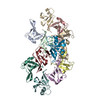


 Z (Sec.)
 Y (Row.)
 X (Col.)


 Sample components
 Processing
 Electron microscopy
 FIELD EMISSION GUN