 Movie
Movie Controller

[English] 日本語
 Yorodumi
- EMDB-2102: Structure of a stalled transfer intermediate of Sm proteins from ... -
+ Open data

- Basic information
| Entry | Database: EMDB / ID: EMD-2102 | |||||||||
|---|---|---|---|---|---|---|---|---|---|---|
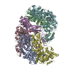
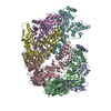
| Title | Structure of a stalled transfer intermediate of Sm proteins from the assembly chaperone pICln to the SMN-complex | |||||||||
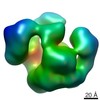
 Map data Map data | Reconstruction of the 8S complex | |||||||||
 Sample Sample |
| |||||||||
 Keywords Keywords | snRNP / spliceosome / assembly chaperone / SMN-complex | |||||||||
| Biological species |  Homo sapiens (human) Homo sapiens (human) | |||||||||
| Method | single particle reconstruction / negative staining / Resolution: 20.0 Å | |||||||||
 Authors Authors | Chari A / Grimm C / Fischer U / Stark H | |||||||||
 Citation Citation | Journal: Mol Cell / Year: 2013 Title: Structural basis of assembly chaperone- mediated snRNP formation. Authors: Clemens Grimm / Ashwin Chari / Jann-Patrick Pelz / Jochen Kuper / Caroline Kisker / Kay Diederichs / Holger Stark / Hermann Schindelin / Utz Fischer /  Abstract: Small nuclear ribonucleoproteins (snRNPs) represent key constituents of major and minor spliceosomes. snRNPs contain a common core, composed of seven Sm proteins bound to snRNA, which forms in a step- ...Small nuclear ribonucleoproteins (snRNPs) represent key constituents of major and minor spliceosomes. snRNPs contain a common core, composed of seven Sm proteins bound to snRNA, which forms in a step-wise and factor-mediated reaction. The assembly chaperone pICln initially mediates the formation of an otherwise unstable pentameric Sm protein unit. This so-called 6S complex docks subsequently onto the SMN complex, which removes pICln and enables the transfer of pre-assembled Sm proteins onto snRNA. X-ray crystallography and electron microscopy was used to investigate the structural basis of snRNP assembly. The 6S complex structure identifies pICln as an Sm protein mimic, which enables the topological organization of the Sm pentamer in a closed ring. A second structure of 6S bound to the SMN complex components SMN and Gemin2 uncovers a plausible mechanism of pICln elimination and Sm protein activation for snRNA binding. Our studies reveal how assembly factors facilitate formation of RNA-protein complexes in vivo. | |||||||||
| History |
|
- Structure visualization
| Movie |
 Movie viewer Movie viewer |
|---|---|
| Structure viewer | EM map: SurfViewMolmilJmol/JSmol |
| Supplemental images |

- Downloads & links
-EMDB archive
| Map data | emd_2102.map.gz | 1.8 MB | EMDB map data format | |
|---|---|---|---|---|
| Header (meta data) | emd-2102-v30.xmlemd-2102.xml | 9.4 KB 9.4 KB | Display Display | EMDB header |
| Images | 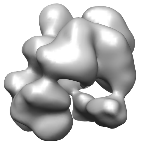 EMD-2102.png EMD-2102.png | 70.2 KB | ||
| Archive directory |  http://ftp.pdbj.org/pub/emdb/structures/EMD-2102ftp://ftp.pdbj.org/pub/emdb/structures/EMD-2102 http://ftp.pdbj.org/pub/emdb/structures/EMD-2102ftp://ftp.pdbj.org/pub/emdb/structures/EMD-2102 | HTTPS FTP |
-Related structure data


-Links
| EMDB pages | EMDB (EBI/PDBe) / EMDataResource |
|---|
-Map
| File | Download / File: emd_2102.map.gz / Format: CCP4 / Size: 1.9 MB / Type: IMAGE STORED AS FLOATING POINT NUMBER (4 BYTES) | ||||||||||||||||||||||||||||||||||||||||||||||||||||||||||||||||||||
|---|---|---|---|---|---|---|---|---|---|---|---|---|---|---|---|---|---|---|---|---|---|---|---|---|---|---|---|---|---|---|---|---|---|---|---|---|---|---|---|---|---|---|---|---|---|---|---|---|---|---|---|---|---|---|---|---|---|---|---|---|---|---|---|---|---|---|---|---|---|
| Annotation | Reconstruction of the 8S complex | ||||||||||||||||||||||||||||||||||||||||||||||||||||||||||||||||||||
| Projections & slices | Image control
Images are generated by Spider. | ||||||||||||||||||||||||||||||||||||||||||||||||||||||||||||||||||||
| Voxel size | X=Y=Z: 2.5 Å | ||||||||||||||||||||||||||||||||||||||||||||||||||||||||||||||||||||
| Density |
| ||||||||||||||||||||||||||||||||||||||||||||||||||||||||||||||||||||
| Symmetry | Space group: 1 | ||||||||||||||||||||||||||||||||||||||||||||||||||||||||||||||||||||
| Details | EMDB XML:
CCP4 map header:
| ||||||||||||||||||||||||||||||||||||||||||||||||||||||||||||||||||||
 Z (Sec.)
 Y (Row.)
 X (Col.)


-Supplemental data
- Sample components
-Entire : 8S complex: a stalled Sm protein transfer intermediate from the a...
| Entire | Name: 8S complex: a stalled Sm protein transfer intermediate from the assembly chaperone pICln to the SMN-complex |
|---|---|
| Components |
|
-Supramolecule #1000: 8S complex: a stalled Sm protein transfer intermediate from the a...
| Supramolecule | Name: 8S complex: a stalled Sm protein transfer intermediate from the assembly chaperone pICln to the SMN-complex type: sample / ID: 1000 / Details: Sample was monodisperse Oligomeric state: One heterooctamer composed of pICln, SmD1, SmD2, SmE, SmF, SmG, SMN and Gemin2 Number unique components: 1 |
|---|---|
| Molecular weight | Experimental: 125 KDa / Theoretical: 125 KDa / Method: Gel filtration, Sedimentation |
-Macromolecule #1: 8S complex
| Macromolecule | Name: 8S complex / type: protein_or_peptide / ID: 1 / Number of copies: 1 / Oligomeric state: monomer / Recombinant expression: Yes |
|---|---|
| Source (natural) | Organism: Homo sapiens (human) / synonym: Human / Organelle: Cytoplasm / Location in cell: Cytosol |
| Molecular weight | Experimental: 125 KDa / Theoretical: 125 KDa |
| Recombinant expression | Organism:  |
-Experimental details
-Structure determination
| Method | negative staining |
|---|---|
 Processing Processing | single particle reconstruction |
| Aggregation state | particle |
-Sample preparation
| Concentration | 10 mg/mL |
|---|---|
| Buffer | pH: 7.5 / Details: 20 mM HEPES, 150 mM NaCl, 5 mM DTT |
| Staining | Type: NEGATIVE Details: Grids were floated on 2 % Uranyl Formate solution for 1 minute. |
| Grid | Details: Custom made holey grid with thin carbon support. |
| Vitrification | Cryogen name: NONE / Instrument: OTHER / Details: negative stain |
- Electron microscopy
| Microscope | FEI/PHILIPS CM200FEG |
|---|---|
| Date | Jan 12, 2012 |
| Image recording | Category: CCD / Film or detector model: GENERIC CCD / Number real images: 48 / Average electron dose: 20 e/Å2 / Bits/pixel: 16 |
| Electron beam | Acceleration voltage: 160 kV / Electron source:  FIELD EMISSION GUN FIELD EMISSION GUN |
| Electron optics | Calibrated magnification: 109000 / Illumination mode: SPOT SCAN / Imaging mode: BRIGHT FIELD / Cs: 2 mm |
| Sample stage | Specimen holder model: SIDE ENTRY, EUCENTRIC |
-Image processing
| CTF correction | Details: Each particle |
|---|---|
| Final reconstruction | Applied symmetry - Point group: C1 (asymmetric) / Algorithm: OTHER / Resolution.type: BY AUTHOR / Resolution: 20.0 Å / Resolution method: FSC 0.5 CUT-OFF / Software - Name: Imagic / Number images used: 10000 |
-Atomic model buiding 1
| Initial model | PDB ID:  4f77 |
|---|---|
| Software | Name: Amira |
| Details | Protocol: Rigid Body |
| Refinement | Space: REAL / Protocol: RIGID BODY FIT |
