National Institutes of Health/National Institute Of Allergy and Infectious Diseases
AI36040
United States
Robert A. Welch Foundation
Q1279
United States
Welch Foundation
Q-1967-20180324
United States
Citation
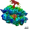
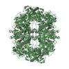
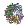
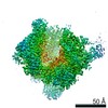
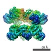
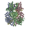
Journal: Sci Adv / Year: 2020 Title: 2.7 Å cryo-EM structure of rotavirus core protein VP3, a unique capping machine with a helicase activity. Authors: Dilip Kumar / Xinzhe Yu / Sue E Crawford / Rodolfo Moreno / Joanita Jakana / Banumathi Sankaran / Ramakrishnan Anish / Soni Kaundal / Liya Hu / Mary K Estes / Zhao Wang / B V Venkataram Prasad / Abstract: In many viruses, including rotavirus (RV), the major pathogen of infantile gastroenteritis, capping of viral messenger RNAs is a pivotal step for efficient translation of the viral genome. In RV, VP3 ...In many viruses, including rotavirus (RV), the major pathogen of infantile gastroenteritis, capping of viral messenger RNAs is a pivotal step for efficient translation of the viral genome. In RV, VP3 caps the nascent transcripts synthesized from the genomic dsRNA segments by the RV polymerase VP1 within the particle core. Here, from cryo-electron microscopy, x-ray crystallography, and biochemical analyses, we show that VP3 forms a stable tetrameric assembly with each subunit having a modular domain organization, which uniquely integrates five distinct enzymatic steps required for capping the transcripts. In addition to the previously known guanylyl- and methyltransferase activities, we show that VP3 exhibits hitherto unsuspected RNA triphosphatase activity necessary for initiating transcript capping and RNA helicase activity likely required for separating the RNA duplex formed transiently during endogenous transcription. From our studies, we propose a new mechanism for how VP3 inside the virion core caps the nascent transcripts exiting from the polymerase.
History
Deposition
Apr 25, 2019
-
Header (metadata) release
Jun 19, 2019
-
Map release
Apr 29, 2020
-
Update
Apr 29, 2020
-
Current status
Apr 29, 2020
Processing site: RCSB / Status: Released
-
Structure visualization
Movie
Surface view with section colored by density value
In the structure databanks used in Yorodumi, some data are registered as the other names, "COVID-19 virus" and "2019-nCoV". Here are the details of the virus and the list of structure data.
Jan 31, 2019. EMDB accession codes are about to change! (news from PDBe EMDB page)
EMDB accession codes are about to change! (news from PDBe EMDB page)
The allocation of 4 digits for EMDB accession codes will soon come to an end. Whilst these codes will remain in use, new EMDB accession codes will include an additional digit and will expand incrementally as the available range of codes is exhausted. The current 4-digit format prefixed with “EMD-” (i.e. EMD-XXXX) will advance to a 5-digit format (i.e. EMD-XXXXX), and so on. It is currently estimated that the 4-digit codes will be depleted around Spring 2019, at which point the 5-digit format will come into force.
The EM Navigator/Yorodumi systems omit the EMD- prefix.
Related info.:Q: What is EMD? / ID/Accession-code notation in Yorodumi/EM Navigator
Yorodumi is a browser for structure data from EMDB, PDB, SASBDB, etc.
This page is also the successor to EM Navigator detail page, and also detail information page/front-end page for Omokage search.
The word "yorodu" (or yorozu) is an old Japanese word meaning "ten thousand". "mi" (miru) is to see.
Related info.:EMDB / PDB / SASBDB / Comparison of 3 databanks / Yorodumi Search / Aug 31, 2016. New EM Navigator & Yorodumi / Yorodumi Papers / Jmol/JSmol / Function and homology information / Changes in new EM Navigator and Yorodumi
 Movie
Movie Controller


 Open data
 Basic information
 Map data
 Sample
 Rotavirus A
 Authors
 United States, 3 items
  Citation
 Structure visualization
 Movie viewer
 Downloads & links
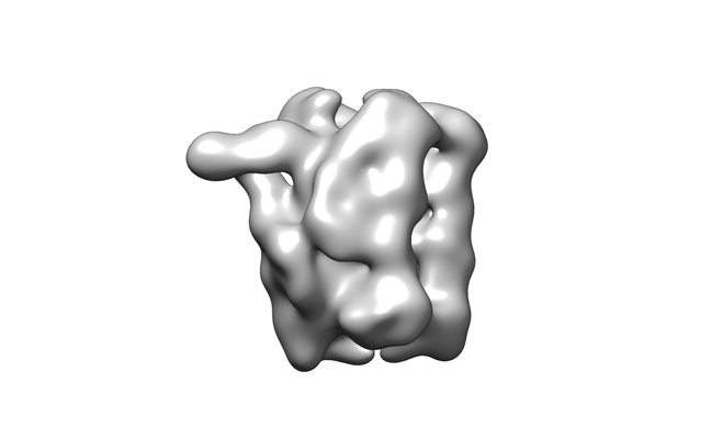 emd_20159.png
 http://ftp.pdbj.org/pub/emdb/structures/EMD-20159
 Z (Sec.)
 Y (Row.)
 X (Col.)


 Sample components
 Spodoptera frugiperda (fall armyworm)
 Processing
 Electron microscopy
 FIELD EMISSION GUN