 Movie
Movie Controller


+ Open data

- Basic information
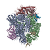
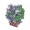
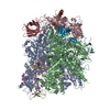
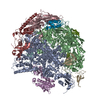
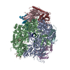
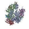
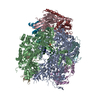
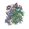
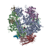
| Entry | Database: PDB / ID: 3ja8 | ||||||
|---|---|---|---|---|---|---|---|
| Title | Cryo-EM structure of the MCM2-7 double hexamer | ||||||
 Components Components | (Minichromosome Maintenance ...) x 6 | ||||||
 Keywords Keywords | HYDROLASE / Cryo-EM / single particle / MCM2-7 / DNA replication | ||||||
| Function / homology |  Function and homology information Function and homology informationMCM core complex / Assembly of the pre-replicative complex / Switching of origins to a post-replicative state / MCM complex binding / nuclear DNA replication / premeiotic DNA replication / pre-replicative complex assembly involved in nuclear cell cycle DNA replication / Activation of the pre-replicative complex / mitotic DNA replication / nuclear pre-replicative complex ...MCM core complex / Assembly of the pre-replicative complex / Switching of origins to a post-replicative state / MCM complex binding / nuclear DNA replication / premeiotic DNA replication / pre-replicative complex assembly involved in nuclear cell cycle DNA replication / Activation of the pre-replicative complex / mitotic DNA replication / nuclear pre-replicative complex / CMG complex / DNA replication preinitiation complex / Activation of ATR in response to replication stress / double-strand break repair via break-induced replication / MCM complex / mitotic DNA replication initiation / silent mating-type cassette heterochromatin formation / single-stranded DNA helicase activity / regulation of DNA-templated DNA replication initiation / DNA strand elongation involved in DNA replication / nuclear replication fork / DNA replication origin binding / DNA replication initiation / subtelomeric heterochromatin formation / DNA helicase activity / helicase activity / transcription elongation by RNA polymerase II / peroxisome / heterochromatin formation / single-stranded DNA binding / DNA helicase / chromosome, telomeric region / DNA replication / chromatin binding / DNA damage response / ATP hydrolysis activity / zinc ion binding / nucleoplasm / ATP binding / nucleus / cytoplasm Similarity search - Function | ||||||
| Biological species |  | ||||||
| Method | ELECTRON MICROSCOPY / single particle reconstruction / cryo EM / Resolution: 3.8 Å | ||||||
 Authors Authors | Li, N. / Zhai, Y. / Zhang, Y. / Li, W. / Yang, M. / Lei, J. / Tye, B.K. / Gao, N. | ||||||
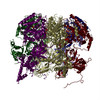
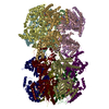
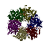
 Citation Citation | Journal: Nature / Year: 2015 Title: Structure of the eukaryotic MCM complex at 3.8 Å. Authors: Ningning Li / Yuanliang Zhai / Yixiao Zhang / Wanqiu Li / Maojun Yang / Jianlin Lei / Bik-Kwoon Tye / Ning Gao /   Abstract: DNA replication in eukaryotes is strictly regulated by several mechanisms. A central step in this replication is the assembly of the heterohexameric minichromosome maintenance (MCM2-7) helicase ...DNA replication in eukaryotes is strictly regulated by several mechanisms. A central step in this replication is the assembly of the heterohexameric minichromosome maintenance (MCM2-7) helicase complex at replication origins during G1 phase as an inactive double hexamer. Here, using cryo-electron microscopy, we report a near-atomic structure of the MCM2-7 double hexamer purified from yeast G1 chromatin. Our structure shows that two single hexamers, arranged in a tilted and twisted fashion through interdigitated amino-terminal domain interactions, form a kinked central channel. Four constricted rings consisting of conserved interior β-hairpins from the two single hexamers create a narrow passageway that tightly fits duplex DNA. This narrow passageway, reinforced by the offset of the two single hexamers at the double hexamer interface, is flanked by two pairs of gate-forming subunits, MCM2 and MCM5. These unusual features of the twisted and tilted single hexamers suggest a concerted mechanism for the melting of origin DNA that requires structural deformation of the intervening DNA. | ||||||
| History |
|
- Structure visualization
| Movie |
Movie viewer |
|---|---|
| Structure viewer | Molecule: MolmilJmol/JSmol |
- Downloads & links
-Download
| PDBx/mmCIF format | 3ja8.cif.gz | 705.8 KB | Display | PDBx/mmCIF format |
|---|---|---|---|---|
| PDB format | pdb3ja8.ent.gz | 554.3 KB | Display | PDB format |
| PDBx/mmJSON format | 3ja8.json.gz | Tree view | PDBx/mmJSON format | |
| Others |  Other downloads Other downloads |
-Validation report
| Arichive directory | https://data.pdbj.org/pub/pdb/validation_reports/ja/3ja8ftp://data.pdbj.org/pub/pdb/validation_reports/ja/3ja8 | HTTPS FTP |
|---|
-Related structure data
| Related structure data | 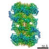 6338MC M: map data used to model this data C: citing same article ( |
|---|---|
| Similar structure data |
-Links
 PDBj
PDBj
- Assembly
| Deposited unit | 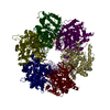
|
|---|---|
| 1 |
|
-Components
-Minichromosome Maintenance ... , 6 types, 6 molecules 234567
| #1: Protein | Mass: 98911.539 Da / Num. of mol.: 1 / Source method: isolated from a natural source / Source: (natural) |
|---|---|
| #2: Protein | Mass: 107653.508 Da / Num. of mol.: 1 / Source method: isolated from a natural source / Source: (natural) |
| #3: Protein | Mass: 105138.375 Da / Num. of mol.: 1 / Source method: isolated from a natural source / Source: (natural) |
| #4: Protein | Mass: 86505.734 Da / Num. of mol.: 1 / Source method: isolated from a natural source / Source: (natural) |
| #5: Protein | Mass: 113110.211 Da / Num. of mol.: 1 / Source method: isolated from a natural source / Source: (natural) |
| #6: Protein | Mass: 95049.875 Da / Num. of mol.: 1 / Source method: isolated from a natural source / Source: (natural) |
-Non-polymers , 1 types, 6 molecules 
| #7: Chemical | ChemComp-ADP /  Mass: 427.201 Da / Num. of mol.: 6 / Source method: obtained synthetically / Formula: C10H15N5O10P2 / Comment: ADP, energy-carrying molecule*YM Mass: 427.201 Da / Num. of mol.: 6 / Source method: obtained synthetically / Formula: C10H15N5O10P2 / Comment: ADP, energy-carrying molecule*YM |
|---|
-Experimental details
-Experiment
| Experiment | Method: ELECTRON MICROSCOPY |
|---|---|
| EM experiment | Aggregation state: PARTICLE / 3D reconstruction method: single particle reconstruction |
- Sample preparation
| Component | Name: Eukaryotic Minichromosome Maintenance Complex / Type: COMPLEX |
|---|---|
| Molecular weight | Value: 1.2 MDa / Experimental value: NO |
| Buffer solution | pH: 7.5 |
| Specimen | Embedding applied: NO / Shadowing applied: NO / Staining applied: NO / Vitrification applied: YES |
| Vitrification | Instrument: FEI VITROBOT MARK IV / Cryogen name: ETHANE / Humidity: 100 % |
- Electron microscopy imaging
| Experimental equipment |  Model: Titan Krios / Image courtesy: FEI Company |
|---|---|
| Microscopy | Model: FEI TITAN KRIOS / Date: Nov 25, 2015 |
| Electron gun | Electron source:  FIELD EMISSION GUN / Accelerating voltage: 300 kV / Illumination mode: FLOOD BEAM FIELD EMISSION GUN / Accelerating voltage: 300 kV / Illumination mode: FLOOD BEAM |
| Electron lens | Mode: BRIGHT FIELD / Nominal magnification: 22500 X / Camera length: 0 mm |
| Specimen holder | Specimen holder model: FEI TITAN KRIOS AUTOGRID HOLDER / Tilt angle max: 0 ° / Tilt angle min: 0 ° |
| Image recording | Electron dose: 22 e/Å2 / Film or detector model: GATAN K2 (4k x 4k) |
- Processing
| EM software | Name: RELION / Category: 3D reconstruction | ||||||||||||
|---|---|---|---|---|---|---|---|---|---|---|---|---|---|
| CTF correction | Details: CTFFIND | ||||||||||||
| Symmetry | Point symmetry: C2 (2 fold cyclic) | ||||||||||||
| 3D reconstruction | Resolution: 3.8 Å / Resolution method: FSC 0.143 CUT-OFF / Num. of particles: 85365 / Details: (Single particle--Applied symmetry: C2) / Symmetry type: POINT | ||||||||||||
| Refinement step | Cycle: LAST
|
