 ムービー
ムービー コントローラー


+ データを開く

- 基本情報
基本情報
| 登録情報 | データベース: EMDB / ID: EMD-2014 | |||||||||
|---|---|---|---|---|---|---|---|---|---|---|
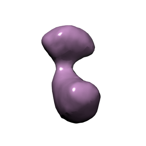
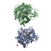
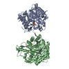
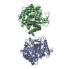
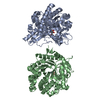
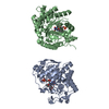
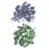
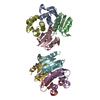
| タイトル | Electron microscopy negative staining map of the cross-linked Ufd1-Npl4 dimer | |||||||||
 マップデータ マップデータ | This corresponds to the EM map of the dimer Ufd1-Npl4 | |||||||||
 試料 試料 |
| |||||||||
 キーワード キーワード | EM / p97 / Ufd1-Npl4 / asymmetric complex / ATPase | |||||||||
| 生物種 |  | |||||||||
| 手法 | 単粒子再構成法 / ネガティブ染色法 / 解像度: 23.0 Å | |||||||||
 データ登録者 データ登録者 | Bebeacua C / Forster A / McKeown C / Meyer HH / Zhang X / Freemont PS | |||||||||
 引用 引用 | ジャーナル: Proc Natl Acad Sci U S A / 年: 2012 タイトル: Distinct conformations of the protein complex p97-Ufd1-Npl4 revealed by electron cryomicroscopy. 著者: Cecilia Bebeacua / Andreas Förster / Ciarán McKeown / Hemmo H Meyer / Xiaodong Zhang / Paul S Freemont /  要旨: p97 is a key regulator of numerous cellular pathways and associates with ubiquitin-binding adaptors to remodel ubiquitin-modified substrate proteins. How adaptor binding to p97 is coordinated and how ...p97 is a key regulator of numerous cellular pathways and associates with ubiquitin-binding adaptors to remodel ubiquitin-modified substrate proteins. How adaptor binding to p97 is coordinated and how adaptors contribute to substrate remodeling is unclear. Here we present the 3D electron cryomicroscopy reconstructions of the major Ufd1-Npl4 adaptor in complex with p97. Our reconstructions show that p97-Ufd1-Npl4 is highly dynamic and that Ufd1-Npl4 assumes distinct positions relative to the p97 ring upon addition of nucleotide. Our results suggest a model for substrate remodeling by p97 and also explains how p97-Ufd1-Npl4 could form other complexes in a hierarchical model of p97-cofactor assembly. | |||||||||
| 履歴 |
|
- 構造の表示
構造の表示
| ムービー |
 ムービービューア ムービービューア |
|---|---|
| 構造ビューア | EMマップ: SurfViewMolmilJmol/JSmol |
| 添付画像 |
- ダウンロードとリンク
-EMDBアーカイブ
| マップデータ | emd_2014.map.gz | 97.2 KB | EMDBマップデータ形式 | |
|---|---|---|---|---|
| ヘッダ (付随情報) | emd-2014-v30.xmlemd-2014.xml | 10.2 KB 10.2 KB | 表示 表示 | EMDBヘッダ |
| 画像 |  emd2014fig.png emd2014fig.png | 51 KB | ||
| アーカイブディレクトリ |  http://ftp.pdbj.org/pub/emdb/structures/EMD-2014ftp://ftp.pdbj.org/pub/emdb/structures/EMD-2014 http://ftp.pdbj.org/pub/emdb/structures/EMD-2014ftp://ftp.pdbj.org/pub/emdb/structures/EMD-2014 | HTTPS FTP |
-関連構造データ


-リンク
| EMDBのページ | EMDB (EBI/PDBe) / EMDataResource |
|---|
-マップ
| ファイル | ダウンロード / ファイル: emd_2014.map.gz / 形式: CCP4 / 大きさ: 1.9 MB / タイプ: IMAGE STORED AS FLOATING POINT NUMBER (4 BYTES) | ||||||||||||||||||||||||||||||||||||||||||||||||||||||||||||
|---|---|---|---|---|---|---|---|---|---|---|---|---|---|---|---|---|---|---|---|---|---|---|---|---|---|---|---|---|---|---|---|---|---|---|---|---|---|---|---|---|---|---|---|---|---|---|---|---|---|---|---|---|---|---|---|---|---|---|---|---|---|
| 注釈 | This corresponds to the EM map of the dimer Ufd1-Npl4 | ||||||||||||||||||||||||||||||||||||||||||||||||||||||||||||
| 投影像・断面図 | 画像のコントロール
画像は Spider により作成 | ||||||||||||||||||||||||||||||||||||||||||||||||||||||||||||
| ボクセルのサイズ | X=Y=Z: 3.53 Å | ||||||||||||||||||||||||||||||||||||||||||||||||||||||||||||
| 密度 |
| ||||||||||||||||||||||||||||||||||||||||||||||||||||||||||||
| 対称性 | 空間群: 1 | ||||||||||||||||||||||||||||||||||||||||||||||||||||||||||||
| 詳細 | EMDB XML:
CCP4マップ ヘッダ情報:
| ||||||||||||||||||||||||||||||||||||||||||||||||||||||||||||
 Z (Sec.)
 Y (Row.)
 X (Col.)


-添付データ
- 試料の構成要素
-全体 : Ufd1-Npl4 cross-linked with glutaraldehyde
| 全体 | 名称: Ufd1-Npl4 cross-linked with glutaraldehyde |
|---|---|
| 要素 |
|
-超分子 #1000: Ufd1-Npl4 cross-linked with glutaraldehyde
| 超分子 | 名称: Ufd1-Npl4 cross-linked with glutaraldehyde / タイプ: sample / ID: 1000 / 集合状態: Ufd1-Npl4 heterodimer / Number unique components: 2 |
|---|---|
| 分子量 | 実験値: 100 KDa / 理論値: 100 KDa |
-分子 #1: Ufd1
| 分子 | 名称: Ufd1 / タイプ: protein_or_peptide / ID: 1 / Name.synonym: Ufd1 / コピー数: 1 / 組換発現: Yes |
|---|---|
| 由来(天然) | 生物種: |
| 組換発現 | 生物種:  |
-分子 #2: Npl4
| 分子 | 名称: Npl4 / タイプ: protein_or_peptide / ID: 2 / Name.synonym: Npl4 / コピー数: 1 / 組換発現: Yes |
|---|---|
| 由来(天然) | 生物種: |
| 組換発現 | 生物種: |
-実験情報
-構造解析
| 手法 | ネガティブ染色法 |
|---|---|
 解析 解析 | 単粒子再構成法 |
| 試料の集合状態 | particle |
-試料調製
| 濃度 | 0.05 mg/mL |
|---|---|
| 緩衝液 | pH: 8 / 詳細: 25 mM HEPES, 500 mM KCl |
| 染色 | タイプ: NEGATIVE 詳細: Grids with adsorbed protein floated on 2% w/v uranyl acetate for 60 seconds. |
| グリッド | 詳細: 200 mesh copper grid |
| 凍結 | 凍結剤: NONE / 装置: OTHER |
- 電子顕微鏡法
| 顕微鏡 | FEI/PHILIPS CM200FEG |
|---|---|
| アライメント法 | Legacy - 非点収差: Objective lens astigmatism was corrected at 100,000 times magnification |
| 特殊光学系 | エネルギーフィルター - 名称: FEI |
| 日付 | 2008年7月1日 |
| 撮影 | カテゴリ: CCD / フィルム・検出器のモデル: GENERIC CCD / 実像数: 1000 / 平均電子線量: 10 e/Å2 |
| 電子線 | 加速電圧: 200 kV / 電子線源:  FIELD EMISSION GUN FIELD EMISSION GUN |
| 電子光学系 | 倍率(補正後): 50000 / 照射モード: SPOT SCAN / 撮影モード: BRIGHT FIELD / Cs: 2.2 mm / 最大 デフォーカス(公称値): 3.0 µm / 最小 デフォーカス(公称値): 1.5 µm / 倍率(公称値): 50000 |
| 試料ステージ | 試料ホルダー: RT / 試料ホルダーモデル: SIDE ENTRY, EUCENTRIC |
-画像解析
| 最終 再構成 | 想定した対称性 - 点群: C1 (非対称) / アルゴリズム: OTHER / 解像度のタイプ: BY AUTHOR / 解像度: 23.0 Å / 解像度の算出法: OTHER / ソフトウェア - 名称: IMAGIC / 使用した粒子像数: 5000 |
|---|
-原子モデル構築 1
| 初期モデル | PDB ID: |
|---|---|
| ソフトウェア | 名称: Chimera |
| 詳細 | Protocol: Rigid Body. The pdb was manually fitted and then automatically refined by the software |
| 精密化 | 空間: REAL / プロトコル: RIGID BODY FIT / 当てはまり具合の基準: Cross Correlation |

