 Movie
Movie Controller

[English] 日本語
 Yorodumi
- EMDB-2014: Electron microscopy negative staining map of the cross-linked Ufd... -
+ Open data

- Basic information
| Entry | Database: EMDB / ID: EMD-2014 | |||||||||
|---|---|---|---|---|---|---|---|---|---|---|
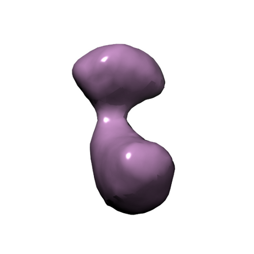
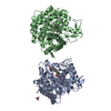
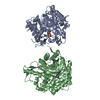
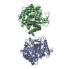
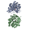
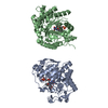
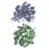
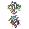
| Title | Electron microscopy negative staining map of the cross-linked Ufd1-Npl4 dimer | |||||||||
 Map data Map data | This corresponds to the EM map of the dimer Ufd1-Npl4 | |||||||||
 Sample Sample |
| |||||||||
 Keywords Keywords | EM / p97 / Ufd1-Npl4 / asymmetric complex / ATPase | |||||||||
| Biological species |  | |||||||||
| Method | single particle reconstruction / negative staining / Resolution: 23.0 Å | |||||||||
 Authors Authors | Bebeacua C / Forster A / McKeown C / Meyer HH / Zhang X / Freemont PS | |||||||||
 Citation Citation | Journal: Proc Natl Acad Sci U S A / Year: 2012 Title: Distinct conformations of the protein complex p97-Ufd1-Npl4 revealed by electron cryomicroscopy. Authors: Cecilia Bebeacua / Andreas Förster / Ciarán McKeown / Hemmo H Meyer / Xiaodong Zhang / Paul S Freemont /  Abstract: p97 is a key regulator of numerous cellular pathways and associates with ubiquitin-binding adaptors to remodel ubiquitin-modified substrate proteins. How adaptor binding to p97 is coordinated and how ...p97 is a key regulator of numerous cellular pathways and associates with ubiquitin-binding adaptors to remodel ubiquitin-modified substrate proteins. How adaptor binding to p97 is coordinated and how adaptors contribute to substrate remodeling is unclear. Here we present the 3D electron cryomicroscopy reconstructions of the major Ufd1-Npl4 adaptor in complex with p97. Our reconstructions show that p97-Ufd1-Npl4 is highly dynamic and that Ufd1-Npl4 assumes distinct positions relative to the p97 ring upon addition of nucleotide. Our results suggest a model for substrate remodeling by p97 and also explains how p97-Ufd1-Npl4 could form other complexes in a hierarchical model of p97-cofactor assembly. | |||||||||
| History |
|
- Structure visualization
| Movie |
 Movie viewer Movie viewer |
|---|---|
| Structure viewer | EM map: SurfViewMolmilJmol/JSmol |
| Supplemental images |
- Downloads & links
-EMDB archive
| Map data | emd_2014.map.gz | 97.2 KB | EMDB map data format | |
|---|---|---|---|---|
| Header (meta data) | emd-2014-v30.xmlemd-2014.xml | 10.2 KB 10.2 KB | Display Display | EMDB header |
| Images |  emd2014fig.png emd2014fig.png | 51 KB | ||
| Archive directory |  http://ftp.pdbj.org/pub/emdb/structures/EMD-2014ftp://ftp.pdbj.org/pub/emdb/structures/EMD-2014 http://ftp.pdbj.org/pub/emdb/structures/EMD-2014ftp://ftp.pdbj.org/pub/emdb/structures/EMD-2014 | HTTPS FTP |
-Related structure data


-Links
| EMDB pages | EMDB (EBI/PDBe) / EMDataResource |
|---|
-Map
| File | Download / File: emd_2014.map.gz / Format: CCP4 / Size: 1.9 MB / Type: IMAGE STORED AS FLOATING POINT NUMBER (4 BYTES) | ||||||||||||||||||||||||||||||||||||||||||||||||||||||||||||
|---|---|---|---|---|---|---|---|---|---|---|---|---|---|---|---|---|---|---|---|---|---|---|---|---|---|---|---|---|---|---|---|---|---|---|---|---|---|---|---|---|---|---|---|---|---|---|---|---|---|---|---|---|---|---|---|---|---|---|---|---|---|
| Annotation | This corresponds to the EM map of the dimer Ufd1-Npl4 | ||||||||||||||||||||||||||||||||||||||||||||||||||||||||||||
| Projections & slices | Image control
Images are generated by Spider. | ||||||||||||||||||||||||||||||||||||||||||||||||||||||||||||
| Voxel size | X=Y=Z: 3.53 Å | ||||||||||||||||||||||||||||||||||||||||||||||||||||||||||||
| Density |
| ||||||||||||||||||||||||||||||||||||||||||||||||||||||||||||
| Symmetry | Space group: 1 | ||||||||||||||||||||||||||||||||||||||||||||||||||||||||||||
| Details | EMDB XML:
CCP4 map header:
| ||||||||||||||||||||||||||||||||||||||||||||||||||||||||||||
 Z (Sec.)
 Y (Row.)
 X (Col.)


-Supplemental data
- Sample components
-Entire : Ufd1-Npl4 cross-linked with glutaraldehyde
| Entire | Name: Ufd1-Npl4 cross-linked with glutaraldehyde |
|---|---|
| Components |
|
-Supramolecule #1000: Ufd1-Npl4 cross-linked with glutaraldehyde
| Supramolecule | Name: Ufd1-Npl4 cross-linked with glutaraldehyde / type: sample / ID: 1000 / Oligomeric state: Ufd1-Npl4 heterodimer / Number unique components: 2 |
|---|---|
| Molecular weight | Experimental: 100 KDa / Theoretical: 100 KDa |
-Macromolecule #1: Ufd1
| Macromolecule | Name: Ufd1 / type: protein_or_peptide / ID: 1 / Name.synonym: Ufd1 / Number of copies: 1 / Recombinant expression: Yes |
|---|---|
| Source (natural) | Organism: |
| Recombinant expression | Organism:  |
-Macromolecule #2: Npl4
| Macromolecule | Name: Npl4 / type: protein_or_peptide / ID: 2 / Name.synonym: Npl4 / Number of copies: 1 / Recombinant expression: Yes |
|---|---|
| Source (natural) | Organism: |
| Recombinant expression | Organism: |
-Experimental details
-Structure determination
| Method | negative staining |
|---|---|
 Processing Processing | single particle reconstruction |
| Aggregation state | particle |
-Sample preparation
| Concentration | 0.05 mg/mL |
|---|---|
| Buffer | pH: 8 / Details: 25 mM HEPES, 500 mM KCl |
| Staining | Type: NEGATIVE Details: Grids with adsorbed protein floated on 2% w/v uranyl acetate for 60 seconds. |
| Grid | Details: 200 mesh copper grid |
| Vitrification | Cryogen name: NONE / Instrument: OTHER |
- Electron microscopy
| Microscope | FEI/PHILIPS CM200FEG |
|---|---|
| Alignment procedure | Legacy - Astigmatism: Objective lens astigmatism was corrected at 100,000 times magnification |
| Specialist optics | Energy filter - Name: FEI |
| Date | Jul 1, 2008 |
| Image recording | Category: CCD / Film or detector model: GENERIC CCD / Number real images: 1000 / Average electron dose: 10 e/Å2 |
| Electron beam | Acceleration voltage: 200 kV / Electron source:  FIELD EMISSION GUN FIELD EMISSION GUN |
| Electron optics | Calibrated magnification: 50000 / Illumination mode: SPOT SCAN / Imaging mode: BRIGHT FIELD / Cs: 2.2 mm / Nominal defocus max: 3.0 µm / Nominal defocus min: 1.5 µm / Nominal magnification: 50000 |
| Sample stage | Specimen holder: RT / Specimen holder model: SIDE ENTRY, EUCENTRIC |
-Image processing
| Final reconstruction | Applied symmetry - Point group: C1 (asymmetric) / Algorithm: OTHER / Resolution.type: BY AUTHOR / Resolution: 23.0 Å / Resolution method: OTHER / Software - Name: IMAGIC / Number images used: 5000 |
|---|
-Atomic model buiding 1
| Initial model | PDB ID: |
|---|---|
| Software | Name: Chimera |
| Details | Protocol: Rigid Body. The pdb was manually fitted and then automatically refined by the software |
| Refinement | Space: REAL / Protocol: RIGID BODY FIT / Target criteria: Cross Correlation |

