 Movie
Movie Controller

[English] 日本語
 Yorodumi
- EMDB-20110: Separating distinct macromolecular assemblies from cryo-EM images -
+ Open data

- Basic information
| Entry | Database: EMDB / ID: EMD-20110 | |||||||||
|---|---|---|---|---|---|---|---|---|---|---|
| Title | Separating distinct macromolecular assemblies from cryo-EM images | |||||||||
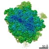
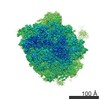
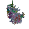
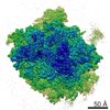
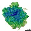
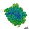
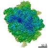
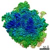
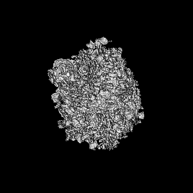
 Map data Map data | Reconstruction of the 60S ribosome | |||||||||
 Sample Sample |
| |||||||||
| Biological species |  | |||||||||
| Method | single particle reconstruction / cryo EM / Resolution: 4.0 Å | |||||||||
 Authors Authors | Verbeke EJ / Zhou Y / Horton AP / Mallam AL / Taylor DW / Marcotte EM | |||||||||
 Citation Citation | Journal: J Struct Biol / Year: 2020 Title: Separating distinct structures of multiple macromolecular assemblies from cryo-EM projections. Authors: Eric J Verbeke / Yi Zhou / Andrew P Horton / Anna L Mallam / David W Taylor / Edward M Marcotte /  Abstract: Single particle analysis for structure determination in cryo-electron microscopy is traditionally applied to samples purified to near homogeneity as current reconstruction algorithms are not designed ...Single particle analysis for structure determination in cryo-electron microscopy is traditionally applied to samples purified to near homogeneity as current reconstruction algorithms are not designed to handle heterogeneous mixtures of structures from many distinct macromolecular complexes. We extend on long established methods and demonstrate that relating two-dimensional projection images by their common lines in a graphical framework is sufficient for partitioning distinct protein and multiprotein complexes within the same data set. The feasibility of this approach is first demonstrated on a large set of synthetic reprojections from 35 unique macromolecular structures spanning a mass range of hundreds to thousands of kilodaltons. We then apply our algorithm on cryo-EM data collected from a mixture of five protein complexes and use existing methods to solve multiple three-dimensional structures ab initio. Incorporating methods to sort single particle cryo-EM data from extremely heterogeneous mixtures will alleviate the need for stringent purification and pave the way toward investigation of samples containing many unique structures. | |||||||||
| History |
|
- Structure visualization
| Movie |
 Movie viewer Movie viewer |
|---|---|
| Structure viewer | EM map: SurfViewMolmilJmol/JSmol |
| Supplemental images |

- Downloads & links
-EMDB archive
| Map data | emd_20110.map.gz | 108.8 MB | EMDB map data format | |
|---|---|---|---|---|
| Header (meta data) | emd-20110-v30.xmlemd-20110.xml | 10.7 KB 10.7 KB | Display Display | EMDB header |
| Images | 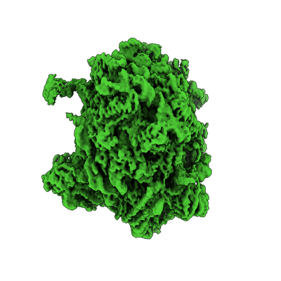 emd_20110.png emd_20110.png | 105.8 KB | ||
| Archive directory |  http://ftp.pdbj.org/pub/emdb/structures/EMD-20110ftp://ftp.pdbj.org/pub/emdb/structures/EMD-20110 http://ftp.pdbj.org/pub/emdb/structures/EMD-20110ftp://ftp.pdbj.org/pub/emdb/structures/EMD-20110 | HTTPS FTP |
-Related structure data
| Related structure data | C: citing same article ( |
|---|---|
| Similar structure data | |
| EM raw data | EMPIAR-10268 (Title: Separating distinct macromolecular assemblies from cryo-EM images Data size: 128.5 Data #1: Drift-corrected and dose-weighted average micographs of a mixture containing 40S, 60S, 80S and apoferritin [micrographs - single frame]) |


-Links
| EMDB pages | EMDB (EBI/PDBe) / EMDataResource |
|---|---|
| Related items in Molecule of the Month |

-Map
| File | Download / File: emd_20110.map.gz / Format: CCP4 / Size: 216 MB / Type: IMAGE STORED AS FLOATING POINT NUMBER (4 BYTES) | ||||||||||||||||||||||||||||||||||||||||||||||||||||||||||||
|---|---|---|---|---|---|---|---|---|---|---|---|---|---|---|---|---|---|---|---|---|---|---|---|---|---|---|---|---|---|---|---|---|---|---|---|---|---|---|---|---|---|---|---|---|---|---|---|---|---|---|---|---|---|---|---|---|---|---|---|---|---|
| Annotation | Reconstruction of the 60S ribosome | ||||||||||||||||||||||||||||||||||||||||||||||||||||||||||||
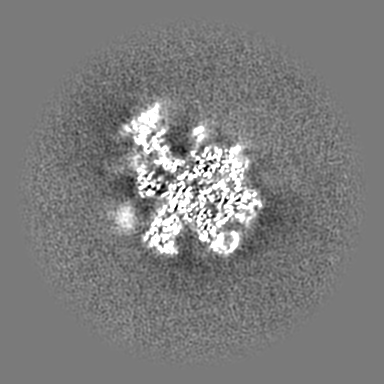
| Projections & slices | Image control
Images are generated by Spider. | ||||||||||||||||||||||||||||||||||||||||||||||||||||||||||||
| Voxel size | X=Y=Z: 1.1 Å | ||||||||||||||||||||||||||||||||||||||||||||||||||||||||||||
| Density |
| ||||||||||||||||||||||||||||||||||||||||||||||||||||||||||||
| Symmetry | Space group: 1 | ||||||||||||||||||||||||||||||||||||||||||||||||||||||||||||
| Details | EMDB XML:
CCP4 map header:
| ||||||||||||||||||||||||||||||||||||||||||||||||||||||||||||
 Z (Sec.)
 Y (Row.)
 X (Col.)


-Supplemental data
- Sample components
-Entire : 60S ribosome
| Entire | Name: 60S ribosome |
|---|---|
| Components |
|
-Supramolecule #1: 60S ribosome
| Supramolecule | Name: 60S ribosome / type: complex / ID: 1 / Parent: 0 / Details: From a complex mixture of defined assemblies |
|---|---|
| Source (natural) | Organism: |
| Molecular weight | Theoretical: 2 MDa |
-Experimental details
-Structure determination
| Method | cryo EM |
|---|---|
 Processing Processing | single particle reconstruction |
| Aggregation state | particle |
-Sample preparation
| Concentration | 1 mg/mL |
|---|---|
| Buffer | pH: 7.4 |
| Grid | Details: unspecified |
| Vitrification | Cryogen name: ETHANE / Chamber humidity: 100 % / Instrument: FEI VITROBOT MARK IV |
| Details | a mixture of 40S, 60S, 80S, and apoferritin |
- Electron microscopy
| Microscope | FEI TITAN KRIOS |
|---|---|
| Image recording | Film or detector model: GATAN K2 SUMMIT (4k x 4k) / Detector mode: COUNTING / Digitization - Frames/image: 1-20 / Number grids imaged: 1 / Number real images: 2400 / Average exposure time: 6.0 sec. / Average electron dose: 40.0 e/Å2 |
| Electron beam | Acceleration voltage: 300 kV / Electron source:  FIELD EMISSION GUN FIELD EMISSION GUN |
| Electron optics | Illumination mode: FLOOD BEAM / Imaging mode: BRIGHT FIELD / Cs: 2.7 mm / Nominal defocus max: -3.0 µm / Nominal defocus min: -2.0 µm |
| Sample stage | Specimen holder model: FEI TITAN KRIOS AUTOGRID HOLDER |
| Experimental equipment |  Model: Titan Krios / Image courtesy: FEI Company |
