 Movie
Movie Controller


+ Open data

- Basic information
| Entry | Database: EMDB / ID: EMD-1869 | |||||||||
|---|---|---|---|---|---|---|---|---|---|---|
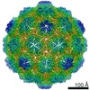
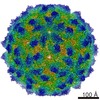
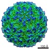
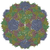
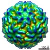
| Title | Procapsid of Staphylococcus aureus Pathogenicity Island 1 | |||||||||
 Map data Map data | Twofold (222) view of isosurface representation of Staphylococcus aureus Pathogenicity Island 1 (SaPI1) procapsid icosahedral reconstruction. | |||||||||
 Sample Sample |
| |||||||||
 Keywords Keywords | Staphylococcus aureus / bacteriophage / capsid / procapsid / size determination / pathogenicity island | |||||||||
| Biological species | Staphylococcus aureus Pathogenicity Island 1 procapsid | |||||||||
| Method | single particle reconstruction / cryo EM / Resolution: 9.99 Å | |||||||||
 Authors Authors | Dearborn AD / Spilman MS / Damle PK / Chang JR / Monroe EB / Saad JS / Christie GE / Dokland T | |||||||||
 Citation Citation | Journal: J Mol Biol / Year: 2011 Title: The Staphylococcus aureus pathogenicity island 1 protein gp6 functions as an internal scaffold during capsid size determination. Authors: Altaira D Dearborn / Michael S Spilman / Priyadarshan K Damle / Jenny R Chang / Eric B Monroe / Jamil S Saad / Gail E Christie / Terje Dokland /  Abstract: Staphylococcus aureus pathogenicity island 1 (SaPI1) is a mobile genetic element that carries genes for several superantigen toxins. SaPI1 is normally stably integrated into the host genome but can ...Staphylococcus aureus pathogenicity island 1 (SaPI1) is a mobile genetic element that carries genes for several superantigen toxins. SaPI1 is normally stably integrated into the host genome but can become mobilized by "helper" bacteriophage 80α, leading to the packaging of SaPI1 genomes into phage-like transducing particles that are composed of structural proteins supplied by the helper phage but having smaller capsids. We show that the SaPI1-encoded protein gp6 is necessary for efficient formation of small capsids. The NMR structure of gp6 reveals a dimeric protein with a helix-loop-helix motif similar to that of bacteriophage scaffolding proteins. The gp6 dimer matches internal densities that bridge capsid subunits in cryo-electron microscopy reconstructions of SaPI1 procapsids, suggesting that gp6 acts as an internal scaffolding protein in capsid size determination. | |||||||||
| History |
|
- Structure visualization
| Movie |
 Movie viewer Movie viewer |
|---|---|
| Structure viewer | EM map: SurfViewMolmilJmol/JSmol |
| Supplemental images |

- Downloads & links
-EMDB archive
| Map data | emd_1869.map.gz | 67.1 MB | EMDB map data format | |
|---|---|---|---|---|
| Header (meta data) | emd-1869-v30.xmlemd-1869.xml | 9.9 KB 9.9 KB | Display Display | EMDB header |
| Images |  emd_1869.png emd_1869.png | 273.3 KB | ||
| Archive directory |  http://ftp.pdbj.org/pub/emdb/structures/EMD-1869ftp://ftp.pdbj.org/pub/emdb/structures/EMD-1869 http://ftp.pdbj.org/pub/emdb/structures/EMD-1869ftp://ftp.pdbj.org/pub/emdb/structures/EMD-1869 | HTTPS FTP |
-Related structure data


-Links
| EMDB pages | EMDB (EBI/PDBe) / EMDataResource |
|---|
-Map
| File | Download / File: emd_1869.map.gz / Format: CCP4 / Size: 89 MB / Type: IMAGE STORED AS FLOATING POINT NUMBER (4 BYTES) | ||||||||||||||||||||||||||||||||||||||||||||||||||||||||||||||||||||
|---|---|---|---|---|---|---|---|---|---|---|---|---|---|---|---|---|---|---|---|---|---|---|---|---|---|---|---|---|---|---|---|---|---|---|---|---|---|---|---|---|---|---|---|---|---|---|---|---|---|---|---|---|---|---|---|---|---|---|---|---|---|---|---|---|---|---|---|---|---|
| Annotation | Twofold (222) view of isosurface representation of Staphylococcus aureus Pathogenicity Island 1 (SaPI1) procapsid icosahedral reconstruction. | ||||||||||||||||||||||||||||||||||||||||||||||||||||||||||||||||||||
| Projections & slices | Image control
Images are generated by Spider. | ||||||||||||||||||||||||||||||||||||||||||||||||||||||||||||||||||||
| Voxel size | X=Y=Z: 2.048 Å | ||||||||||||||||||||||||||||||||||||||||||||||||||||||||||||||||||||
| Density |
| ||||||||||||||||||||||||||||||||||||||||||||||||||||||||||||||||||||
| Symmetry | Space group: 1 | ||||||||||||||||||||||||||||||||||||||||||||||||||||||||||||||||||||
| Details | EMDB XML:
CCP4 map header:
| ||||||||||||||||||||||||||||||||||||||||||||||||||||||||||||||||||||
 Z (Sec.)
 Y (Row.)
 X (Col.)


-Supplemental data
- Sample components
-Entire : Staphylococcus aureus ST63 (Bacteriophage 80alpha delta(terS) SaP...
| Entire | Name: Staphylococcus aureus ST63 (Bacteriophage 80alpha delta(terS) SaPI1 delta(terS)) |
|---|---|
| Components |
|
-Supramolecule #1000: Staphylococcus aureus ST63 (Bacteriophage 80alpha delta(terS) SaP...
| Supramolecule | Name: Staphylococcus aureus ST63 (Bacteriophage 80alpha delta(terS) SaPI1 delta(terS)) type: sample / ID: 1000 Details: Purified by CsCl and sucrose gradient centrifugation Number unique components: 1 |
|---|---|
| Molecular weight | Theoretical: 8.4 MDa |
-Supramolecule #1: Staphylococcus aureus Pathogenicity Island 1 procapsid
| Supramolecule | Name: Staphylococcus aureus Pathogenicity Island 1 procapsid type: virus / ID: 1 / Name.synonym: SaPI1 procapsid Sci species name: Staphylococcus aureus Pathogenicity Island 1 procapsid Virus type: VIRUS-LIKE PARTICLE / Virus isolate: SPECIES / Virus enveloped: No / Virus empty: Yes / Syn species name: SaPI1 procapsid |
|---|---|
| Host (natural) | Organism:   Staphylococcus aureus (bacteria) / synonym: BACTERIA(EUBACTERIA) Staphylococcus aureus (bacteria) / synonym: BACTERIA(EUBACTERIA) |
| Molecular weight | Experimental: 8.4 MDa / Theoretical: 8.4 MDa |
| Virus shell | Shell ID: 1 / Name: procapsid / Diameter: 370 Å / T number (triangulation number): 4 |
-Experimental details
-Structure determination
| Method | cryo EM |
|---|---|
 Processing Processing | single particle reconstruction |
| Aggregation state | particle |
-Sample preparation
| Concentration | 1.0 mg/mL |
|---|---|
| Buffer | pH: 7.8 / Details: 20 mM Tris-HCl, 50 mM NaCl, 1 mM MgCl2, 2 mM CaCl2 |
| Grid | Details: 400 mesh holey film C-flat R 2/1 |
| Vitrification | Cryogen name: ETHANE / Chamber temperature: 108 K / Instrument: HOMEMADE PLUNGER / Details: Vitrification instrument: manual / Method: Blotted for 2 seconds before plunging |
- Electron microscopy
| Microscope | FEI TECNAI F20 |
|---|---|
| Temperature | Average: 97 K |
| Date | Nov 18, 2008 |
| Image recording | Category: FILM / Film or detector model: KODAK SO-163 FILM / Digitization - Scanner: NIKON SUPER COOLSCAN 9000 / Digitization - Sampling interval: 6.3 µm / Number real images: 83 / Average electron dose: 20 e/Å2 Details: Scanned images were binned to 12.6 microns per pixel Od range: 2 / Bits/pixel: 16 |
| Electron beam | Acceleration voltage: 200 kV / Electron source:  FIELD EMISSION GUN FIELD EMISSION GUN |
| Electron optics | Calibrated magnification: 62000 / Illumination mode: FLOOD BEAM / Imaging mode: BRIGHT FIELD / Cs: 2.0 mm / Nominal defocus max: 2.4 µm / Nominal defocus min: 0.8 µm / Nominal magnification: 62000 |
| Sample stage | Specimen holder: Eucentric / Specimen holder model: GATAN LIQUID NITROGEN |
| Experimental equipment |  Model: Tecnai F20 / Image courtesy: FEI Company |
-Image processing
| CTF correction | Details: Each micrograph |
|---|---|
| Final reconstruction | Applied symmetry - Point group: I (icosahedral) / Algorithm: OTHER / Resolution.type: BY AUTHOR / Resolution: 9.99 Å / Resolution method: FSC 0.5 CUT-OFF / Software - Name: EMAN Details: Final maps were calculated to 8A and filtered at 5A resolution Number images used: 3944 |
