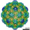
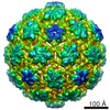
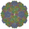
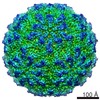
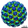
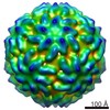
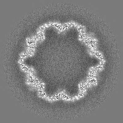
Journal: ACS Nano / Year: 2019 Title: Iron-Sequestering Nanocompartments as Multiplexed Electron Microscopy Gene Reporters. Authors: Felix Sigmund / Susanne Pettinger / Massimo Kube / Fabian Schneider / Martina Schifferer / Steffen Schneider / Maria V Efremova / Jesús Pujol-Martí / Michaela Aichler / Axel Walch / Thomas ...Authors: Felix Sigmund / Susanne Pettinger / Massimo Kube / Fabian Schneider / Martina Schifferer / Steffen Schneider / Maria V Efremova / Jesús Pujol-Martí / Michaela Aichler / Axel Walch / Thomas Misgeld / Hendrik Dietz / Gil G Westmeyer / Abstract: Multicolored gene reporters for light microscopy are indispensable for biomedical research, but equivalent genetic tools for electron microscopy (EM) are still rare despite the increasing importance ...Multicolored gene reporters for light microscopy are indispensable for biomedical research, but equivalent genetic tools for electron microscopy (EM) are still rare despite the increasing importance of nanometer resolution for reverse engineering of molecular machinery and reliable mapping of cellular circuits. We here introduce the fully genetic encapsulin/cargo system of (Qt), which in combination with the recently characterized encapsulin system from (Mx) enables multiplexed gene reporter imaging conventional transmission electron microscopy (TEM) in mammalian cells. Cryo-electron reconstructions revealed that the Qt encapsulin shell self-assembles to nanospheres with = 4 icosahedral symmetry and a diameter of ∼43 nm harboring two putative pore regions at the 5-fold and 3-fold axes. We also found that upon heterologous expression in mammalian cells, the native cargo is autotargeted to the inner surface of the shell and exhibits ferroxidase activity leading to efficient intraluminal iron biomineralization, which enhances cellular TEM contrast. We furthermore demonstrate that the two differently sized encapsulins of Qt and Mx do not intermix and can be robustly differentiated by conventional TEM a deep learning classifier to enable automated multiplexed EM gene reporter imaging.
History
Deposition
Apr 17, 2019
-
Header (metadata) release
Jun 19, 2019
-
Map release
Jun 26, 2019
-
Update
Nov 25, 2020
-
Current status
Nov 25, 2020
Processing site: PDBe / Status: Released
-
Structure visualization
Movie
Surface view with section colored by density value
In the structure databanks used in Yorodumi, some data are registered as the other names, "COVID-19 virus" and "2019-nCoV". Here are the details of the virus and the list of structure data.
Jan 31, 2019. EMDB accession codes are about to change! (news from PDBe EMDB page)
EMDB accession codes are about to change! (news from PDBe EMDB page)
The allocation of 4 digits for EMDB accession codes will soon come to an end. Whilst these codes will remain in use, new EMDB accession codes will include an additional digit and will expand incrementally as the available range of codes is exhausted. The current 4-digit format prefixed with “EMD-” (i.e. EMD-XXXX) will advance to a 5-digit format (i.e. EMD-XXXXX), and so on. It is currently estimated that the 4-digit codes will be depleted around Spring 2019, at which point the 5-digit format will come into force.
The EM Navigator/Yorodumi systems omit the EMD- prefix.
Related info.:Q: What is EMD? / ID/Accession-code notation in Yorodumi/EM Navigator
Yorodumi is a browser for structure data from EMDB, PDB, SASBDB, etc.
This page is also the successor to EM Navigator detail page, and also detail information page/front-end page for Omokage search.
The word "yorodu" (or yorozu) is an old Japanese word meaning "ten thousand". "mi" (miru) is to see.
Related info.:EMDB / PDB / SASBDB / Comparison of 3 databanks / Yorodumi Search / Aug 31, 2016. New EM Navigator & Yorodumi / Yorodumi Papers / Jmol/JSmol / Function and homology information / Changes in new EM Navigator and Yorodumi
 Movie
Movie Controller


 Open data
 Basic information
 Map data
 Sample
 Authors
 Citation

 Structure visualization
 Movie viewer
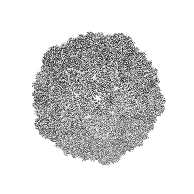
 Downloads & links
 emd_4879.png
 http://ftp.pdbj.org/pub/emdb/structures/EMD-4879

 Z (Sec.)
 Y (Row.)
 X (Col.)


 Sample components
 Processing
 Electron microscopy
 FIELD EMISSION GUN

