 Movie
Movie Controller

[English] 日本語
 Yorodumi
- EMDB-1813: Structural Comparison of HIV-1 Envelope Spikes with and without t... -
+ Open data

- Basic information
| Entry | Database: EMDB / ID: EMD-1813 | |||||||||
|---|---|---|---|---|---|---|---|---|---|---|
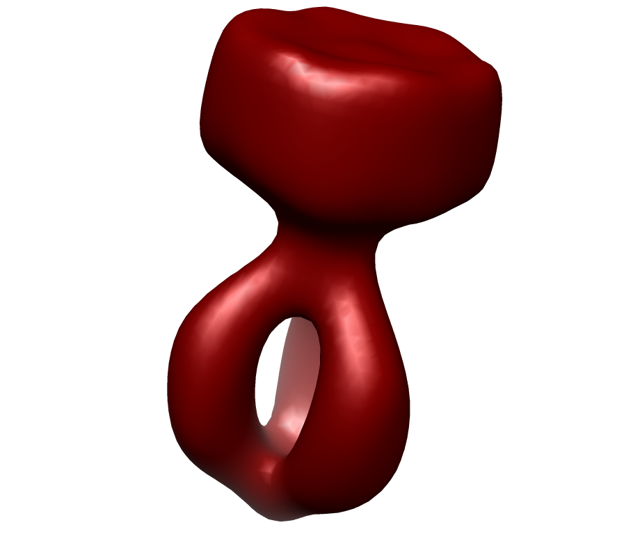
| Title | Structural Comparison of HIV-1 Envelope Spikes with and without the V1V2 Loop. | |||||||||
 Map data Map data | This is the map of the envelope glycoprotein of HIV-1. | |||||||||
 Sample Sample |
| |||||||||
 Keywords Keywords | Virus envelope. | |||||||||
| Biological species |   Human immunodeficiency virus 1 Human immunodeficiency virus 1 | |||||||||
| Method | subtomogram averaging / cryo EM / Resolution: 33.0 Å | |||||||||
 Authors Authors | Hu G / Liu J / Taylor KA / Roux KH | |||||||||
 Citation Citation | Journal: J Virol / Year: 2011 Title: Structural comparison of HIV-1 envelope spikes with and without the V1/V2 loop. Authors: Guiqing Hu / Jun Liu / Kenneth A Taylor / Kenneth H Roux /  Abstract: We have used cryoelectron tomography of vitreous-ice-embedded HIV-1 virions to compare the envelope (Env) spikes of a wild-type strain with those of a mutant strain in which the V1/V2 loop has been ...We have used cryoelectron tomography of vitreous-ice-embedded HIV-1 virions to compare the envelope (Env) spikes of a wild-type strain with those of a mutant strain in which the V1/V2 loop has been deleted. Deletion of V1/V2 results in a spike with far more structural heterogeneity than is observed in the wild type, likely reflecting greatly enhanced gp120 protomer flexibility. A major difference between the two forms is a pronounced loss of mass from the "peak" of the native Env spike. The apparent loss of contact among three gp120 protomers likely accounts for the more open structure, heterogeneity in configuration, and previous observations that broadly neutralizing epitopes and reactive sites on other structural elements are more exposed in such constructs. | |||||||||
| History |
|
- Structure visualization
| Movie |
 Movie viewer Movie viewer |
|---|---|
| Structure viewer | EM map: SurfViewMolmilJmol/JSmol |
| Supplemental images |
- Downloads & links
-EMDB archive
| Map data | emd_1813.map.gz | 215 KB | EMDB map data format | |
|---|---|---|---|---|
| Header (meta data) | emd-1813-v30.xmlemd-1813.xml | 9 KB 9 KB | Display Display | EMDB header |
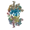
| Images |  emd_1813.png emd_1813.png | 91 KB | ||
| Archive directory |  http://ftp.pdbj.org/pub/emdb/structures/EMD-1813ftp://ftp.pdbj.org/pub/emdb/structures/EMD-1813 http://ftp.pdbj.org/pub/emdb/structures/EMD-1813ftp://ftp.pdbj.org/pub/emdb/structures/EMD-1813 | HTTPS FTP |
-Related structure data


-Links
| EMDB pages | EMDB (EBI/PDBe) / EMDataResource |
|---|
-Map
| File | Download / File: emd_1813.map.gz / Format: CCP4 / Size: 230.5 KB / Type: IMAGE STORED AS FLOATING POINT NUMBER (4 BYTES) | ||||||||||||||||||||||||||||||||||||||||||||||||||||||||||||||||||||
|---|---|---|---|---|---|---|---|---|---|---|---|---|---|---|---|---|---|---|---|---|---|---|---|---|---|---|---|---|---|---|---|---|---|---|---|---|---|---|---|---|---|---|---|---|---|---|---|---|---|---|---|---|---|---|---|---|---|---|---|---|---|---|---|---|---|---|---|---|---|
| Annotation | This is the map of the envelope glycoprotein of HIV-1. | ||||||||||||||||||||||||||||||||||||||||||||||||||||||||||||||||||||
| Projections & slices | Image control
Images are generated by Spider. generated in cubic-lattice coordinate | ||||||||||||||||||||||||||||||||||||||||||||||||||||||||||||||||||||
| Voxel size | X=Y=Z: 4.6 Å | ||||||||||||||||||||||||||||||||||||||||||||||||||||||||||||||||||||
| Density |
| ||||||||||||||||||||||||||||||||||||||||||||||||||||||||||||||||||||
| Symmetry | Space group: 1 | ||||||||||||||||||||||||||||||||||||||||||||||||||||||||||||||||||||
| Details | EMDB XML:
CCP4 map header:
| ||||||||||||||||||||||||||||||||||||||||||||||||||||||||||||||||||||
 Z (Sec.)
 Y (Row.)
 X (Col.)


-Supplemental data
- Sample components
-Entire : Envelope glycoprotein
| Entire | Name: Envelope glycoprotein |
|---|---|
| Components |
|
-Supramolecule #1000: Envelope glycoprotein
| Supramolecule | Name: Envelope glycoprotein / type: sample / ID: 1000 / Details: The sample was ice-embedded. / Number unique components: 1 |
|---|
-Macromolecule #1: Envelope glycoprotein gp160
| Macromolecule | Name: Envelope glycoprotein gp160 / type: protein_or_peptide / ID: 1 / Name.synonym: Envelope glycoprotein / Recombinant expression: Yes / Database: NCBI |
|---|---|
| Source (natural) | Organism: Human immunodeficiency virus 1 |
-Experimental details
-Structure determination
| Method | cryo EM |
|---|---|
 Processing Processing | subtomogram averaging |
-Sample preparation
| Grid | Details: Holey carbon grid |
|---|---|
| Vitrification | Cryogen name: ETHANE / Instrument: OTHER |
- Electron microscopy
| Microscope | FEI POLARA 300 |
|---|---|
| Image recording | Category: CCD / Film or detector model: GENERIC CCD / Average electron dose: 100 e/Å2 |
| Electron beam | Acceleration voltage: 300 kV / Electron source:  FIELD EMISSION GUN FIELD EMISSION GUN |
| Electron optics | Illumination mode: FLOOD BEAM / Imaging mode: BRIGHT FIELD / Nominal defocus max: 5.0 µm / Nominal defocus min: 4.0 µm / Nominal magnification: 39000 |
| Sample stage | Specimen holder: Multi-specimen holer / Specimen holder model: OTHER / Tilt series - Axis1 - Min angle: -65 ° / Tilt series - Axis1 - Max angle: 65 ° |
| Experimental equipment |  Model: Tecnai Polara / Image courtesy: FEI Company |
-Image processing
| Final reconstruction | Algorithm: OTHER / Resolution.type: BY AUTHOR / Resolution: 33.0 Å / Resolution method: FSC 0.5 CUT-OFF / Software - Name: Protomo and I3 |
|---|
-Atomic model buiding 1
| Initial model | PDB ID: Chain - Chain ID: A |
|---|---|
| Software | Name: Manual |
| Details | PDBEntryID_givenInChain. Protocol: Manual. The fitting was conducted manually in chimera. The V3 loop is built from the V3 loop of PDB 2B4C. |
| Refinement | Space: REAL / Protocol: RIGID BODY FIT |

-Atomic model buiding 2
| Initial model | PDB ID: Chain - Chain ID: A |
|---|---|
| Software | Name: Manual |
| Details | PDBEntryID_givenInChain. Protocol: Manual. The fitting was conducted manually in chimera. The V3 loop is built from the V3 loop of PDB 2B4C. |
| Refinement | Space: REAL / Protocol: RIGID BODY FIT |

