 Movie
Movie Controller

[English] 日本語
 Yorodumi
- EMDB-1807: Saccharomyces cerevisiae ribonucleotide reductase hole complex at... -
+ Open data

- Basic information
| Entry | Database: EMDB / ID: EMD-1807 | |||||||||
|---|---|---|---|---|---|---|---|---|---|---|
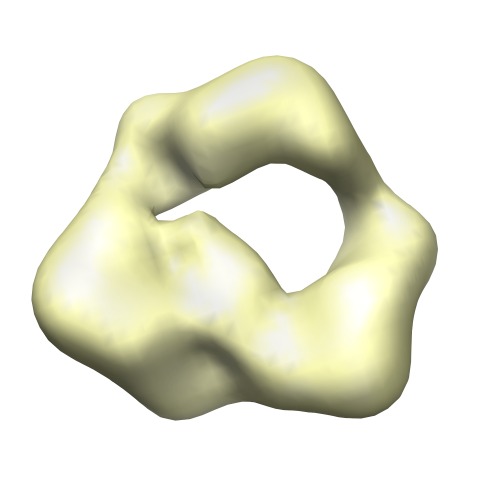
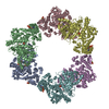
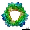
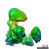
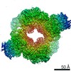
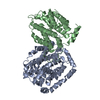
| Title | Saccharomyces cerevisiae ribonucleotide reductase hole complex at the presence of dATP | |||||||||
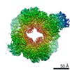
 Map data Map data | This is an EM map of Yeast dATP Ribonucleotide Reductase complex | |||||||||
 Sample Sample |
| |||||||||
 Keywords Keywords | Ribonucleotide reductase / hexamer / dimer | |||||||||
| Biological species |  | |||||||||
| Method | single particle reconstruction / cryo EM / negative staining / Resolution: 28.0 Å | |||||||||
 Authors Authors | Fairman JW / Wijerathna SR / Ahmad MF / Xu H / Nakano R / Jha S / Prendergast J / Welin RM / Flodin S / Roos A ...Fairman JW / Wijerathna SR / Ahmad MF / Xu H / Nakano R / Jha S / Prendergast J / Welin RM / Flodin S / Roos A / Nordlund P / Li Z / Walz T / Dealwis CG | |||||||||
 Citation Citation | Journal: Nat Struct Mol Biol / Year: 2011 Title: Structural basis for allosteric regulation of human ribonucleotide reductase by nucleotide-induced oligomerization. Authors: James Wesley Fairman / Sanath Ranjan Wijerathna / Md Faiz Ahmad / Hai Xu / Ryo Nakano / Shalini Jha / Jay Prendergast / R Martin Welin / Susanne Flodin / Annette Roos / Pär Nordlund / ...Authors: James Wesley Fairman / Sanath Ranjan Wijerathna / Md Faiz Ahmad / Hai Xu / Ryo Nakano / Shalini Jha / Jay Prendergast / R Martin Welin / Susanne Flodin / Annette Roos / Pär Nordlund / Zongli Li / Thomas Walz / Chris Godfrey Dealwis /  Abstract: Ribonucleotide reductase (RR) is an α(n)β(n) (RR1-RR2) complex that maintains balanced dNTP pools by reducing NDPs to dNDPs. RR1 is the catalytic subunit, and RR2 houses the free radical required ...Ribonucleotide reductase (RR) is an α(n)β(n) (RR1-RR2) complex that maintains balanced dNTP pools by reducing NDPs to dNDPs. RR1 is the catalytic subunit, and RR2 houses the free radical required for catalysis. RR is allosterically regulated by its activator ATP and its inhibitor dATP, which regulate RR activity by inducing oligomerization of RR1. Here, we report the first X-ray structures of human RR1 bound to TTP alone, dATP alone, TTP-GDP, TTP-ATP, and TTP-dATP. These structures provide insights into regulation of RR by ATP or dATP. At physiological dATP concentrations, RR1 forms inactive hexamers. We determined the first X-ray structure of the RR1-dATP hexamer and used single-particle electron microscopy to visualize the α(6)-ββ'-dATP holocomplex. Site-directed mutagenesis and functional assays confirm that hexamerization is a prerequisite for inhibition by dATP. Our data indicate a mechanism for regulating RR activity by dATP-induced oligomerization. | |||||||||
| History |
|
- Structure visualization
| Movie |
 Movie viewer Movie viewer |
|---|---|
| Structure viewer | EM map: SurfViewMolmilJmol/JSmol |
| Supplemental images |
- Downloads & links
-EMDB archive
| Map data | emd_1807.map.gz | 7.5 MB | EMDB map data format | |
|---|---|---|---|---|
| Header (meta data) | emd-1807-v30.xmlemd-1807.xml | 12.1 KB 12.1 KB | Display Display | EMDB header |
| Images | 1807.tif | 107.6 KB | ||
| Archive directory |  http://ftp.pdbj.org/pub/emdb/structures/EMD-1807ftp://ftp.pdbj.org/pub/emdb/structures/EMD-1807 http://ftp.pdbj.org/pub/emdb/structures/EMD-1807ftp://ftp.pdbj.org/pub/emdb/structures/EMD-1807 | HTTPS FTP |
-Validation report
| Summary document | emd_1807_validation.pdf.gz | 195.2 KB | Display | EMDB validaton report |
|---|---|---|---|---|
| Full document | emd_1807_full_validation.pdf.gz | 194.4 KB | Display | |
| Data in XML | emd_1807_validation.xml.gz | 5.6 KB | Display | |
| Arichive directory | https://ftp.pdbj.org/pub/emdb/validation_reports/EMD-1807ftp://ftp.pdbj.org/pub/emdb/validation_reports/EMD-1807 | HTTPS FTP |
-Related structure data
| Related structure data | 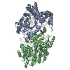 2wghC 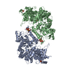 3hncC 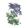 3hndC 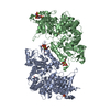 3hneC  3hnfC 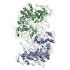 3pawC C: citing same article ( |
|---|---|
| Similar structure data |
-Links
| EMDB pages | EMDB (EBI/PDBe) / EMDataResource |
|---|
-Map
| File | Download / File: emd_1807.map.gz / Format: CCP4 / Size: 7.8 MB / Type: IMAGE STORED AS FLOATING POINT NUMBER (4 BYTES) | ||||||||||||||||||||||||||||||||||||||||||||||||||||||||||||||||||||
|---|---|---|---|---|---|---|---|---|---|---|---|---|---|---|---|---|---|---|---|---|---|---|---|---|---|---|---|---|---|---|---|---|---|---|---|---|---|---|---|---|---|---|---|---|---|---|---|---|---|---|---|---|---|---|---|---|---|---|---|---|---|---|---|---|---|---|---|---|---|
| Annotation | This is an EM map of Yeast dATP Ribonucleotide Reductase complex | ||||||||||||||||||||||||||||||||||||||||||||||||||||||||||||||||||||
| Projections & slices | Image control
Images are generated by Spider. | ||||||||||||||||||||||||||||||||||||||||||||||||||||||||||||||||||||
| Voxel size | X=Y=Z: 3.1 Å | ||||||||||||||||||||||||||||||||||||||||||||||||||||||||||||||||||||
| Density |
| ||||||||||||||||||||||||||||||||||||||||||||||||||||||||||||||||||||
| Symmetry | Space group: 1 | ||||||||||||||||||||||||||||||||||||||||||||||||||||||||||||||||||||
| Details | EMDB XML:
CCP4 map header:
| ||||||||||||||||||||||||||||||||||||||||||||||||||||||||||||||||||||
 Z (Sec.)
 Y (Row.)
 X (Col.)


-Supplemental data
- Sample components
-Entire : Yeast ribonucleotide reductase complex
| Entire | Name: Yeast ribonucleotide reductase complex |
|---|---|
| Components |
|
-Supramolecule #1000: Yeast ribonucleotide reductase complex
| Supramolecule | Name: Yeast ribonucleotide reductase complex / type: sample / ID: 1000 Oligomeric state: One homohexamer of yeast RR1 binds to one hetero-dimer of yeast RR2.RR4 Number unique components: 2 |
|---|---|
| Molecular weight | Experimental: 700 KDa / Theoretical: 680 KDa / Method: Size exclusion chromatography |
-Macromolecule #1: Yeast ribonucleotide reductase RR1
| Macromolecule | Name: Yeast ribonucleotide reductase RR1 / type: protein_or_peptide / ID: 1 / Name.synonym: RR1 / Recombinant expression: Yes |
|---|---|
| Source (natural) | Organism: |
-Macromolecule #2: Yeast ribonucleotide reductase RR2.RR4
| Macromolecule | Name: Yeast ribonucleotide reductase RR2.RR4 / type: protein_or_peptide / ID: 2 / Name.synonym: RR2.RR4 / Recombinant expression: Yes |
|---|---|
| Source (natural) | Organism: |
-Experimental details
-Structure determination
| Method | negative staining, cryo EM |
|---|---|
 Processing Processing | single particle reconstruction |
| Aggregation state | particle |
-Sample preparation
| Concentration | 0.1 mg/mL |
|---|---|
| Buffer | pH: 8.5 Details: 50mM Ammonium actate,5mM MgCl2, 0.1M KCl, 50uM dATP,100uM hydroxyurea with 5% glycerol |
| Staining | Type: NEGATIVE Details: Grids with adsorbed protein was stained on 0.75% w/v uranyl formate for 30 seconds. |
| Grid | Details: Quantifoil R2/1 Coppor grid |
| Vitrification | Cryogen name: NITROGEN / Instrument: OTHER |
- Electron microscopy
| Microscope | FEI TECNAI F20 |
|---|---|
| Alignment procedure | Legacy - Astigmatism: Objective lens astigmatism was corrected at 150,000 times magnificatioin |
| Image recording | Category: FILM / Film or detector model: KODAK SO-163 FILM / Digitization - Scanner: ZEISS SCAI / Digitization - Sampling interval: 4.2 µm / Number real images: 52 |
| Electron beam | Acceleration voltage: 200 kV / Electron source:  FIELD EMISSION GUN FIELD EMISSION GUN |
| Electron optics | Calibrated magnification: 49883 / Illumination mode: FLOOD BEAM / Imaging mode: BRIGHT FIELD / Cs: 2 mm / Nominal magnification: 50000 |
| Sample stage | Specimen holder: Side entry liquid nitrogen-cooled cryo specimen holder Specimen holder model: GATAN LIQUID NITROGEN / Tilt angle max: 50 |
| Experimental equipment |  Model: Tecnai F20 / Image courtesy: FEI Company |
-Image processing
| Details | The quantifoil grids are pre-coated with continuous carbon film. 5 ul sample is applied onto the carbon film grid,wait for 30 seconds, blot from side, wash it in a drop of water, blot from side, stain in a drop of 0.75% uranyl formate for 30 seconds, with the sample side facing up insert into a drop of 0.75% uranyl formate where a small piece of carbon film has been floated, pick up the carbon film from underneath, gently blot from both side. Monitor the thickness of the remaining sample between two carbon films . When the thickness is becoming right (milky in color), plunge it into liquid nitrogen to freeze. Look at the grid with cryo EM procedure. |
|---|---|
| Final reconstruction | Applied symmetry - Point group: C1 (asymmetric) / Algorithm: OTHER / Resolution.type: BY AUTHOR / Resolution: 28.0 Å / Resolution method: FSC 0.5 CUT-OFF / Software - Name: SPIDER Details: The final map was calculated from a selected sub-data-set based on 2D classification using Back Projection and Angular Refinement routines in Spider. Number images used: 829 |
| Final two d classification | Number classes: 50 |
-Atomic model buiding 1
| Initial model | PDB ID: |
|---|---|
| Software | Name: Camera |
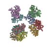
| Details | Protocol: Rigid Body. The Yeast RR1.dATP hexamer (3PAW) was fitted into the EM density map and the Yeast RR2.RR4 heterodimer (1JK0) was fitted into the difference map using camera fit in map function. |
| Refinement | Space: REAL / Protocol: RIGID BODY FIT |
-Atomic model buiding 2
| Initial model | PDB ID: |
|---|---|
| Software | Name: Camera |
| Details | Protocol: Rigid Body. The Yeast RR1.dATP hexamer (3PAW) was fitted into the EM density map and the Yeast RR2.RR4 heterodimer (1JK0) was fitted into the difference map using camera fit in map function. |
| Refinement | Space: REAL / Protocol: RIGID BODY FIT |
