 Movie
Movie Controller


+ Open data

- Basic information
| Entry | Database: PDB / ID: 1jk0 | ||||||
|---|---|---|---|---|---|---|---|
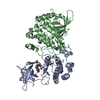
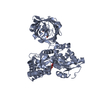
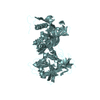
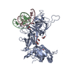
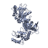
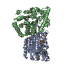
| Title | Ribonucleotide reductase Y2Y4 heterodimer | ||||||
 Components Components |
| ||||||
 Keywords Keywords | OXIDOREDUCTASE / ribonucleotide reductase / R2 | ||||||
| Function / homology |  Function and homology information Function and homology informationInterconversion of nucleotide di- and triphosphates / ribonucleoside-diphosphate reductase complex / ribonucleoside-diphosphate reductase / ribonucleoside-diphosphate reductase activity, thioredoxin disulfide as acceptor / deoxyribonucleotide biosynthetic process / ferrous iron binding / molecular adaptor activity / protein heterodimerization activity / zinc ion binding / nucleus / cytoplasm Similarity search - Function | ||||||
| Biological species |  | ||||||
| Method |  X-RAY DIFFRACTION / SYNCHROTRON / MIR / Resolution: 2.8 Å X-RAY DIFFRACTION / SYNCHROTRON / MIR / Resolution: 2.8 Å | ||||||
 Authors Authors | Voegtli, W.C. / Perlstein, D.L. / Ge, J. / Stubbe, J. / Rosenzweig, A.C. | ||||||
 Citation Citation | Journal: Proc.Natl.Acad.Sci.USA / Year: 2001 Title: Structure of the yeast ribonucleotide reductase Y2Y4 heterodimer. Authors: Voegtli, W.C. / Ge, J. / Perlstein, D.L. / Stubbe, J. / Rosenzweig, A.C. | ||||||
| History |
|
- Structure visualization
| Structure viewer | Molecule: MolmilJmol/JSmol |
|---|
- Downloads & links
-Download
| PDBx/mmCIF format | 1jk0.cif.gz | 138.5 KB | Display | PDBx/mmCIF format |
|---|---|---|---|---|
| PDB format | pdb1jk0.ent.gz | 106 KB | Display | PDB format |
| PDBx/mmJSON format | 1jk0.json.gz | Tree view | PDBx/mmJSON format | |
| Others |  Other downloads Other downloads |
-Validation report
| Arichive directory | https://data.pdbj.org/pub/pdb/validation_reports/jk/1jk0ftp://data.pdbj.org/pub/pdb/validation_reports/jk/1jk0 | HTTPS FTP |
|---|
-Related structure data
| Similar structure data |
|---|


-Links
 PDBj
PDBj
- Assembly
| Deposited unit | 
| ||||||||
|---|---|---|---|---|---|---|---|---|---|
| 1 |
| ||||||||
| Unit cell |
| ||||||||
| Details | Biological assembly is a heterodimer of monomers of Y2 and Y4 |
-Components
| #1: Protein | Mass: 48378.656 Da / Num. of mol.: 1 Source method: isolated from a genetically manipulated source Source: (gene. exp.) Gene: RNR2 / Plasmid: pET-14b / Species (production host): Escherichia coli / Production host:  References: UniProt: P09938, ribonucleoside-diphosphate reductase |
|---|---|
| #2: Protein | Mass: 40102.500 Da / Num. of mol.: 1 Source method: isolated from a genetically manipulated source Source: (gene. exp.) Gene: RNR4 / Plasmid: pET-14b / Species (production host): Escherichia coli / Production host: References: UniProt: P49723, ribonucleoside-diphosphate reductase |
| #3: Chemical | ChemComp-ZN /   Mass: 65.409 Da / Num. of mol.: 1 / Source method: obtained synthetically / Formula: Zn Mass: 65.409 Da / Num. of mol.: 1 / Source method: obtained synthetically / Formula: Zn |
| #4: Water | ChemComp-HOH /  Mass: 18.015 Da / Num. of mol.: 58 / Source method: isolated from a natural source / Formula: H2O Mass: 18.015 Da / Num. of mol.: 58 / Source method: isolated from a natural source / Formula: H2O |
-Experimental details
-Experiment
| Experiment | Method: X-RAY DIFFRACTION / Number of used crystals: 1 |
|---|
- Sample preparation
| Crystal | Density Matthews: 2.38 Å3/Da / Density % sol: 48.25 % | ||||||||||||||||||||||||||||||||||||
|---|---|---|---|---|---|---|---|---|---|---|---|---|---|---|---|---|---|---|---|---|---|---|---|---|---|---|---|---|---|---|---|---|---|---|---|---|---|
| Crystal grow | Temperature: 293 K / Method: sandwich drop / pH: 4.9 Details: 100 mM acetate, 14% w/v polyethyleneglycol 4000, 200 mM NaCl, pH 4.9, sandwich drop, temperature 293K | ||||||||||||||||||||||||||||||||||||
| Crystal grow | *PLUS pH: 5.3 | ||||||||||||||||||||||||||||||||||||
| Components of the solutions | *PLUS
|
-Data collection
| Diffraction | Mean temperature: 110 K |
|---|---|
| Diffraction source | Source: SYNCHROTRON / Site: SSRL  / Beamline: BL9-1 / Wavelength: 0.946 Å / Beamline: BL9-1 / Wavelength: 0.946 Å |
| Detector | Type: MARRESEARCH / Detector: IMAGE PLATE / Date: Jun 30, 2000 |
| Radiation | Monochromator: Si double crystal / Protocol: SINGLE WAVELENGTH / Monochromatic (M) / Laue (L): M / Scattering type: x-ray |
| Radiation wavelength | Wavelength: 0.946 Å / Relative weight: 1 |
| Reflection | Resolution: 2.8→9 Å / Num. all: 20545 / Num. obs: 19844 / % possible obs: 96.6 % / Observed criterion σ(F): 1 / Observed criterion σ(I): 1 / Redundancy: 3.1 % / Biso Wilson estimate: 41.3 Å2 / Rmerge(I) obs: 0.061 / Rsym value: 0.061 / Net I/σ(I): 17.7 |
| Reflection shell | Resolution: 2.8→2.87 Å / Redundancy: 3.1 % / Rmerge(I) obs: 0.286 / Mean I/σ(I) obs: 2.7 / Num. unique all: 1530 / Rsym value: 0.286 / % possible all: 99.7 |
| Reflection | *PLUS Lowest resolution: 15 Å / Num. obs: 20645 / % possible obs: 99.9 % / Num. measured all: 65155 / Rmerge(I) obs: 0.063 |
| Reflection shell | *PLUS Lowest resolution: 2.85 Å / % possible obs: 99.8 % / Rmerge(I) obs: 0.285 |
- Processing
| Software |
| |||||||||||||||||||||||||
|---|---|---|---|---|---|---|---|---|---|---|---|---|---|---|---|---|---|---|---|---|---|---|---|---|---|---|
| Refinement | Method to determine structure: MIR / Resolution: 2.8→9 Å / Rfactor Rfree error: 0.008 / Data cutoff high absF: 564143.99 / Data cutoff low absF: 0 / Isotropic thermal model: RESTRAINED / Cross valid method: THROUGHOUT / σ(F): 1 / Stereochemistry target values: Engh & Huber
| |||||||||||||||||||||||||
| Solvent computation | Solvent model: FLAT MODEL / Bsol: 56.5531 Å2 / ksol: 0.443753 e/Å3 | |||||||||||||||||||||||||
| Displacement parameters | Biso mean: 46.9 Å2
| |||||||||||||||||||||||||
| Refine analyze |
| |||||||||||||||||||||||||
| Refinement step | Cycle: LAST / Resolution: 2.8→9 Å
| |||||||||||||||||||||||||
| Refine LS restraints |
| |||||||||||||||||||||||||
| LS refinement shell | Resolution: 2.8→2.97 Å / Rfactor Rfree error: 0.024 / Total num. of bins used: 6
| |||||||||||||||||||||||||
| Xplor file |
| |||||||||||||||||||||||||
| Software | *PLUS Name: CNS / Version: 1 / Classification: refinement | |||||||||||||||||||||||||
| Refinement | *PLUS σ(F): 1 / % reflection Rfree: 7.4 % | |||||||||||||||||||||||||
| Solvent computation | *PLUS | |||||||||||||||||||||||||
| Displacement parameters | *PLUS Biso mean: 46.9 Å2 | |||||||||||||||||||||||||
| Refine LS restraints | *PLUS
| |||||||||||||||||||||||||
| LS refinement shell | *PLUS Rfactor Rfree: 0.374 / % reflection Rfree: 8 % / Rfactor Rwork: 0.317 |
