 Movie
Movie Controller

[English] 日本語
 Yorodumi
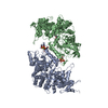
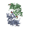
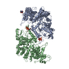
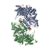
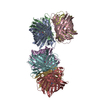
- PDB-3paw: Low resolution X-ray crystal structure of Yeast Rnr1p with dATP b... -
+ Open data

- Basic information
| Entry | Database: PDB / ID: 3paw | ||||||
|---|---|---|---|---|---|---|---|
| Title | Low resolution X-ray crystal structure of Yeast Rnr1p with dATP bound in the A-site | ||||||
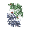
 Components Components | Ribonucleoside-diphosphate reductase large chain 1 | ||||||
 Keywords Keywords | OXIDOREDUCTASE / Ribonucleotide reductase / Nucleotide binding | ||||||
| Function / homology |  Function and homology information Function and homology informationribonucleoside-diphosphate reductase complex / ribonucleoside-diphosphate reductase / ribonucleoside-diphosphate reductase activity, thioredoxin disulfide as acceptor / deoxyribonucleotide biosynthetic process / nucleotide binding / ATP binding / identical protein binding / nucleus / cytoplasm Similarity search - Function | ||||||
| Biological species |  | ||||||
| Method |  X-RAY DIFFRACTION / SYNCHROTRON / MOLECULAR REPLACEMENT / Resolution: 6.61 Å X-RAY DIFFRACTION / SYNCHROTRON / MOLECULAR REPLACEMENT / Resolution: 6.61 Å | ||||||
 Authors Authors | Fairman, J.W. / Wijerathna, S.R. / Dealwis, C.G. | ||||||
 Citation Citation | Journal: Nat Struct Mol Biol / Year: 2011 Title: Structural basis for allosteric regulation of human ribonucleotide reductase by nucleotide-induced oligomerization. Authors: James Wesley Fairman / Sanath Ranjan Wijerathna / Md Faiz Ahmad / Hai Xu / Ryo Nakano / Shalini Jha / Jay Prendergast / R Martin Welin / Susanne Flodin / Annette Roos / Pär Nordlund / ...Authors: James Wesley Fairman / Sanath Ranjan Wijerathna / Md Faiz Ahmad / Hai Xu / Ryo Nakano / Shalini Jha / Jay Prendergast / R Martin Welin / Susanne Flodin / Annette Roos / Pär Nordlund / Zongli Li / Thomas Walz / Chris Godfrey Dealwis /  Abstract: Ribonucleotide reductase (RR) is an α(n)β(n) (RR1-RR2) complex that maintains balanced dNTP pools by reducing NDPs to dNDPs. RR1 is the catalytic subunit, and RR2 houses the free radical required ...Ribonucleotide reductase (RR) is an α(n)β(n) (RR1-RR2) complex that maintains balanced dNTP pools by reducing NDPs to dNDPs. RR1 is the catalytic subunit, and RR2 houses the free radical required for catalysis. RR is allosterically regulated by its activator ATP and its inhibitor dATP, which regulate RR activity by inducing oligomerization of RR1. Here, we report the first X-ray structures of human RR1 bound to TTP alone, dATP alone, TTP-GDP, TTP-ATP, and TTP-dATP. These structures provide insights into regulation of RR by ATP or dATP. At physiological dATP concentrations, RR1 forms inactive hexamers. We determined the first X-ray structure of the RR1-dATP hexamer and used single-particle electron microscopy to visualize the α(6)-ββ'-dATP holocomplex. Site-directed mutagenesis and functional assays confirm that hexamerization is a prerequisite for inhibition by dATP. Our data indicate a mechanism for regulating RR activity by dATP-induced oligomerization. | ||||||
| History |
|
- Structure visualization
| Structure viewer | Molecule: MolmilJmol/JSmol |
|---|
- Downloads & links
-Download
| PDBx/mmCIF format | 3paw.cif.gz | 448.5 KB | Display | PDBx/mmCIF format |
|---|---|---|---|---|
| PDB format | pdb3paw.ent.gz | 305.8 KB | Display | PDB format |
| PDBx/mmJSON format | 3paw.json.gz | Tree view | PDBx/mmJSON format | |
| Others |  Other downloads Other downloads |
-Validation report
| Arichive directory | https://data.pdbj.org/pub/pdb/validation_reports/pa/3pawftp://data.pdbj.org/pub/pdb/validation_reports/pa/3paw | HTTPS FTP |
|---|
-Related structure data
| Related structure data | 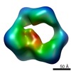 1807C 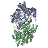 2wghC 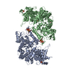 3hncC  3hndC  3hneC  3hnfC C: citing same article ( |
|---|---|
| Similar structure data |


-Links
 PDBj
PDBj
- Assembly
| Deposited unit | 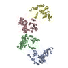
| ||||||||
|---|---|---|---|---|---|---|---|---|---|
| 1 | 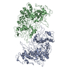
| ||||||||
| 2 | 
| ||||||||
| Unit cell |
|
-Components
| #1: Protein | Mass: 99672.984 Da / Num. of mol.: 4 Source method: isolated from a genetically manipulated source Source: (gene. exp.) Gene: RNR1, CRT7, RIR1, SDS12, YER070W / Plasmid: pET21 / Production host:  References: UniProt: P21524, ribonucleoside-diphosphate reductase |
|---|
-Experimental details
-Experiment
| Experiment | Method: X-RAY DIFFRACTION / Number of used crystals: 1 |
|---|
- Sample preparation
| Crystal | Density Matthews: 3.83 Å3/Da / Density % sol: 67.9 % |
|---|
-Data collection
| Diffraction source | Source: SYNCHROTRON / Site: APS / Beamline: 14-ID-B / Wavelength: 0.9 Å |
|---|---|
| Detector | Type: MAR scanner 345 mm plate / Detector: IMAGE PLATE |
| Radiation | Protocol: SINGLE WAVELENGTH / Monochromatic (M) / Laue (L): M / Scattering type: x-ray |
| Radiation wavelength | Wavelength: 0.9 Å / Relative weight: 1 |
| Reflection | Resolution: 6.61→192.45 Å / Num. obs: 9893 |
- Processing
| Software | Name: REFMAC / Version: 5.5.0109 / Classification: refinement | ||||||||||||||||||||||||||||||||||||||||||||||||||||||||||||||||||||||||||||||||||||||||||||||||||||||||||||||||||||||||||||||||||||||||||||||||||||||||||||||||||||||||||
|---|---|---|---|---|---|---|---|---|---|---|---|---|---|---|---|---|---|---|---|---|---|---|---|---|---|---|---|---|---|---|---|---|---|---|---|---|---|---|---|---|---|---|---|---|---|---|---|---|---|---|---|---|---|---|---|---|---|---|---|---|---|---|---|---|---|---|---|---|---|---|---|---|---|---|---|---|---|---|---|---|---|---|---|---|---|---|---|---|---|---|---|---|---|---|---|---|---|---|---|---|---|---|---|---|---|---|---|---|---|---|---|---|---|---|---|---|---|---|---|---|---|---|---|---|---|---|---|---|---|---|---|---|---|---|---|---|---|---|---|---|---|---|---|---|---|---|---|---|---|---|---|---|---|---|---|---|---|---|---|---|---|---|---|---|---|---|---|---|---|---|---|
| Refinement | Method to determine structure: MOLECULAR REPLACEMENT / Resolution: 6.61→192.45 Å / Cor.coef. Fo:Fc: 0.674 / Cor.coef. Fo:Fc free: 0.531 / SU B: 0.006 / SU ML: 0 / Cross valid method: THROUGHOUT / ESU R: 3.423 / ESU R Free: 3.869 / Stereochemistry target values: MAXIMUM LIKELIHOOD / Details: HYDROGENS HAVE BEEN ADDED IN THE RIDING POSITIONS
| ||||||||||||||||||||||||||||||||||||||||||||||||||||||||||||||||||||||||||||||||||||||||||||||||||||||||||||||||||||||||||||||||||||||||||||||||||||||||||||||||||||||||||
| Solvent computation | Ion probe radii: 0.8 Å / Shrinkage radii: 0.8 Å / VDW probe radii: 1.4 Å / Solvent model: MASK | ||||||||||||||||||||||||||||||||||||||||||||||||||||||||||||||||||||||||||||||||||||||||||||||||||||||||||||||||||||||||||||||||||||||||||||||||||||||||||||||||||||||||||
| Displacement parameters | Biso mean: 88.172 Å2
| ||||||||||||||||||||||||||||||||||||||||||||||||||||||||||||||||||||||||||||||||||||||||||||||||||||||||||||||||||||||||||||||||||||||||||||||||||||||||||||||||||||||||||
| Refinement step | Cycle: LAST / Resolution: 6.61→192.45 Å
| ||||||||||||||||||||||||||||||||||||||||||||||||||||||||||||||||||||||||||||||||||||||||||||||||||||||||||||||||||||||||||||||||||||||||||||||||||||||||||||||||||||||||||
| Refine LS restraints |
| ||||||||||||||||||||||||||||||||||||||||||||||||||||||||||||||||||||||||||||||||||||||||||||||||||||||||||||||||||||||||||||||||||||||||||||||||||||||||||||||||||||||||||
| LS refinement shell | Resolution: 6.612→6.784 Å / Total num. of bins used: 20
|
