 Movie
Movie Controller

[English] 日本語
 Yorodumi
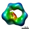
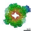
- PDB-6aui: Human ribonucleotide reductase large subunit (alpha) with dATP and CDP -
+ Open data

- Basic information
| Entry | Database: PDB / ID: 6aui | ||||||||||||
|---|---|---|---|---|---|---|---|---|---|---|---|---|---|
| Title | Human ribonucleotide reductase large subunit (alpha) with dATP and CDP | ||||||||||||
 Components Components | Ribonucleoside-diphosphate reductase large subunit | ||||||||||||
 Keywords Keywords | OXIDOREDUCTASE / Ribonucleotide Reductase Electron transfer Radical chemistry Thiyl radical | ||||||||||||
| Function / homology |  Function and homology information Function and homology informationribonucleoside-diphosphate reductase activity / pyrimidine nucleobase metabolic process / cell proliferation in forebrain / ribonucleoside diphosphate metabolic process / positive regulation of G0 to G1 transition / 2'-deoxyribonucleotide biosynthetic process / mitochondrial DNA replication / Interconversion of nucleotide di- and triphosphates / ribonucleoside-diphosphate reductase complex / ribonucleoside-diphosphate reductase ...ribonucleoside-diphosphate reductase activity / pyrimidine nucleobase metabolic process / cell proliferation in forebrain / ribonucleoside diphosphate metabolic process / positive regulation of G0 to G1 transition / 2'-deoxyribonucleotide biosynthetic process / mitochondrial DNA replication / Interconversion of nucleotide di- and triphosphates / ribonucleoside-diphosphate reductase complex / ribonucleoside-diphosphate reductase / ribonucleoside-diphosphate reductase activity, thioredoxin disulfide as acceptor / deoxyribonucleotide biosynthetic process / response to ionizing radiation / protein heterotetramerization / DNA synthesis involved in DNA repair / positive regulation of G2/M transition of mitotic cell cycle / positive regulation of G1/S transition of mitotic cell cycle / male gonad development / centriolar satellite / disordered domain specific binding / nuclear envelope / retina development in camera-type eye / ciliary basal body / DNA repair / neuronal cell body / mitochondrion / ATP binding / identical protein binding / cytosol Similarity search - Function | ||||||||||||
| Biological species |  Homo sapiens (human) Homo sapiens (human) | ||||||||||||
| Method | ELECTRON MICROSCOPY / single particle reconstruction / cryo EM / Resolution: 3.3 Å | ||||||||||||
 Authors Authors | Brignole, E.J. / Drennan, C.L. / Asturias, F.J. / Tsai, K.L. / Penczek, P.A. | ||||||||||||
| Funding support |  United States, 3items United States, 3items
| ||||||||||||
 Citation Citation | Journal: Elife / Year: 2018 Title: 3.3-Å resolution cryo-EM structure of human ribonucleotide reductase with substrate and allosteric regulators bound. Authors: Edward J Brignole / Kuang-Lei Tsai / Johnathan Chittuluru / Haoran Li / Yimon Aye / Pawel A Penczek / JoAnne Stubbe / Catherine L Drennan / Francisco Asturias / Abstract: Ribonucleotide reductases (RNRs) convert ribonucleotides into deoxyribonucleotides, a reaction essential for DNA replication and repair. Human RNR requires two subunits for activity, the α subunit ...Ribonucleotide reductases (RNRs) convert ribonucleotides into deoxyribonucleotides, a reaction essential for DNA replication and repair. Human RNR requires two subunits for activity, the α subunit contains the active site, and the β subunit houses the radical cofactor. Here, we present a 3.3-Å resolution structure by cryo-electron microscopy (EM) of a dATP-inhibited state of human RNR. This structure, which was determined in the presence of substrate CDP and allosteric regulators ATP and dATP, has three α units arranged in an α ring. At near-atomic resolution, these data provide insight into the molecular basis for CDP recognition by allosteric specificity effectors dATP/ATP. Additionally, we present lower-resolution EM structures of human α in the presence of both the anticancer drug clofarabine triphosphate and β. Together, these structures support a model for RNR inhibition in which β is excluded from binding in a radical transfer competent position when α exists as a stable hexamer. | ||||||||||||
| History |
|
- Structure visualization
| Movie |
Movie viewer |
|---|---|
| Structure viewer | Molecule: MolmilJmol/JSmol |
- Downloads & links
-Download
| PDBx/mmCIF format | 6aui.cif.gz | 782.7 KB | Display | PDBx/mmCIF format |
|---|---|---|---|---|
| PDB format | pdb6aui.ent.gz | 651.9 KB | Display | PDB format |
| PDBx/mmJSON format | 6aui.json.gz | Tree view | PDBx/mmJSON format | |
| Others |  Other downloads Other downloads |
-Validation report
| Arichive directory | https://data.pdbj.org/pub/pdb/validation_reports/au/6auiftp://data.pdbj.org/pub/pdb/validation_reports/au/6aui | HTTPS FTP |
|---|
-Related structure data
| Related structure data | 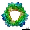 7006MC M: map data used to model this data C: citing same article ( |
|---|---|
| Similar structure data |


-Links
 PDBj
PDBj


- Assembly
| Deposited unit | 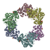
|
|---|---|
| 1 |
|
-Components
| #1: Protein | Mass: 92350.391 Da / Num. of mol.: 6 Source method: isolated from a genetically manipulated source Source: (gene. exp.) Homo sapiens (human) / Gene: RRM1, RR1 / Plasmid: pET-28aDetails (production host): Gene inserted between NdeI and NotI sites Production host:  References: UniProt: P23921, ribonucleoside-diphosphate reductase #2: Chemical | ChemComp-DTP /   Mass: 491.182 Da / Num. of mol.: 12 / Source method: obtained synthetically / Formula: C10H16N5O12P3 Mass: 491.182 Da / Num. of mol.: 12 / Source method: obtained synthetically / Formula: C10H16N5O12P3#3: Chemical | ChemComp-MG /   Mass: 24.305 Da / Num. of mol.: 12 / Source method: obtained synthetically / Formula: Mg Mass: 24.305 Da / Num. of mol.: 12 / Source method: obtained synthetically / Formula: Mg#4: Chemical | ChemComp-CDP / 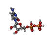  Mass: 403.176 Da / Num. of mol.: 6 / Source method: obtained synthetically / Formula: C9H15N3O11P2 Mass: 403.176 Da / Num. of mol.: 6 / Source method: obtained synthetically / Formula: C9H15N3O11P2#5: Water | ChemComp-HOH / |  Mass: 18.015 Da / Num. of mol.: 36 / Source method: isolated from a natural source / Formula: H2O Mass: 18.015 Da / Num. of mol.: 36 / Source method: isolated from a natural source / Formula: H2O |
|---|
-Experimental details
-Experiment
| Experiment | Method: ELECTRON MICROSCOPY |
|---|---|
| EM experiment | Aggregation state: PARTICLE / 3D reconstruction method: single particle reconstruction |
- Sample preparation
| Component | Name: Human ribonucleotide reductase large subnunit (alpha) / Type: COMPLEX / Entity ID: #1 / Source: RECOMBINANT | |||||||||||||||||||||||||
|---|---|---|---|---|---|---|---|---|---|---|---|---|---|---|---|---|---|---|---|---|---|---|---|---|---|---|
| Molecular weight | Units: MEGADALTONS / Experimental value: NO | |||||||||||||||||||||||||
| Source (natural) | Organism: Homo sapiens (human) | |||||||||||||||||||||||||
| Source (recombinant) | Organism: | |||||||||||||||||||||||||
| Buffer solution | pH: 7.6 | |||||||||||||||||||||||||
| Buffer component |
| |||||||||||||||||||||||||
| Specimen | Conc.: 1.3 mg/ml / Embedding applied: NO / Shadowing applied: NO / Staining applied: NO / Vitrification applied: YES Details: 14 microM alpha and 0.05 mM dATP, 3 mM ATP, 1 mM CDP in 50 mM HEPES, pH 7.6, 15 mM MgCl2, 1 mM EDTA, 5 mM DTT, and 50 mM KCl | |||||||||||||||||||||||||
| Specimen support | Details: glow discharged at 20 mA in an EMITech K100X Grid was first cleaned in a Solarus 950 (Gatan) at 25 W for 10 s in 75/25 Ar/O2 gas mixture Grid material: COPPER / Grid mesh size: 400 divisions/in. / Grid type: Protochips C-Flat | |||||||||||||||||||||||||
| Vitrification | Instrument: HOMEMADE PLUNGER / Cryogen name: ETHANE / Humidity: 75 % / Chamber temperature: 277 K Details: manual blot with a strip of Whatman paper until drop stops wicking determined visually |
- Electron microscopy imaging
| Experimental equipment |  Model: Titan Krios / Image courtesy: FEI Company |
|---|---|
| Microscopy | Model: FEI TITAN KRIOS |
| Electron gun | Electron source:  FIELD EMISSION GUN / Accelerating voltage: 300 kV / Illumination mode: FLOOD BEAM FIELD EMISSION GUN / Accelerating voltage: 300 kV / Illumination mode: FLOOD BEAM |
| Electron lens | Mode: BRIGHT FIELD / Nominal magnification: 22500 X / Nominal defocus max: 2800 nm / Nominal defocus min: 800 nm |
| Specimen holder | Cryogen: NITROGEN / Specimen holder model: FEI TITAN KRIOS AUTOGRID HOLDER |
| Image recording | Average exposure time: 7.6 sec. / Electron dose: 44 e/Å2 / Detector mode: COUNTING / Film or detector model: GATAN K2 SUMMIT (4k x 4k) / Num. of real images: 2144 |
| Image scans | Movie frames/image: 38 / Used frames/image: 5-38 |
- Processing
| Software | Name: PHENIX / Version: dev_2650: / Classification: refinement | |||||||||||||||||||||||||||
|---|---|---|---|---|---|---|---|---|---|---|---|---|---|---|---|---|---|---|---|---|---|---|---|---|---|---|---|---|
| EM software |
| |||||||||||||||||||||||||||
| CTF correction | Type: PHASE FLIPPING AND AMPLITUDE CORRECTION | |||||||||||||||||||||||||||
| Symmetry | Point symmetry: D3 (2x3 fold dihedral) | |||||||||||||||||||||||||||
| 3D reconstruction | Resolution: 3.3 Å / Resolution method: FSC 0.143 CUT-OFF / Num. of particles: 43885 / Symmetry type: POINT | |||||||||||||||||||||||||||
| Atomic model building | Protocol: FLEXIBLE FIT / Space: REAL | |||||||||||||||||||||||||||
| Refine LS restraints |
|
