 Movie
Movie Controller


+ Open data

- Basic information
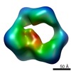
| Entry | Database: PDB / ID: 5zrv | |||||||||||||||||||||||||||||||||
|---|---|---|---|---|---|---|---|---|---|---|---|---|---|---|---|---|---|---|---|---|---|---|---|---|---|---|---|---|---|---|---|---|---|---|
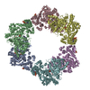
| Title | Structure of human mitochondrial trifunctional protein, octamer | |||||||||||||||||||||||||||||||||
 Components Components |
| |||||||||||||||||||||||||||||||||
 Keywords Keywords | Liase / Oxidoreductase/Transferase / fatty acid beta-oxidation / cryo-EM single-particle reconstruction / mitochondrial trifunctional protein / Oxidoreductase-Transferase complex | |||||||||||||||||||||||||||||||||
| Function / homology |  Function and homology information Function and homology informationlong-chain-3-hydroxyacyl-CoA dehydrogenase / cardiolipin acyl-chain remodeling / Acyl chain remodeling of CL / Beta oxidation of myristoyl-CoA to lauroyl-CoA / Beta oxidation of palmitoyl-CoA to myristoyl-CoA / acetyl-CoA C-myristoyltransferase / acetyl-CoA C-myristoyltransferase activity / mitochondrial fatty acid beta-oxidation multienzyme complex / mitochondrial fatty acid beta-oxidation of unsaturated fatty acids / Beta oxidation of lauroyl-CoA to decanoyl-CoA-CoA ...long-chain-3-hydroxyacyl-CoA dehydrogenase / cardiolipin acyl-chain remodeling / Acyl chain remodeling of CL / Beta oxidation of myristoyl-CoA to lauroyl-CoA / Beta oxidation of palmitoyl-CoA to myristoyl-CoA / acetyl-CoA C-myristoyltransferase / acetyl-CoA C-myristoyltransferase activity / mitochondrial fatty acid beta-oxidation multienzyme complex / mitochondrial fatty acid beta-oxidation of unsaturated fatty acids / Beta oxidation of lauroyl-CoA to decanoyl-CoA-CoA / Beta oxidation of hexanoyl-CoA to butanoyl-CoA / Beta oxidation of octanoyl-CoA to hexanoyl-CoA / Beta oxidation of decanoyl-CoA to octanoyl-CoA-CoA / acetyl-CoA C-acyltransferase / acetyl-CoA C-acyltransferase activity / long-chain fatty acyl-CoA hydrolase activity / long-chain (3S)-3-hydroxyacyl-CoA dehydrogenase (NAD+) activity / 3-hydroxyacyl-CoA dehydratase activity / enoyl-CoA hydratase / (3S)-3-hydroxyacyl-CoA dehydrogenase (NAD+) activity / acetyl-CoA C-acetyltransferase activity / enoyl-CoA hydratase activity / mitochondrial envelope / fatty acid beta-oxidation / lncRNA binding / mitochondrial nucleoid / NAD+ binding / Transferases; Acyltransferases; Transferring groups other than aminoacyl groups / response to insulin / sperm principal piece / cellular response to lipopolysaccharide / gene expression / mitochondrial outer membrane / mitochondrial inner membrane / protein-containing complex binding / endoplasmic reticulum / mitochondrion / RNA binding / nucleoplasm Similarity search - Function | |||||||||||||||||||||||||||||||||
| Biological species |  Homo sapiens (human) Homo sapiens (human) | |||||||||||||||||||||||||||||||||
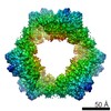
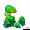
| Method | ELECTRON MICROSCOPY / single particle reconstruction / cryo EM / Resolution: 7.7 Å | |||||||||||||||||||||||||||||||||
 Authors Authors | Liang, K. / Li, N. / Dai, J. / Wang, X. / Liu, P. / Chen, X. / Wang, C. / Gao, N. / Xiao, J. | |||||||||||||||||||||||||||||||||
| Funding support |  China, 1items China, 1items
| |||||||||||||||||||||||||||||||||
 Citation Citation | Journal: Proc Natl Acad Sci U S A / Year: 2018 Title: Cryo-EM structure of human mitochondrial trifunctional protein. Authors: Kai Liang / Ningning Li / Xiao Wang / Jianye Dai / Pulan Liu / Chu Wang / Xiao-Wei Chen / Ning Gao / Junyu Xiao / Abstract: The mitochondrial trifunctional protein (TFP) catalyzes three reactions in the fatty acid β-oxidation process. Mutations in the two TFP subunits cause mitochondrial trifunctional protein deficiency ...The mitochondrial trifunctional protein (TFP) catalyzes three reactions in the fatty acid β-oxidation process. Mutations in the two TFP subunits cause mitochondrial trifunctional protein deficiency and acute fatty liver of pregnancy that can lead to death. Here we report a 4.2-Å cryo-electron microscopy α2β2 tetrameric structure of the human TFP. The tetramer has a V-shaped architecture that displays a distinct assembly compared with the bacterial TFPs. A concave surface of the TFP tetramer interacts with the detergent molecules in the structure, suggesting that this region is involved in associating with the membrane. Deletion of a helical hairpin in TFPβ decreases its binding to the liposomes in vitro and reduces its membrane targeting in cells. Our results provide the structural basis for TFP function and have important implications for fatty acid oxidation related diseases. | |||||||||||||||||||||||||||||||||
| History |
|
- Structure visualization
| Movie |
Movie viewer |
|---|---|
| Structure viewer | Molecule: MolmilJmol/JSmol |
- Downloads & links
-Download
| PDBx/mmCIF format | 5zrv.cif.gz | 562.4 KB | Display | PDBx/mmCIF format |
|---|---|---|---|---|
| PDB format | pdb5zrv.ent.gz | 366.8 KB | Display | PDB format |
| PDBx/mmJSON format | 5zrv.json.gz | Tree view | PDBx/mmJSON format | |
| Others |  Other downloads Other downloads |
-Validation report
| Arichive directory | https://data.pdbj.org/pub/pdb/validation_reports/zr/5zrvftp://data.pdbj.org/pub/pdb/validation_reports/zr/5zrv | HTTPS FTP |
|---|
-Related structure data
| Related structure data |  6944MC  6940C  6945C 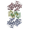 5zqzC C: citing same article ( M: map data used to model this data |
|---|---|
| Similar structure data |
-Links
 PDBj
PDBj


- Assembly
| Deposited unit | 
|
|---|---|
| 1 |
|
-Components
| #1: Protein | Mass: 83112.625 Da / Num. of mol.: 4 Source method: isolated from a genetically manipulated source Source: (gene. exp.) Homo sapiens (human) / Gene: HADHA, HADH / Production host:  References: UniProt: P40939, enoyl-CoA hydratase, long-chain-3-hydroxyacyl-CoA dehydrogenase #2: Protein | Mass: 51360.359 Da / Num. of mol.: 4 Source method: isolated from a genetically manipulated source Source: (gene. exp.) Homo sapiens (human) / Gene: HADHB, MSTP029 / Production host: Has protein modification | N | |
|---|
-Experimental details
-Experiment
| Experiment | Method: ELECTRON MICROSCOPY |
|---|---|
| EM experiment | Aggregation state: PARTICLE / 3D reconstruction method: single particle reconstruction |
- Sample preparation
| Component | Name: human mitochondrial trifunctional protein / Type: COMPLEX / Entity ID: all / Source: RECOMBINANT |
|---|---|
| Molecular weight | Units: MEGADALTONS / Experimental value: NO |
| Source (natural) | Organism: Homo sapiens (human) |
| Source (recombinant) | Organism: |
| Buffer solution | pH: 8 |
| Specimen | Embedding applied: NO / Shadowing applied: NO / Staining applied: NO / Vitrification applied: YES |
| Specimen support | Grid material: GOLD |
| Vitrification | Cryogen name: ETHANE |
- Electron microscopy imaging
| Experimental equipment |  Model: Titan Krios / Image courtesy: FEI Company |
|---|---|
| Microscopy | Model: FEI TITAN KRIOS |
| Electron gun | Electron source:  FIELD EMISSION GUN / Accelerating voltage: 300 kV / Illumination mode: FLOOD BEAM FIELD EMISSION GUN / Accelerating voltage: 300 kV / Illumination mode: FLOOD BEAM |
| Electron lens | Mode: BRIGHT FIELD |
| Image recording | Electron dose: 50 e/Å2 / Film or detector model: GATAN K2 SUMMIT (4k x 4k) |
- Processing
| Software | Name: PHENIX / Version: 1.11.1_2575: / Classification: refinement | ||||||||||||||||||||||||
|---|---|---|---|---|---|---|---|---|---|---|---|---|---|---|---|---|---|---|---|---|---|---|---|---|---|
| EM software | Name: PHENIX / Category: model refinement | ||||||||||||||||||||||||
| CTF correction | Type: PHASE FLIPPING AND AMPLITUDE CORRECTION | ||||||||||||||||||||||||
| 3D reconstruction | Resolution: 7.7 Å / Resolution method: FSC 0.143 CUT-OFF / Num. of particles: 48564 / Details: Gold Standard / Symmetry type: POINT | ||||||||||||||||||||||||
| Refine LS restraints |
|
