 Movie
Movie Controller

[English] 日本語
 Yorodumi
- EMDB-1801: Single particle cryo-electron microscopy analysis of unliganded H... -
+ Open data

- Basic information
| Entry | Database: EMDB / ID: EMD-1801 | |||||||||
|---|---|---|---|---|---|---|---|---|---|---|
| Title | Single particle cryo-electron microscopy analysis of unliganded HIV-1 Env | |||||||||
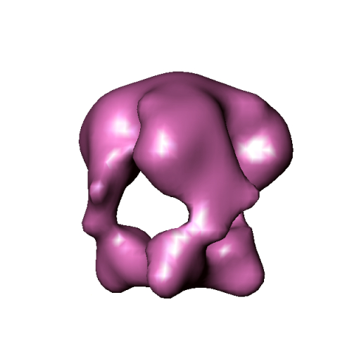
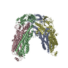
 Map data Map data | This is an image of a surface rendered unliganded HIV-1 Env | |||||||||
 Sample Sample |
| |||||||||
 Keywords Keywords | HIV-1 spike / Membrane Fusion / Structural Transition / Cryo-EM / Single Particle | |||||||||
| Function / homology | Human immunodeficiency virus 1, envelope glycoprotein Gp120 / viral envelope Function and homology information Function and homology information | |||||||||
| Biological species |   Human immunodeficiency virus 1 Human immunodeficiency virus 1 | |||||||||
| Method | single particle reconstruction / cryo EM / negative staining / Resolution: 18.0 Å | |||||||||
 Authors Authors | Wu SR / Loving R / Lindqvist B / Hebert H / Koeck P / Sjoberg M / Garoff H | |||||||||
 Citation Citation | Journal: Proc Natl Acad Sci U S A / Year: 2010 Title: Single-particle cryoelectron microscopy analysis reveals the HIV-1 spike as a tripod structure. Authors: Shang-Rung Wu / Robin Löving / Birgitta Lindqvist / Hans Hebert / Philip J B Koeck / Mathilda Sjöberg / Henrik Garoff /  Abstract: The HIV-1 spike is a trimer of the transmembrane gp41 and the peripheral gp120 subunit pair. It is activated for virus-cell membrane fusion by binding sequentially to CD4 and to a chemokine receptor. ...The HIV-1 spike is a trimer of the transmembrane gp41 and the peripheral gp120 subunit pair. It is activated for virus-cell membrane fusion by binding sequentially to CD4 and to a chemokine receptor. Here we have studied the structural transition of the trimeric spike during the activation process. We solubilized and isolated unliganded and CD4-bound spikes from virus-like particles and used cryoelectron microscopy to reconstruct their 3D structures. In order to increase the yield and stability of the spike, we used an endodomain deleted and gp120-gp41 disulfide-linked variant. The unliganded spike displayed a hollow cage-like structure where the gp120-gp41 protomeric units formed a roof and bottom, and separated lobes and legs on the sides. The tripod structure was verified by fitting the recent atomic core structure of gp120 with intact N- and C-terminal ends into the spike density map. This defined the lobe as gp120 core, showed that the legs contained the polypeptide termini, and suggested the deleted variable loops V1/V2 and V3 to occupy the roof and gp41 the bottom. CD4 binding shifted the roof density peripherally and condensed the bottom density centrally. Fitting with a V3 containing gp120 core suggested that the V1/V2 loops in the roof were displaced laterally and the V3 lifted up, while the core and leg were kept in place. The loop displacements probably prepared the spike for coreceptor interaction and roof opening so that a new fusion-active gp41 structure, assembled at the center of the cage bottom, could reach the target membrane. | |||||||||
| History |
|
- Structure visualization
| Movie |
Movie viewer |
|---|---|
| Structure viewer | EM map: SurfViewMolmilJmol/JSmol |
| Supplemental images |
- Downloads & links
-EMDB archive
| Map data | emd_1801.map.gz | 734.6 KB | EMDB map data format | |
|---|---|---|---|---|
| Header (meta data) | emd-1801-v30.xmlemd-1801.xml | 10.6 KB 10.6 KB | Display Display | EMDB header |
| Images |  EMD_1801.jpg EMD_1801.jpg | 76.2 KB | ||
| Archive directory |  http://ftp.pdbj.org/pub/emdb/structures/EMD-1801ftp://ftp.pdbj.org/pub/emdb/structures/EMD-1801 http://ftp.pdbj.org/pub/emdb/structures/EMD-1801ftp://ftp.pdbj.org/pub/emdb/structures/EMD-1801 | HTTPS FTP |
-Related structure data


-Links
| EMDB pages | EMDB (EBI/PDBe) / EMDataResource |
|---|
-Map
| File | Download / File: emd_1801.map.gz / Format: CCP4 / Size: 1.4 MB / Type: IMAGE STORED AS FLOATING POINT NUMBER (4 BYTES) | ||||||||||||||||||||||||||||||||||||||||||||||||||||||||||||||||||||
|---|---|---|---|---|---|---|---|---|---|---|---|---|---|---|---|---|---|---|---|---|---|---|---|---|---|---|---|---|---|---|---|---|---|---|---|---|---|---|---|---|---|---|---|---|---|---|---|---|---|---|---|---|---|---|---|---|---|---|---|---|---|---|---|---|---|---|---|---|---|
| Annotation | This is an image of a surface rendered unliganded HIV-1 Env | ||||||||||||||||||||||||||||||||||||||||||||||||||||||||||||||||||||
| Projections & slices | Image control
Images are generated by Spider. | ||||||||||||||||||||||||||||||||||||||||||||||||||||||||||||||||||||
| Voxel size | X=Y=Z: 3.5 Å | ||||||||||||||||||||||||||||||||||||||||||||||||||||||||||||||||||||
| Density |
| ||||||||||||||||||||||||||||||||||||||||||||||||||||||||||||||||||||
| Symmetry | Space group: 1 | ||||||||||||||||||||||||||||||||||||||||||||||||||||||||||||||||||||
| Details | EMDB XML:
CCP4 map header:
| ||||||||||||||||||||||||||||||||||||||||||||||||||||||||||||||||||||
 Z (Sec.)
 Y (Row.)
 X (Col.)


-Supplemental data
- Sample components
-Entire : Unliganded HIV-1 Env
| Entire | Name: Unliganded HIV-1 Env |
|---|---|
| Components |
|
-Supramolecule #1000: Unliganded HIV-1 Env
| Supramolecule | Name: Unliganded HIV-1 Env / type: sample / ID: 1000 / Details: The sample was monodisperse / Oligomeric state: Trimer / Number unique components: 1 |
|---|---|
| Molecular weight | Experimental: 550 KDa / Theoretical: 420 KDa / Method: Blue Native PAGE |
-Macromolecule #1: gp160deltaCTSOS
| Macromolecule | Name: gp160deltaCTSOS / type: protein_or_peptide / ID: 1 / Name.synonym: HIV-1 Env / Number of copies: 3 / Oligomeric state: Trimer / Recombinant expression: Yes |
|---|---|
| Source (natural) | Organism: Human immunodeficiency virus 1 / Strain: JR-FL / synonym: HIV-1 |
| Molecular weight | Experimental: 550 KDa / Theoretical: 420 KDa |
| Recombinant expression | Organism: 293T / Recombinant plasmid: p C AGGS JRFL 160deltaCTSOS |
| Sequence | GO: viral envelope InterPro: Human immunodeficiency virus 1, envelope glycoprotein Gp120 |
-Experimental details
-Structure determination
| Method | negative staining, cryo EM |
|---|---|
 Processing Processing | single particle reconstruction |
| Aggregation state | particle |
-Sample preparation
| Buffer | pH: 7.4 / Details: 50 mM HEPES, 100 mM NaCl, 1.8 mM CaCl2 |
|---|---|
| Staining | Type: NEGATIVE / Details: No stain was applied |
| Grid | Details: 200 mesh Cu Holey carbon grid |
| Vitrification | Cryogen name: ETHANE / Chamber humidity: 99 % / Chamber temperature: 77 K / Instrument: OTHER / Details: Vitrification instrument: Vitrobot / Method: Blot for 3 seconds before plunging |
- Electron microscopy
| Microscope | JEOL 2100F |
|---|---|
| Temperature | Min: 93 K / Max: 96 K / Average: 95 K |
| Alignment procedure | Legacy - Astigmatism: Objective lens astigmatism was corrected using online FFT Legacy - Electron beam tilt params: No Tilt |
| Date | Nov 18, 2008 |
| Image recording | Category: CCD / Film or detector model: GENERIC CCD / Digitization - Sampling interval: 3.5 µm / Number real images: 148 / Average electron dose: 9 e/Å2 / Bits/pixel: 16 |
| Electron beam | Acceleration voltage: 200 kV / Electron source:  FIELD EMISSION GUN FIELD EMISSION GUN |
| Electron optics | Illumination mode: SPOT SCAN / Imaging mode: BRIGHT FIELD / Cs: 2.0 mm / Nominal defocus max: 6.0 µm / Nominal defocus min: 2.5 µm / Nominal magnification: 43300 |
| Sample stage | Specimen holder: Eucentric / Specimen holder model: GATAN LIQUID NITROGEN |
-Image processing
| CTF correction | Details: Each Digitized Image |
|---|---|
| Final reconstruction | Applied symmetry - Point group: C3 (3 fold cyclic) / Algorithm: OTHER / Resolution.type: BY AUTHOR / Resolution: 18.0 Å / Resolution method: FSC 0.5 CUT-OFF / Software - Name: EMAN / Number images used: 11213 |
| Final two d classification | Number classes: 131 |
-Atomic model buiding 1
| Initial model | PDB ID: Chain - Chain ID: A |
|---|---|
| Software | Name: O |
| Details | PDBEntryID_givenInChain. Protocol: Rigid Body |
| Refinement | Space: REAL / Protocol: RIGID BODY FIT |

