ムービー
ムービー コントローラー


+ データを開く

- 基本情報
基本情報
| 登録情報 | データベース: EMDB / ID: EMD-1752 | |||||||||
|---|---|---|---|---|---|---|---|---|---|---|
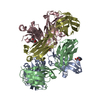
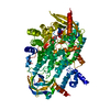
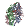
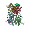
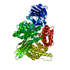
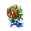
| タイトル | 3D reconstruction of the rotavirus VP6 trimer using the Fast Projection Matching (FPM) algorithm. | |||||||||
 マップデータ マップデータ | Map of rotavirus VP6 trimer (from EMD-1461) after icosahedral averaging and 13-fold non-icosahedral averaging | |||||||||
 試料 試料 |
| |||||||||
 キーワード キーワード | Rotavirus VP6 protein / Methods / Projection Matching | |||||||||
| 生物種 |  Bovine rotavirus (ウイルス) Bovine rotavirus (ウイルス) | |||||||||
| 手法 | 単粒子再構成法 / クライオ電子顕微鏡法 / 解像度: 3.5 Å | |||||||||
 データ登録者 データ登録者 | Estrozi LF / Navaza J | |||||||||
 引用 引用 | ジャーナル: J Struct Biol / 年: 2010 タイトル: Ab initio high-resolution single-particle 3D reconstructions: the symmetry adapted functions way. 著者: Leandro F Estrozi / Jorge Navaza /  要旨: A protocol to attain high-resolution single-particle reconstructions is presented. The protocol is the concatenation of two procedures: one to obtain an ab initio low-resolution reconstruction, the ...A protocol to attain high-resolution single-particle reconstructions is presented. The protocol is the concatenation of two procedures: one to obtain an ab initio low-resolution reconstruction, the other to determine a fixed point of the consecutive applications of fast projection matching and 3D reconstruction. It is a reciprocal space formulation where the Fourier coefficients of the 3D scattering density are expressed in terms of symmetry adapted functions and the 2D particle images are represented by their Fourier-Bessel transforms. The new protocol shows advantages in terms of speed and accuracy when compared to other methods currently in use. We illustrate its performance as applied to high-resolution cryo-electron micrographs of rotavirus. | |||||||||
| 履歴 |
|
- 構造の表示
構造の表示
| ムービー |
 ムービービューア ムービービューア |
|---|---|
| 構造ビューア | EMマップ: SurfViewMolmilJmol/JSmol |
| 添付画像 |

- ダウンロードとリンク
-EMDBアーカイブ
| マップデータ | emd_1752.map.gz | 2.2 MB | EMDBマップデータ形式 | |
|---|---|---|---|---|
| ヘッダ (付随情報) | emd-1752-v30.xmlemd-1752.xml | 11.4 KB 11.4 KB | 表示 表示 | EMDBヘッダ |
| 画像 |  emd_1752.png emd_1752.png | 65 KB | ||
| アーカイブディレクトリ |  http://ftp.pdbj.org/pub/emdb/structures/EMD-1752ftp://ftp.pdbj.org/pub/emdb/structures/EMD-1752 http://ftp.pdbj.org/pub/emdb/structures/EMD-1752ftp://ftp.pdbj.org/pub/emdb/structures/EMD-1752 | HTTPS FTP |
-関連構造データ


-リンク
| EMDBのページ | EMDB (EBI/PDBe) / EMDataResource |
|---|
-マップ
| ファイル | ダウンロード / ファイル: emd_1752.map.gz / 形式: CCP4 / 大きさ: 5.5 MB / タイプ: IMAGE STORED AS FLOATING POINT NUMBER (4 BYTES) | ||||||||||||||||||||||||||||||||||||||||||||||||||||||||||||||||||||
|---|---|---|---|---|---|---|---|---|---|---|---|---|---|---|---|---|---|---|---|---|---|---|---|---|---|---|---|---|---|---|---|---|---|---|---|---|---|---|---|---|---|---|---|---|---|---|---|---|---|---|---|---|---|---|---|---|---|---|---|---|---|---|---|---|---|---|---|---|---|
| 注釈 | Map of rotavirus VP6 trimer (from EMD-1461) after icosahedral averaging and 13-fold non-icosahedral averaging | ||||||||||||||||||||||||||||||||||||||||||||||||||||||||||||||||||||
| 投影像・断面図 | 画像のコントロール
画像は Spider により作成 | ||||||||||||||||||||||||||||||||||||||||||||||||||||||||||||||||||||
| ボクセルのサイズ | X=Y=Z: 1.14 Å | ||||||||||||||||||||||||||||||||||||||||||||||||||||||||||||||||||||
| 密度 |
| ||||||||||||||||||||||||||||||||||||||||||||||||||||||||||||||||||||
| 対称性 | 空間群: 1 | ||||||||||||||||||||||||||||||||||||||||||||||||||||||||||||||||||||
| 詳細 | EMDB XML:
CCP4マップ ヘッダ情報:
| ||||||||||||||||||||||||||||||||||||||||||||||||||||||||||||||||||||
 Z (Sec.)
 Y (Row.)
 X (Col.)


-添付データ
- 試料の構成要素
-全体 : Rotavirus VP6 protein
| 全体 | 名称: Rotavirus VP6 protein |
|---|---|
| 要素 |
|
-超分子 #1000: Rotavirus VP6 protein
| 超分子 | 名称: Rotavirus VP6 protein / タイプ: sample / ID: 1000 集合状態: 780 molecules of VP6 form a DLP particle with 12 molecules of VP1, 120 molecules of VP2, 12 molecules of VP3 and 11 dsRNA molecules Number unique components: 5 |
|---|
-分子 #1: VP6
| 分子 | 名称: VP6 / タイプ: protein_or_peptide / ID: 1 / Name.synonym: VP6 / 組換発現: No |
|---|---|
| 由来(天然) | 生物種: Bovine rotavirus (ウイルス) / 別称: Rotavirus |
| 分子量 | 理論値: 41 KDa |
-分子 #2: VP1
| 分子 | 名称: VP1 / タイプ: protein_or_peptide / ID: 2 / Name.synonym: VP1 / 組換発現: No |
|---|---|
| 由来(天然) | 生物種: Bovine rotavirus (ウイルス) / 別称: Rotavirus |
-分子 #3: VP2
| 分子 | 名称: VP2 / タイプ: protein_or_peptide / ID: 3 / Name.synonym: VP2 / 組換発現: No |
|---|---|
| 由来(天然) | 生物種: Bovine rotavirus (ウイルス) / 別称: Rotavirus |
-分子 #4: VP3
| 分子 | 名称: VP3 / タイプ: protein_or_peptide / ID: 4 / Name.synonym: VP3 / 組換発現: No |
|---|---|
| 由来(天然) | 生物種: Bovine rotavirus (ウイルス) / 別称: Rotavirus |
-分子 #5: dsRNA
| 分子 | 名称: dsRNA / タイプ: rna / ID: 5 / Name.synonym: dsRNA / 分類: OTHER / Structure: OTHER / Synthetic?: No |
|---|---|
| 由来(天然) | 生物種: Bovine rotavirus (ウイルス) / 別称: Rotavirus |
-実験情報
-構造解析
| 手法 | クライオ電子顕微鏡法 |
|---|---|
 解析 解析 | 単粒子再構成法 |
| 試料の集合状態 | particle |
-試料調製
| 濃度 | 5 mg/mL |
|---|---|
| 緩衝液 | pH: 7.4 |
| グリッド | 詳細: Lacy carbon and C-flat |
| 凍結 | 凍結剤: ETHANE / チャンバー内湿度: 30 % / 装置: HOMEMADE PLUNGER 詳細: Vitrification instrument: Home-made. Vitrification carried out in air at room temperature 手法: Blot for 3 seconds before plunging |
- 電子顕微鏡法
| 顕微鏡 | FEI TECNAI F30 |
|---|---|
| 温度 | 平均: 90 K |
| アライメント法 | Legacy - 非点収差: Objective lens astigmatism was corrected |
| 日付 | 2007年6月1日 |
| 撮影 | カテゴリ: FILM / フィルム・検出器のモデル: KODAK SO-163 FILM / デジタル化 - スキャナー: ZEISS SCAI / デジタル化 - サンプリング間隔: 7 µm / 実像数: 386 / 平均電子線量: 15 e/Å2 / Od range: 1 / ビット/ピクセル: 8 |
| 電子線 | 加速電圧: 300 kV / 電子線源:  FIELD EMISSION GUN FIELD EMISSION GUN |
| 電子光学系 | 倍率(補正後): 56540 / 照射モード: FLOOD BEAM / 撮影モード: BRIGHT FIELD / Cs: 2 mm / 最大 デフォーカス(公称値): 3.5 µm / 最小 デフォーカス(公称値): 1.1 µm / 倍率(公称値): 59000 |
| 試料ステージ | 試料ホルダー: Eucentric, side-entry / 試料ホルダーモデル: GATAN LIQUID NITROGEN |
| 実験機器 |  モデル: Tecnai F30 / 画像提供: FEI Company |
-画像解析
| CTF補正 | 詳細: Phase flipping for each particle |
|---|---|
| 最終 再構成 | 想定した対称性 - 点群: I (正20面体型対称) / アルゴリズム: OTHER / 解像度のタイプ: BY AUTHOR / 解像度: 3.5 Å / 解像度の算出法: OTHER / ソフトウェア - 名称: RIco / 使用した粒子像数: 7000 |
-原子モデル構築 1
| 初期モデル | PDB ID: Chain - Chain ID: A |
|---|---|
| ソフトウェア | 名称: URO |
| 詳細 | PDBEntryID_givenInChain. Protocol: Rigid body |
| 精密化 | 空間: RECIPROCAL / プロトコル: RIGID BODY FIT / 当てはまり具合の基準: Correlation coefficient |