Movie
Movie Controller

[English] 日本語
 Yorodumi
- EMDB-1752: 3D reconstruction of the rotavirus VP6 trimer using the Fast Proj... -
+ Open data

- Basic information
| Entry | Database: EMDB / ID: EMD-1752 | |||||||||
|---|---|---|---|---|---|---|---|---|---|---|
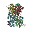
| Title | 3D reconstruction of the rotavirus VP6 trimer using the Fast Projection Matching (FPM) algorithm. | |||||||||
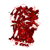
 Map data Map data | Map of rotavirus VP6 trimer (from EMD-1461) after icosahedral averaging and 13-fold non-icosahedral averaging | |||||||||
 Sample Sample |
| |||||||||
 Keywords Keywords | Rotavirus VP6 protein / Methods / Projection Matching | |||||||||
| Biological species |  Bovine rotavirus Bovine rotavirus | |||||||||
| Method | single particle reconstruction / cryo EM / Resolution: 3.5 Å | |||||||||
 Authors Authors | Estrozi LF / Navaza J | |||||||||
 Citation Citation | Journal: J Struct Biol / Year: 2010 Title: Ab initio high-resolution single-particle 3D reconstructions: the symmetry adapted functions way. Authors: Leandro F Estrozi / Jorge Navaza /  Abstract: A protocol to attain high-resolution single-particle reconstructions is presented. The protocol is the concatenation of two procedures: one to obtain an ab initio low-resolution reconstruction, the ...A protocol to attain high-resolution single-particle reconstructions is presented. The protocol is the concatenation of two procedures: one to obtain an ab initio low-resolution reconstruction, the other to determine a fixed point of the consecutive applications of fast projection matching and 3D reconstruction. It is a reciprocal space formulation where the Fourier coefficients of the 3D scattering density are expressed in terms of symmetry adapted functions and the 2D particle images are represented by their Fourier-Bessel transforms. The new protocol shows advantages in terms of speed and accuracy when compared to other methods currently in use. We illustrate its performance as applied to high-resolution cryo-electron micrographs of rotavirus. | |||||||||
| History |
|
- Structure visualization
| Movie |
 Movie viewer Movie viewer |
|---|---|
| Structure viewer | EM map: SurfViewMolmilJmol/JSmol |
| Supplemental images |

- Downloads & links
-EMDB archive
| Map data | emd_1752.map.gz | 2.2 MB | EMDB map data format | |
|---|---|---|---|---|
| Header (meta data) | emd-1752-v30.xmlemd-1752.xml | 11.4 KB 11.4 KB | Display Display | EMDB header |
| Images |  emd_1752.png emd_1752.png | 65 KB | ||
| Archive directory |  http://ftp.pdbj.org/pub/emdb/structures/EMD-1752ftp://ftp.pdbj.org/pub/emdb/structures/EMD-1752 http://ftp.pdbj.org/pub/emdb/structures/EMD-1752ftp://ftp.pdbj.org/pub/emdb/structures/EMD-1752 | HTTPS FTP |
-Related structure data
| Similar structure data |
|---|


-Links
| EMDB pages | EMDB (EBI/PDBe) / EMDataResource |
|---|
-Map
| File | Download / File: emd_1752.map.gz / Format: CCP4 / Size: 5.5 MB / Type: IMAGE STORED AS FLOATING POINT NUMBER (4 BYTES) | ||||||||||||||||||||||||||||||||||||||||||||||||||||||||||||||||||||
|---|---|---|---|---|---|---|---|---|---|---|---|---|---|---|---|---|---|---|---|---|---|---|---|---|---|---|---|---|---|---|---|---|---|---|---|---|---|---|---|---|---|---|---|---|---|---|---|---|---|---|---|---|---|---|---|---|---|---|---|---|---|---|---|---|---|---|---|---|---|
| Annotation | Map of rotavirus VP6 trimer (from EMD-1461) after icosahedral averaging and 13-fold non-icosahedral averaging | ||||||||||||||||||||||||||||||||||||||||||||||||||||||||||||||||||||
| Projections & slices | Image control
Images are generated by Spider. | ||||||||||||||||||||||||||||||||||||||||||||||||||||||||||||||||||||
| Voxel size | X=Y=Z: 1.14 Å | ||||||||||||||||||||||||||||||||||||||||||||||||||||||||||||||||||||
| Density |
| ||||||||||||||||||||||||||||||||||||||||||||||||||||||||||||||||||||
| Symmetry | Space group: 1 | ||||||||||||||||||||||||||||||||||||||||||||||||||||||||||||||||||||
| Details | EMDB XML:
CCP4 map header:
| ||||||||||||||||||||||||||||||||||||||||||||||||||||||||||||||||||||
 Z (Sec.)
 Y (Row.)
 X (Col.)


-Supplemental data
- Sample components
-Entire : Rotavirus VP6 protein
| Entire | Name: Rotavirus VP6 protein |
|---|---|
| Components |
|
-Supramolecule #1000: Rotavirus VP6 protein
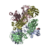
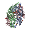
| Supramolecule | Name: Rotavirus VP6 protein / type: sample / ID: 1000 Oligomeric state: 780 molecules of VP6 form a DLP particle with 12 molecules of VP1, 120 molecules of VP2, 12 molecules of VP3 and 11 dsRNA molecules Number unique components: 5 |
|---|
-Macromolecule #1: VP6
| Macromolecule | Name: VP6 / type: protein_or_peptide / ID: 1 / Name.synonym: VP6 / Recombinant expression: No |
|---|---|
| Source (natural) | Organism: Bovine rotavirus / synonym: Rotavirus |
| Molecular weight | Theoretical: 41 KDa |
-Macromolecule #2: VP1
| Macromolecule | Name: VP1 / type: protein_or_peptide / ID: 2 / Name.synonym: VP1 / Recombinant expression: No |
|---|---|
| Source (natural) | Organism: Bovine rotavirus / synonym: Rotavirus |
-Macromolecule #3: VP2
| Macromolecule | Name: VP2 / type: protein_or_peptide / ID: 3 / Name.synonym: VP2 / Recombinant expression: No |
|---|---|
| Source (natural) | Organism: Bovine rotavirus / synonym: Rotavirus |
-Macromolecule #4: VP3
| Macromolecule | Name: VP3 / type: protein_or_peptide / ID: 4 / Name.synonym: VP3 / Recombinant expression: No |
|---|---|
| Source (natural) | Organism: Bovine rotavirus / synonym: Rotavirus |
-Macromolecule #5: dsRNA
| Macromolecule | Name: dsRNA / type: rna / ID: 5 / Name.synonym: dsRNA / Classification: OTHER / Structure: OTHER / Synthetic?: No |
|---|---|
| Source (natural) | Organism: Bovine rotavirus / synonym: Rotavirus |
-Experimental details
-Structure determination
| Method | cryo EM |
|---|---|
 Processing Processing | single particle reconstruction |
| Aggregation state | particle |
-Sample preparation
| Concentration | 5 mg/mL |
|---|---|
| Buffer | pH: 7.4 |
| Grid | Details: Lacy carbon and C-flat |
| Vitrification | Cryogen name: ETHANE / Chamber humidity: 30 % / Instrument: HOMEMADE PLUNGER Details: Vitrification instrument: Home-made. Vitrification carried out in air at room temperature Method: Blot for 3 seconds before plunging |
- Electron microscopy
| Microscope | FEI TECNAI F30 |
|---|---|
| Temperature | Average: 90 K |
| Alignment procedure | Legacy - Astigmatism: Objective lens astigmatism was corrected |
| Date | Jun 1, 2007 |
| Image recording | Category: FILM / Film or detector model: KODAK SO-163 FILM / Digitization - Scanner: ZEISS SCAI / Digitization - Sampling interval: 7 µm / Number real images: 386 / Average electron dose: 15 e/Å2 / Od range: 1 / Bits/pixel: 8 |
| Electron beam | Acceleration voltage: 300 kV / Electron source:  FIELD EMISSION GUN FIELD EMISSION GUN |
| Electron optics | Calibrated magnification: 56540 / Illumination mode: FLOOD BEAM / Imaging mode: BRIGHT FIELD / Cs: 2 mm / Nominal defocus max: 3.5 µm / Nominal defocus min: 1.1 µm / Nominal magnification: 59000 |
| Sample stage | Specimen holder: Eucentric, side-entry / Specimen holder model: GATAN LIQUID NITROGEN |
| Experimental equipment |  Model: Tecnai F30 / Image courtesy: FEI Company |
-Image processing
| CTF correction | Details: Phase flipping for each particle |
|---|---|
| Final reconstruction | Applied symmetry - Point group: I (icosahedral) / Algorithm: OTHER / Resolution.type: BY AUTHOR / Resolution: 3.5 Å / Resolution method: OTHER / Software - Name: RIco / Number images used: 7000 |
-Atomic model buiding 1
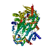
| Initial model | PDB ID: Chain - Chain ID: A |
|---|---|
| Software | Name: URO |
| Details | PDBEntryID_givenInChain. Protocol: Rigid body |
| Refinement | Space: RECIPROCAL / Protocol: RIGID BODY FIT / Target criteria: Correlation coefficient |