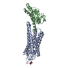
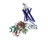
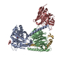
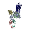
[日本語] English
- EMDB-17344: Cryo-EM structure of Rhodopsin-Gi bound with antibody fragments s... -

+
データを開く


IDまたはキーワード:

読み込み中...

-
基本情報

登録情報
データベース: EMDB / ID: EMD-17344
タイトルCryo-EM structure of Rhodopsin-Gi bound with antibody fragments scFv16 and Fab79, conformation 1
マップデータ
試料
  • 複合体: Rhodopsin-Gi-scFv16-Fab79 complex
    • 複合体: Rhodopsin
      • タンパク質・ペプチド: Rhodopsin
    • 複合体: Guanine nucleotide-binding protein G(i) subunit alpha-1
      • タンパク質・ペプチド: Guanine nucleotide-binding protein G(i) subunit alpha-1
    • 複合体: Guanine nucleotide-binding protein G(I)/G(S)/G(T) subunit beta-1 and Guanine nucleotide-binding protein G(T) subunit gamma-T1
      • タンパク質・ペプチド: Guanine nucleotide-binding protein G(I)/G(S)/G(T) subunit beta-1
      • タンパク質・ペプチド: Guanine nucleotide-binding protein G(T) subunit gamma-T1
    • 複合体: single-chain Fv 16
      • タンパク質・ペプチド: single-chain Fv 16
    • 複合体: Immunoglobin G heavy chain FAB fragment and Immunoglobin G light chain
      • タンパク質・ペプチド: Immunoglobin G heavy chain FAB fragment
      • タンパク質・ペプチド: Immunoglobin G light chain
キーワードGPCR / Rhodopsin / G protein / Fab / SIGNALING PROTEIN
機能・相同性
機能・相同性情報


Opsins / VxPx cargo-targeting to cilium / sperm head plasma membrane / rod bipolar cell differentiation / absorption of visible light / opsin binding / The canonical retinoid cycle in rods (twilight vision) / G protein-coupled opsin signaling pathway / Olfactory Signaling Pathway / Sensory perception of sweet, bitter, and umami (glutamate) taste ...Opsins / VxPx cargo-targeting to cilium / sperm head plasma membrane / rod bipolar cell differentiation / absorption of visible light / opsin binding / The canonical retinoid cycle in rods (twilight vision) / G protein-coupled opsin signaling pathway / Olfactory Signaling Pathway / Sensory perception of sweet, bitter, and umami (glutamate) taste / 11-cis retinal binding / podosome assembly / Synthesis, secretion, and inactivation of Glucagon-like Peptide-1 (GLP-1) / G protein-coupled photoreceptor activity / photoreceptor inner segment membrane / rod photoreceptor outer segment / cellular response to light stimulus / eye photoreceptor cell development / G protein-coupled receptor complex / Inactivation, recovery and regulation of the phototransduction cascade / thermotaxis / Activation of the phototransduction cascade / outer membrane / detection of temperature stimulus involved in thermoception / response to light intensity / photoreceptor cell maintenance / arrestin family protein binding / Activation of G protein gated Potassium channels / G-protein activation / G beta:gamma signalling through PI3Kgamma / Prostacyclin signalling through prostacyclin receptor / G beta:gamma signalling through PLC beta / ADP signalling through P2Y purinoceptor 1 / Thromboxane signalling through TP receptor / Presynaptic function of Kainate receptors / G beta:gamma signalling through CDC42 / Inhibition of voltage gated Ca2+ channels via Gbeta/gamma subunits / G alpha (12/13) signalling events / Glucagon-type ligand receptors / G beta:gamma signalling through BTK / ADP signalling through P2Y purinoceptor 12 / Adrenaline,noradrenaline inhibits insulin secretion / Cooperation of PDCL (PhLP1) and TRiC/CCT in G-protein beta folding / Ca2+ pathway / Thrombin signalling through proteinase activated receptors (PARs) / G alpha (z) signalling events / Extra-nuclear estrogen signaling / photoreceptor outer segment membrane / G alpha (s) signalling events / G alpha (q) signalling events / G alpha (i) signalling events / Glucagon-like Peptide-1 (GLP1) regulates insulin secretion / High laminar flow shear stress activates signaling by PIEZO1 and PECAM1:CDH5:KDR in endothelial cells / Vasopressin regulates renal water homeostasis via Aquaporins / response to light stimulus / phototransduction, visible light / G-protein alpha-subunit binding / phototransduction / photoreceptor outer segment / adenylate cyclase inhibitor activity / positive regulation of protein localization to cell cortex / T cell migration / Adenylate cyclase inhibitory pathway / D2 dopamine receptor binding / response to prostaglandin E / sperm midpiece / adenylate cyclase regulator activity / G protein-coupled serotonin receptor binding / adenylate cyclase-inhibiting serotonin receptor signaling pathway / visual perception / cellular response to forskolin / regulation of mitotic spindle organization / guanyl-nucleotide exchange factor activity / Regulation of insulin secretion / positive regulation of cholesterol biosynthetic process / negative regulation of insulin secretion / G protein-coupled receptor binding / response to peptide hormone / adenylate cyclase-inhibiting G protein-coupled receptor signaling pathway / adenylate cyclase-modulating G protein-coupled receptor signaling pathway / microtubule cytoskeleton organization / centriolar satellite / G-protein beta/gamma-subunit complex binding / ADP signalling through P2Y purinoceptor 12 / photoreceptor disc membrane / Adrenaline,noradrenaline inhibits insulin secretion / cell-cell junction / GDP binding / G alpha (z) signalling events / cellular response to catecholamine stimulus / ADORA2B mediated anti-inflammatory cytokines production / intracellular protein localization / adenylate cyclase-activating dopamine receptor signaling pathway / GPER1 signaling / cellular response to prostaglandin E stimulus / G-protein beta-subunit binding / heterotrimeric G-protein complex / sensory perception of taste / signaling receptor complex adaptor activity / retina development in camera-type eye
類似検索 - 分子機能
Rhodopsin, N-terminal / Amino terminal of the G-protein receptor rhodopsin / Rhodopsin / Opsin / Visual pigments (opsins) retinal binding site / Visual pigments (opsins) retinal binding site. / : / G-protein alpha subunit, group I / Serpentine type 7TM GPCR chemoreceptor Srsx / Guanine nucleotide binding protein (G-protein), alpha subunit ...Rhodopsin, N-terminal / Amino terminal of the G-protein receptor rhodopsin / Rhodopsin / Opsin / Visual pigments (opsins) retinal binding site / Visual pigments (opsins) retinal binding site. / : / G-protein alpha subunit, group I / Serpentine type 7TM GPCR chemoreceptor Srsx / Guanine nucleotide binding protein (G-protein), alpha subunit / G protein alpha subunit, helical insertion / G-protein alpha subunit / G-alpha domain profile. / G protein alpha subunit / G-protein, gamma subunit / G-protein gamma subunit domain profile. / G-protein gamma-like domain / G-protein gamma-like domain superfamily / GGL domain / G protein gamma subunit-like motifs / GGL domain / G protein beta WD-40 repeat protein / Guanine nucleotide-binding protein, beta subunit / G-protein, beta subunit / G-protein coupled receptors family 1 signature. / G protein-coupled receptor, rhodopsin-like / GPCR, rhodopsin-like, 7TM / G-protein coupled receptors family 1 profile. / 7 transmembrane receptor (rhodopsin family) / G-protein beta WD-40 repeat / WD40 repeat, conserved site / Trp-Asp (WD) repeats signature. / Trp-Asp (WD) repeats profile. / Trp-Asp (WD) repeats circular profile. / WD40 repeats / WD40 repeat / WD40-repeat-containing domain superfamily / WD40/YVTN repeat-like-containing domain superfamily / P-loop containing nucleoside triphosphate hydrolase
類似検索 - ドメイン・相同性
Guanine nucleotide-binding protein G(T) subunit gamma-T1 / Rhodopsin / Guanine nucleotide-binding protein G(I)/G(S)/G(T) subunit beta-1 / Guanine nucleotide-binding protein G(i) subunit alpha-1
類似検索 - 構成要素
生物種Bos taurus (ウシ) / Homo sapiens (ヒト) / Mus musculus (ハツカネズミ)
手法単粒子再構成法 / クライオ電子顕微鏡法 / 解像度: 5.2 Å
データ登録者Pamula F / Tejero O / Muehle J / Thoma R / Schertler GFX / Marino J / Tsai C-J
資金援助 スイス, European Union, 4件
OrganizationGrant number
Swiss National Science Foundation173335 スイス
Swiss National Science Foundation153145 スイス
Swiss National Science Foundation192760 スイス
European Research Council (ERC)951644European Union
引用ジャーナル: Biophys J / : 2025
タイトル: Tool antibody fragments reveal multiple conformations of the rhodopsin-Gi signaling complex.
著者: Filip Pamula / Oliver Tejero / Jonas Mühle / Ralf Thoma / Gebhard F X Schertler / Jacopo Marino / Ching-Ju Tsai /
要旨: Antibody Fab fragments are widely used protein binders that assist in structural studies of G-protein-coupled receptor (GPCR) signaling complexes. Expanding the repertoire of such binders to target ...Antibody Fab fragments are widely used protein binders that assist in structural studies of G-protein-coupled receptor (GPCR) signaling complexes. Expanding the repertoire of such binders to target distinct components of the signaling complex offers opportunities to probe conformational regulation and dynamics. Here, we report the biochemical and cryo-EM characterization of two Fab fragments, Fab79 and Fab13, raised against the rhodopsin-Gαiβγ complex. Fab79 binds to the flexible α-helical domain (AHD) of the Gαi subunit and prevents complex dissociation in the presence of the nonhydrolyzable GTP analog, GTPγS, likely by hindering AHD closure, a step necessary for complex dissociation. In contrast, Fab13 binds rigidly to Gβ without directly contacting Gα or the receptor. These findings show that Fab79 and Fab13 reveal functionally relevant conformational states of G-protein activation and serve as practical tools to stabilize or modulate GPCR signaling complexes.
履歴
登録2023年5月11日-
ヘッダ(付随情報) 公開2024年11月20日-
マップ公開2024年11月20日-
更新2025年10月29日-
現状2025年10月29日処理サイト: PDBe / 状態: 公開

-
構造の表示

添付画像

ダウンロードとリンク

-
マップ

ファイルダウンロード / ファイル: emd_17344.map.gz / 形式: CCP4 / 大きさ: 209.3 MB / タイプ: IMAGE STORED AS FLOATING POINT NUMBER (4 BYTES)
投影像・断面図

画像のコントロール

大きさ
明度
コントラスト
その他
Z (Sec.)Y (Row.)X (Col.)
0.85 Å/pix.
x 380 pix.
= 324.52 Å
0.85 Å/pix.
x 380 pix.
= 324.52 Å
0.85 Å/pix.
x 380 pix.
= 324.52 Å

表面

投影像

断面 (1/3)

断面 (1/2)

断面 (2/3)

画像は Spider により作成

ボクセルのサイズX=Y=Z: 0.854 Å
密度
表面レベル登録者による: 0.006
最小 - 最大-0.011423453 - 0.027867006
平均 (標準偏差)0.00007048641 (±0.0008808457)
対称性空間群: 1
詳細

EMDB XML:

マップ形状
Axis orderXYZ
Origin000
サイズ380380380
Spacing380380380
セルA=B=C: 324.52 Å
α=β=γ: 90.0 °

-
添付データ

-
ハーフマップ: #1

ファイルemd_17344_half_map_1.map
投影像・断面図
ZYX

投影像

断面 (1/2)
密度ヒストグラム

-
ハーフマップ: #2

ファイルemd_17344_half_map_2.map
投影像・断面図
ZYX

投影像

断面 (1/2)
密度ヒストグラム

-
試料の構成要素

+
全体 : Rhodopsin-Gi-scFv16-Fab79 complex

全体名称: Rhodopsin-Gi-scFv16-Fab79 complex
要素
  • 複合体: Rhodopsin-Gi-scFv16-Fab79 complex
    • 複合体: Rhodopsin
      • タンパク質・ペプチド: Rhodopsin
    • 複合体: Guanine nucleotide-binding protein G(i) subunit alpha-1
      • タンパク質・ペプチド: Guanine nucleotide-binding protein G(i) subunit alpha-1
    • 複合体: Guanine nucleotide-binding protein G(I)/G(S)/G(T) subunit beta-1 and Guanine nucleotide-binding protein G(T) subunit gamma-T1
      • タンパク質・ペプチド: Guanine nucleotide-binding protein G(I)/G(S)/G(T) subunit beta-1
      • タンパク質・ペプチド: Guanine nucleotide-binding protein G(T) subunit gamma-T1
    • 複合体: single-chain Fv 16
      • タンパク質・ペプチド: single-chain Fv 16
    • 複合体: Immunoglobin G heavy chain FAB fragment and Immunoglobin G light chain
      • タンパク質・ペプチド: Immunoglobin G heavy chain FAB fragment
      • タンパク質・ペプチド: Immunoglobin G light chain

+
超分子 #1: Rhodopsin-Gi-scFv16-Fab79 complex

超分子名称: Rhodopsin-Gi-scFv16-Fab79 complex / タイプ: complex / ID: 1 / 親要素: 0 / 含まれる分子: all / 詳細: bovine rhodopsin N2C/M257Y/D282C
分子量理論値: 195 KDa

+
超分子 #2: Rhodopsin

超分子名称: Rhodopsin / タイプ: complex / ID: 2 / 親要素: 1 / 含まれる分子: #1
由来(天然)生物種: Bos taurus (ウシ)

+
超分子 #3: Guanine nucleotide-binding protein G(i) subunit alpha-1

超分子名称: Guanine nucleotide-binding protein G(i) subunit alpha-1
タイプ: complex / ID: 3 / 親要素: 1 / 含まれる分子: #2
由来(天然)生物種: Homo sapiens (ヒト)

+
超分子 #4: Guanine nucleotide-binding protein G(I)/G(S)/G(T) subunit beta-1 ...

超分子名称: Guanine nucleotide-binding protein G(I)/G(S)/G(T) subunit beta-1 and Guanine nucleotide-binding protein G(T) subunit gamma-T1
タイプ: complex / ID: 4 / 親要素: 1 / 含まれる分子: #3-#4
由来(天然)生物種: Bos taurus (ウシ)

+
超分子 #5: single-chain Fv 16

超分子名称: single-chain Fv 16 / タイプ: complex / ID: 5 / 親要素: 1 / 含まれる分子: #5
由来(天然)生物種: Mus musculus (ハツカネズミ)

+
超分子 #6: Immunoglobin G heavy chain FAB fragment and Immunoglobin G light chain

超分子名称: Immunoglobin G heavy chain FAB fragment and Immunoglobin G light chain
タイプ: complex / ID: 6 / 親要素: 1 / 含まれる分子: #6-#7
由来(天然)生物種: Mus musculus (ハツカネズミ)

+
分子 #1: Rhodopsin

分子名称: Rhodopsin / タイプ: protein_or_peptide / ID: 1 / コピー数: 1 / 光学異性体: LEVO
由来(天然)生物種: Bos taurus (ウシ) / 細胞: rod photoreceptor cell
分子量理論値: 39.040527 KDa
組換発現生物種: Homo sapiens (ヒト)
配列文字列: MCGTEGPNFY VPFSNKTGVV RSPFEAPQYY LAEPWQFSML AAYMFLLIML GFPINFLTLY VTVQHKKLRT PLNYILLNLA VADLFMVFG GFTTTLYTSL HGYFVFGPTG CNLEGFFATL GGEIALWSLV VLAIERYVVV CKPMSNFRFG ENHAIMGVAF T WVMALACA ...文字列:
MCGTEGPNFY VPFSNKTGVV RSPFEAPQYY LAEPWQFSML AAYMFLLIML GFPINFLTLY VTVQHKKLRT PLNYILLNLA VADLFMVFG GFTTTLYTSL HGYFVFGPTG CNLEGFFATL GGEIALWSLV VLAIERYVVV CKPMSNFRFG ENHAIMGVAF T WVMALACA APPLVGWSRY IPEGMQCSCG IDYYTPHEET NNESFVIYMF VVHFIIPLIV IFFCYGQLVF TVKEAAAQQQ ES ATTQKAE KEVTRMVIIY VIAFLICWLP YAGVAFYIFT HQGSCFGPIF MTIPAFFAKT SAVYNPVIYI MMNKQFRNCM VTT LCCGKN PLGDDEASTT VSKTETSQVA PA

UniProtKB: Rhodopsin

+
分子 #2: Guanine nucleotide-binding protein G(i) subunit alpha-1

分子名称: Guanine nucleotide-binding protein G(i) subunit alpha-1
タイプ: protein_or_peptide / ID: 2 / コピー数: 1 / 光学異性体: LEVO
由来(天然)生物種: Homo sapiens (ヒト)
分子量理論値: 43.182078 KDa
組換発現生物種: Escherichia coli BL21(DE3) (大腸菌)
配列文字列: MKKHHHHHHH HHHENLYFQG GSMGCTLSAE DKAAVERSKM IDRNLREDGE KAAREVKLLL LGAGESGKST IVKQMKIIHE AGYSEEECK QYKAVVYSNT IQSIIAIIRA MGRLKIDFGD SARADDARQL FVLAGAAEEG FMTAELAGVI KRLWKDSGVQ A CFNRSREY ...文字列:
MKKHHHHHHH HHHENLYFQG GSMGCTLSAE DKAAVERSKM IDRNLREDGE KAAREVKLLL LGAGESGKST IVKQMKIIHE AGYSEEECK QYKAVVYSNT IQSIIAIIRA MGRLKIDFGD SARADDARQL FVLAGAAEEG FMTAELAGVI KRLWKDSGVQ A CFNRSREY QLNDSAAYYL NDLDRIAQPN YIPTQQDVLR TRVKTTGIVE THFTFKDLHF KMFDVGGQRS ERKKWIHCFE GV TAIIFCV ALSDYDLVLA EDEEMNRMHE SMKLFDSICN NKWFTDTSII LFLNKKDLFE EKIKKSPLTI CYPEYAGSNT YEE AAAYIQ CQFEDLNKRK DTKEIYTHFT CATDTKNVQF VFDAVTDVII KNNLKDCGLF

UniProtKB: Guanine nucleotide-binding protein G(i) subunit alpha-1

+
分子 #3: Guanine nucleotide-binding protein G(I)/G(S)/G(T) subunit beta-1

分子名称: Guanine nucleotide-binding protein G(I)/G(S)/G(T) subunit beta-1
タイプ: protein_or_peptide / ID: 3 / コピー数: 1 / 光学異性体: LEVO
由来(天然)生物種: Bos taurus (ウシ) / 器官: Eye / 組織: retina
分子量理論値: 37.41693 KDa
配列文字列: MSELDQLRQE AEQLKNQIRD ARKACADATL SQITNNIDPV GRIQMRTRRT LRGHLAKIYA MHWGTDSRLL VSASQDGKLI IWDSYTTNK VHAIPLRSSW VMTCAYAPSG NYVACGGLDN ICSIYNLKTR EGNVRVSREL AGHTGYLSCC RFLDDNQIVT S SGDTTCAL ...文字列:
MSELDQLRQE AEQLKNQIRD ARKACADATL SQITNNIDPV GRIQMRTRRT LRGHLAKIYA MHWGTDSRLL VSASQDGKLI IWDSYTTNK VHAIPLRSSW VMTCAYAPSG NYVACGGLDN ICSIYNLKTR EGNVRVSREL AGHTGYLSCC RFLDDNQIVT S SGDTTCAL WDIETGQQTT TFTGHTGDVM SLSLAPDTRL FVSGACDASA KLWDVREGMC RQTFTGHESD INAICFFPNG NA FATGSDD ATCRLFDLRA DQELMTYSHD NIICGITSVS FSKSGRLLLA GYDDFNCNVW DALKADRAGV LAGHDNRVSC LGV TDDGMA VATGSWDSFL KIWN

UniProtKB: Guanine nucleotide-binding protein G(I)/G(S)/G(T) subunit beta-1

+
分子 #4: Guanine nucleotide-binding protein G(T) subunit gamma-T1

分子名称: Guanine nucleotide-binding protein G(T) subunit gamma-T1
タイプ: protein_or_peptide / ID: 4 / コピー数: 1 / 光学異性体: LEVO
由来(天然)生物種: Bos taurus (ウシ) / 器官: Eye / 組織: retina
分子量理論値: 8.556918 KDa
配列文字列:
MPVINIEDLT EKDKLKMEVD QLKKEVTLER MLVSKCCEEF RDYVEERSGE DPLVKGIPED KNPFKELKGG CVIS

UniProtKB: Guanine nucleotide-binding protein G(T) subunit gamma-T1

+
分子 #5: single-chain Fv 16

分子名称: single-chain Fv 16 / タイプ: protein_or_peptide / ID: 5 / コピー数: 1 / 光学異性体: LEVO
由来(天然)生物種: Mus musculus (ハツカネズミ) / 細胞: B cell
分子量理論値: 27.784896 KDa
組換発現生物種: Trichoplusia ni (イラクサキンウワバ)
配列文字列: DVQLVESGGG LVQPGGSRKL SCSASGFAFS SFGMHWVRQA PEKGLEWVAY ISSGSGTIYY ADTVKGRFTI SRDDPKNTLF LQMTSLRSE DTAMYYCVRS IYYYGSSPFD FWGQGTTLTV SSGGGGSGGG GSGGGGSDIV MTQATSSVPV TPGESVSISC R SSKSLLHS ...文字列:
DVQLVESGGG LVQPGGSRKL SCSASGFAFS SFGMHWVRQA PEKGLEWVAY ISSGSGTIYY ADTVKGRFTI SRDDPKNTLF LQMTSLRSE DTAMYYCVRS IYYYGSSPFD FWGQGTTLTV SSGGGGSGGG GSGGGGSDIV MTQATSSVPV TPGESVSISC R SSKSLLHS NGNTYLYWFL QRPGQSPQLL IYRMSNLASG VPDRFSGSGS GTAFTLTISR LEAEDVGVYY CMQHLEYPLT FG AGTKLEL KAAAHHHHHH HH

+
分子 #6: Immunoglobin G heavy chain FAB fragment

分子名称: Immunoglobin G heavy chain FAB fragment / タイプ: protein_or_peptide / ID: 6 / コピー数: 1 / 光学異性体: LEVO
由来(天然)生物種: Mus musculus (ハツカネズミ)
分子量理論値: 27.971973 KDa
配列文字列: MNFVLSLIFL ALILKGVQCE VQLVESGGGL VKPGGSLKLS CAASGFTFSS YAMSWVRQTP EKRLEWVATI SSRGLYTYFP DSMKGRFTI SRDNAKNTLS LQMSSLRSED TAMYYCLRGG GYDADYWGQG TTLTVSSAKT TAPSVYPLAP VCGDTTGSSV T LGCLVKGY ...文字列:
MNFVLSLIFL ALILKGVQCE VQLVESGGGL VKPGGSLKLS CAASGFTFSS YAMSWVRQTP EKRLEWVATI SSRGLYTYFP DSMKGRFTI SRDNAKNTLS LQMSSLRSED TAMYYCLRGG GYDADYWGQG TTLTVSSAKT TAPSVYPLAP VCGDTTGSSV T LGCLVKGY FPEPVTLTWN SGSLSSGVHT FPAVLQSDLY TLSSSVTVTS STWPSQSITC NVAHPASSTK VDKKIEPRGP TI KPCPPCK CPAPNLLGGP SVFIF

+
分子 #7: Immunoglobin G light chain

分子名称: Immunoglobin G light chain / タイプ: protein_or_peptide / ID: 7 / コピー数: 1 / 光学異性体: LEVO
由来(天然)生物種: Mus musculus (ハツカネズミ)
分子量理論値: 26.37258 KDa
配列文字列: MMSPAQFLFL LVLWIRETNG DVVMTQTPLT LSVTIGQPAS ISCKSSQSLL DSDGETSLNW LLQRPGQSPK RLIYLVSKLD SGVPDRFTG SGSGTDFTLK ISRVEAADLG VYYCWQGTHF PLTFGAGTKL ELKRADAAPT VSIFPPSSEQ LTSGGASVVC F LNNFYPKD ...文字列:
MMSPAQFLFL LVLWIRETNG DVVMTQTPLT LSVTIGQPAS ISCKSSQSLL DSDGETSLNW LLQRPGQSPK RLIYLVSKLD SGVPDRFTG SGSGTDFTLK ISRVEAADLG VYYCWQGTHF PLTFGAGTKL ELKRADAAPT VSIFPPSSEQ LTSGGASVVC F LNNFYPKD INVKWKIDGS ERQNGVLNSW TDQDSKDSTY SMSSTLTLTK DEYERHNSYT CEATHKTSTS PIVKSFNRNE C

-
実験情報

-
構造解析

手法クライオ電子顕微鏡法
解析単粒子再構成法
試料の集合状態particle

-
試料調製

濃度0.7 mg/mL
緩衝液pH: 7.5
構成要素:
濃度名称
20.0 mMC8H18N2O4SHEPES
100.0 mMNaClsodium chloride

詳細: 20 mM HEPES (pH 7.5), 100 mM NaCl
グリッドモデル: Quantifoil R1.2/1.3 / 材質: COPPER / メッシュ: 200 / 前処理 - タイプ: GLOW DISCHARGE
凍結凍結剤: ETHANE / チャンバー内湿度: 100 % / チャンバー内温度: 277 K / 装置: FEI VITROBOT MARK IV
詳細Monodisperse

-
電子顕微鏡法

顕微鏡FEI TITAN KRIOS
撮影フィルム・検出器のモデル: GATAN K2 SUMMIT (4k x 4k)
検出モード: COUNTING / 平均電子線量: 60.0 e/Å2
電子線加速電圧: 300 kV / 電子線源: FIELD EMISSION GUN
電子光学系照射モード: FLOOD BEAM / 撮影モード: BRIGHT FIELD / 最大 デフォーカス(公称値): 2.4 µm / 最小 デフォーカス(公称値): 0.8 µm / 倍率(公称値): 165000
試料ステージ試料ホルダーモデル: FEI TITAN KRIOS AUTOGRID HOLDER
ホルダー冷却材: NITROGEN
実験機器
モデル: Titan Krios / 画像提供: FEI Company

+
画像解析

粒子像選択選択した数: 7026029
CTF補正タイプ: PHASE FLIPPING AND AMPLITUDE CORRECTION
初期モデルモデルのタイプ: NONE
最終 再構成解像度のタイプ: BY AUTHOR / 解像度: 5.2 Å / 解像度の算出法: FSC 0.143 CUT-OFF / ソフトウェア - 名称: RELION (ver. 3.1) / 使用した粒子像数: 98000
初期 角度割当タイプ: MAXIMUM LIKELIHOOD / ソフトウェア - 名称: cryoSPARC (ver. 2.13)
最終 角度割当タイプ: MAXIMUM LIKELIHOOD

-
原子モデル構築 1

初期モデル
PDB IDChain詳細

chain_id: R, residue_range: 1-322, source_name: PDB, initial_model_type: experimental model

chain_id: A, residue_range: 1-42, source_name: PDB, initial_model_type: experimental model

chain_id: A, residue_range: 181-354, source_name: PDB, initial_model_type: experimental model
chain_id: A, residue_range: 43-180, source_name: AlphaFold, initial_model_type: in silico model

chain_id: B, source_name: PDB, initial_model_type: experimental model

chain_id: G, source_name: PDB, initial_model_type: experimental model

chain_id: S, source_name: PDB, initial_model_type: experimental modelchain D in 6CRK.pdb
chain_id: H, source_name: AlphaFold, initial_model_type: in silico model
chain_id: L, source_name: AlphaFold, initial_model_type: in silico model
精密化空間: REAL / プロトコル: AB INITIO MODEL / 温度因子: 360
当てはまり具合の基準: cross-correlation coefficient
得られたモデル

PDB-8p13:
Cryo-EM structure of Rhodopsin-Gi bound with antibody fragments scFv16 and Fab79, conformation 1

+
万見について

-
お知らせ

-
2022年2月9日: EMDBエントリの付随情報ファイルのフォーマットが新しくなりました

EMDBエントリの付随情報ファイルのフォーマットが新しくなりました

  • EMDBのヘッダファイルのバージョン3が、公式のフォーマットとなりました。
  • これまでは公式だったバージョン1.9は、アーカイブから削除されます。

関連情報:EMDBヘッダ

外部リンク:wwPDBはEMDBデータモデルのバージョン3へ移行します

-
2020年8月12日: 新型コロナ情報

新型コロナ情報

URL: https://pdbj.org/emnavi/covid19.php

新ページ: EM Navigatorに新型コロナウイルスの特設ページを開設しました。

関連情報:Covid-19情報 / 2020年3月5日: 新型コロナウイルスの構造データ

+
2020年3月5日: 新型コロナウイルスの構造データ

新型コロナウイルスの構造データ

関連情報:万見生物種 / 2020年8月12日: 新型コロナ情報

外部リンク:COVID-19特集ページ - PDBj / 今月の分子2020年2月:コロナウイルスプロテーアーゼ

+
2019年1月31日: EMDBのIDの桁数の変更

EMDBのIDの桁数の変更

  • EMDBエントリに付与されているアクセスコード(EMDB-ID)は4桁の数字(例、EMD-1234)でしたが、間もなく枯渇します。これまでの4桁のID番号は4桁のまま変更されませんが、4桁の数字を使い切った後に発行されるIDは5桁以上の数字(例、EMD-12345)になります。5桁のIDは2019年の春頃から発行される見通しです。
  • EM Navigator/万見では、接頭語「EMD-」は省略されています。

関連情報:Q: 「EMD」とは何ですか? / 万見/EM NavigatorにおけるID/アクセスコードの表記

外部リンク:EMDB Accession Codes are Changing Soon! / PDBjへお問い合わせ

+
2017年7月12日: PDB大規模アップデート

PDB大規模アップデート

  • 新バージョンのPDBx/mmCIF辞書形式に基づくデータがリリースされました。
  • 今回の更新はバージョン番号が4から5になる大規模なもので、全エントリデータの書き換えが行われる「Remediation」というアップデートに該当します。
  • このバージョンアップで、電子顕微鏡の実験手法に関する多くの項目の書式が改定されました(例:em_softwareなど)。
  • EM NavigatorとYorodumiでも、この改定に基づいた表示内容になります。

外部リンク:wwPDB Remediation / OneDepデータ基準に準拠した、より強化された内容のモデル構造ファイルが、PDBアーカイブで公開されました。

-
万見 (Yorodumi)

幾万の構造データを、幾万の視点から

  • 万見(Yorodumi)は、EMDB/PDB/SASBDBなどの構造データを閲覧するためのページです。
  • EM Navigatorの詳細ページの後継、Omokage検索のフロントエンドも兼ねています。

関連情報:EMDB / PDB / SASBDB / 3つのデータバンクの比較 / 万見検索 / 2016年8月31日: 新しいEM Navigatorと万見 / 万見文献 / Jmol/JSmol / 機能・相同性情報 / 新しいEM Navigatorと万見の変更点

他の情報も見る