Movie
Movie Controller

[English] 日本語
 Yorodumi
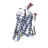
- PDB-8p12: Cryo-EM structure of Rhodopsin-Gi bound to antibody fragment Fab13 -
+ Open data

- Basic information
| Entry | Database: PDB / ID: 8p12 | |||||||||||||||
|---|---|---|---|---|---|---|---|---|---|---|---|---|---|---|---|---|
| Title | Cryo-EM structure of Rhodopsin-Gi bound to antibody fragment Fab13 | |||||||||||||||
 Components Components |
| |||||||||||||||
 Keywords Keywords | SIGNALING PROTEIN / GPCR / Rhodopsin / G protein / Fab | |||||||||||||||
| Function / homology |  Function and homology information Function and homology informationOpsins / VxPx cargo-targeting to cilium / sperm head plasma membrane / transducin-mediated opsin signaling pathway / opsin binding / The canonical retinoid cycle in rods (twilight vision) / absorption of visible light / G protein-coupled opsin signaling pathway / Olfactory Signaling Pathway / 11-cis retinal binding ...Opsins / VxPx cargo-targeting to cilium / sperm head plasma membrane / transducin-mediated opsin signaling pathway / opsin binding / The canonical retinoid cycle in rods (twilight vision) / absorption of visible light / G protein-coupled opsin signaling pathway / Olfactory Signaling Pathway / 11-cis retinal binding / Sensory perception of sweet, bitter, and umami (glutamate) taste / Synthesis, secretion, and inactivation of Glucagon-like Peptide-1 (GLP-1) / G protein-coupled photoreceptor activity / photoreceptor inner segment membrane / cellular response to light stimulus / G protein-coupled receptor complex / Inactivation, recovery and regulation of the phototransduction cascade / Activation of the phototransduction cascade / outer membrane / arrestin family protein binding / Activation of G protein gated Potassium channels / G-protein activation / G beta:gamma signalling through PI3Kgamma / Prostacyclin signalling through prostacyclin receptor / G beta:gamma signalling through PLC beta / ADP signalling through P2Y purinoceptor 1 / Thromboxane signalling through TP receptor / Presynaptic function of Kainate receptors / G beta:gamma signalling through CDC42 / Inhibition of voltage gated Ca2+ channels via Gbeta/gamma subunits / G alpha (12/13) signalling events / Glucagon-type ligand receptors / G beta:gamma signalling through BTK / ADP signalling through P2Y purinoceptor 12 / Adrenaline,noradrenaline inhibits insulin secretion / Cooperation of PDCL (PhLP1) and TRiC/CCT in G-protein beta folding / Ca2+ pathway / G alpha (z) signalling events / Thrombin signalling through proteinase activated receptors (PARs) / Extra-nuclear estrogen signaling / G alpha (s) signalling events / G alpha (q) signalling events / photoreceptor outer segment membrane / Glucagon-like Peptide-1 (GLP1) regulates insulin secretion / G alpha (i) signalling events / High laminar flow shear stress activates signaling by PIEZO1 and PECAM1:CDH5:KDR in endothelial cells / Vasopressin regulates renal water homeostasis via Aquaporins / phototransduction, visible light / phototransduction / response to light stimulus / photoreceptor outer segment / G-protein alpha-subunit binding / regulation of eating behavior / adenylate cyclase inhibitor activity / positive regulation of protein localization to cell cortex / T cell migration / positive regulation of relaxation of smooth muscle / Adenylate cyclase inhibitory pathway / visual perception / D2 dopamine receptor binding / guanyl-nucleotide exchange factor activity / adenylate cyclase-inhibiting serotonin receptor signaling pathway / G protein-coupled serotonin receptor binding / cellular response to forskolin / mast cell degranulation / regulation of mitotic spindle organization / chemokine-mediated signaling pathway / Regulation of insulin secretion / neuropeptide signaling pathway / response to prostaglandin E / positive regulation of cholesterol biosynthetic process / G protein-coupled receptor binding / response to peptide hormone / G-protein beta/gamma-subunit complex binding / adenylate cyclase-modulating G protein-coupled receptor signaling pathway / adenylate cyclase-inhibiting G protein-coupled receptor signaling pathway / cell-cell junction / photoreceptor disc membrane / GDP binding / ADP signalling through P2Y purinoceptor 12 / Adrenaline,noradrenaline inhibits insulin secretion / G alpha (z) signalling events / cellular response to catecholamine stimulus / ADORA2B mediated anti-inflammatory cytokines production / adenylate cyclase-activating dopamine receptor signaling pathway / GPER1 signaling / sperm midpiece / cellular response to prostaglandin E stimulus / heterotrimeric G-protein complex / G-protein beta-subunit binding / sensory perception of taste / signaling receptor complex adaptor activity / adenylate cyclase-activating G protein-coupled receptor signaling pathway / retina development in camera-type eye / GTPase binding / G protein activity / midbody / cell cortex / G alpha (i) signalling events / G alpha (s) signalling events Similarity search - Function | |||||||||||||||
| Biological species |   Homo sapiens (human) Homo sapiens (human) | |||||||||||||||
| Method | ELECTRON MICROSCOPY / single particle reconstruction / cryo EM / Resolution: 3.21 Å | |||||||||||||||
 Authors Authors | Pamula, F. / Tejero, O. / Muehle, J. / Thoma, R. / Schertler, G.F.X. / Marino, J. / Tsai, C.-J. | |||||||||||||||
| Funding support |  Switzerland, European Union, 4items Switzerland, European Union, 4items
| |||||||||||||||
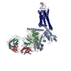
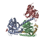
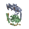
 Citation Citation | Journal: Biophys J / Year: 2026 Title: Tool antibody fragments reveal multiple conformations of the rhodopsin-Gi signaling complex. Authors: Filip Pamula / Oliver Tejero / Jonas Mühle / Ralf Thoma / Gebhard F X Schertler / Jacopo Marino / Ching-Ju Tsai / Abstract: Antibody Fab fragments are widely used protein binders that assist in structural studies of G-protein-coupled receptor (GPCR) signaling complexes. Expanding the repertoire of such binders to target ...Antibody Fab fragments are widely used protein binders that assist in structural studies of G-protein-coupled receptor (GPCR) signaling complexes. Expanding the repertoire of such binders to target distinct components of the signaling complex offers opportunities to probe conformational regulation and dynamics. Here, we report the biochemical and cryo-EM characterization of two Fab fragments, Fab79 and Fab13, raised against the rhodopsin-Gαiβγ complex. Fab79 binds to the flexible α-helical domain (AHD) of the Gαi subunit and prevents complex dissociation in the presence of the nonhydrolyzable GTP analog, GTPγS, likely by hindering AHD closure, a step necessary for complex dissociation. In contrast, Fab13 binds rigidly to Gβ without directly contacting Gα or the receptor. These findings show that Fab79 and Fab13 reveal functionally relevant conformational states of G-protein activation and serve as practical tools to stabilize or modulate GPCR signaling complexes. | |||||||||||||||
| History |
|
- Structure visualization
| Structure viewer | Molecule: MolmilJmol/JSmol |
|---|
- Downloads & links
-Download
| PDBx/mmCIF format | 8p12.cif.gz | 271.4 KB | Display | PDBx/mmCIF format |
|---|---|---|---|---|
| PDB format | pdb8p12.ent.gz | 211.1 KB | Display | PDB format |
| PDBx/mmJSON format | 8p12.json.gz | Tree view | PDBx/mmJSON format | |
| Others |  Other downloads Other downloads |
-Validation report
| Arichive directory | https://data.pdbj.org/pub/pdb/validation_reports/p1/8p12ftp://data.pdbj.org/pub/pdb/validation_reports/p1/8p12 | HTTPS FTP |
|---|
-Related structure data
| Related structure data |  17343MC  8p13C 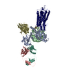 8p15C M: map data used to model this data C: citing same article ( |
|---|---|
| Similar structure data |


-Links
 PDBj
PDBj


- Assembly
| Deposited unit | 
|
|---|---|
| 1 |
|
-Components
-Protein , 1 types, 1 molecules R
| #1: Protein | Mass: 39040.527 Da / Num. of mol.: 1 / Mutation: N2C, M257Y, D282C Source method: isolated from a genetically manipulated source Source: (gene. exp.) Homo sapiens (human) / Strain (production host): HEK293 / References: UniProt: P02699 |
|---|
-Guanine nucleotide-binding protein ... , 3 types, 3 molecules ABG
| #2: Protein | Mass: 43182.078 Da / Num. of mol.: 1 Source method: isolated from a genetically manipulated source Source: (gene. exp.) Homo sapiens (human) / Gene: GNAI1 / Production host:  |
|---|---|
| #3: Protein | Mass: 37416.930 Da / Num. of mol.: 1 / Source method: isolated from a natural source / Source: (natural) |
| #4: Protein | Mass: 8556.918 Da / Num. of mol.: 1 / Source method: isolated from a natural source / Source: (natural) |
-Antibody , 2 types, 2 molecules LH
| #5: Antibody | Mass: 26349.555 Da / Num. of mol.: 1 / Source method: isolated from a natural source / Source: (natural) |
|---|---|
| #6: Antibody | Mass: 27448.336 Da / Num. of mol.: 1 / Source method: isolated from a natural source / Source: (natural) |
-Details
| Has protein modification | Y |
|---|
-Experimental details
-Experiment
| Experiment | Method: ELECTRON MICROSCOPY |
|---|---|
| EM experiment | Aggregation state: PARTICLE / 3D reconstruction method: single particle reconstruction |
- Sample preparation
| Component |
| ||||||||||||||||||||||||||||||||||||||||||
|---|---|---|---|---|---|---|---|---|---|---|---|---|---|---|---|---|---|---|---|---|---|---|---|---|---|---|---|---|---|---|---|---|---|---|---|---|---|---|---|---|---|---|---|
| Molecular weight | Value: 0.17 MDa / Experimental value: NO | ||||||||||||||||||||||||||||||||||||||||||
| Source (natural) |
| ||||||||||||||||||||||||||||||||||||||||||
| Source (recombinant) |
| ||||||||||||||||||||||||||||||||||||||||||
| Buffer solution | pH: 7.5 / Details: 20 mM HEPES (pH 7.5), 100 mM NaCl | ||||||||||||||||||||||||||||||||||||||||||
| Buffer component |
| ||||||||||||||||||||||||||||||||||||||||||
| Specimen | Conc.: 4 mg/ml / Embedding applied: NO / Shadowing applied: NO / Staining applied: NO / Vitrification applied: YES / Details: Monodispersed | ||||||||||||||||||||||||||||||||||||||||||
| Specimen support | Grid material: COPPER / Grid mesh size: 200 divisions/in. / Grid type: Quantifoil R1.2/1.3 | ||||||||||||||||||||||||||||||||||||||||||
| Vitrification | Instrument: FEI VITROBOT MARK IV / Cryogen name: ETHANE / Humidity: 100 % / Chamber temperature: 277 K |
- Electron microscopy imaging
| Experimental equipment |  Model: Titan Krios / Image courtesy: FEI Company |
|---|---|
| Microscopy | Model: FEI TITAN KRIOS |
| Electron gun | Electron source:  FIELD EMISSION GUN / Accelerating voltage: 300 kV / Illumination mode: FLOOD BEAM FIELD EMISSION GUN / Accelerating voltage: 300 kV / Illumination mode: FLOOD BEAM |
| Electron lens | Mode: BRIGHT FIELD / Nominal defocus max: 2000 nm / Nominal defocus min: 800 nm / Alignment procedure: COMA FREE |
| Specimen holder | Cryogen: NITROGEN / Specimen holder model: FEI TITAN KRIOS AUTOGRID HOLDER |
| Image recording | Electron dose: 66 e/Å2 / Film or detector model: GATAN K3 BIOQUANTUM (6k x 4k) |
- Processing
| Software | Name: PHENIX / Version: 1.20.1_4487: / Classification: refinement | ||||||||||||||||||||||||||||||||||||||||||||||||||||||||||||||||||||||||||||||||||||||||||
|---|---|---|---|---|---|---|---|---|---|---|---|---|---|---|---|---|---|---|---|---|---|---|---|---|---|---|---|---|---|---|---|---|---|---|---|---|---|---|---|---|---|---|---|---|---|---|---|---|---|---|---|---|---|---|---|---|---|---|---|---|---|---|---|---|---|---|---|---|---|---|---|---|---|---|---|---|---|---|---|---|---|---|---|---|---|---|---|---|---|---|---|
| EM software |
| ||||||||||||||||||||||||||||||||||||||||||||||||||||||||||||||||||||||||||||||||||||||||||
| CTF correction | Type: PHASE FLIPPING AND AMPLITUDE CORRECTION | ||||||||||||||||||||||||||||||||||||||||||||||||||||||||||||||||||||||||||||||||||||||||||
| Particle selection | Num. of particles selected: 3752145 | ||||||||||||||||||||||||||||||||||||||||||||||||||||||||||||||||||||||||||||||||||||||||||
| 3D reconstruction | Resolution: 3.21 Å / Resolution method: FSC 0.143 CUT-OFF / Num. of particles: 587113 / Symmetry type: POINT | ||||||||||||||||||||||||||||||||||||||||||||||||||||||||||||||||||||||||||||||||||||||||||
| Atomic model building | B value: 150 / Protocol: AB INITIO MODEL / Space: REAL / Target criteria: cross-correlation coefficient | ||||||||||||||||||||||||||||||||||||||||||||||||||||||||||||||||||||||||||||||||||||||||||
| Atomic model building | 3D fitting-ID: 1
| ||||||||||||||||||||||||||||||||||||||||||||||||||||||||||||||||||||||||||||||||||||||||||
| Refine LS restraints |
|