ムービー
ムービー コントローラー


+ データを開く

- 基本情報
基本情報
| 登録情報 | データベース: EMDB / ID: EMD-1646 | |||||||||
|---|---|---|---|---|---|---|---|---|---|---|
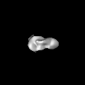
| タイトル | Structure of the Human Dicer-TRBP Complex by Electron Microscopy | |||||||||
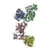
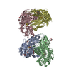
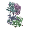
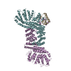
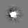
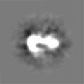
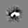
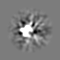
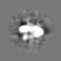
 マップデータ マップデータ | This is the 3D volume of human Dicer-TRBP complex | |||||||||
 試料 試料 |
| |||||||||
 キーワード キーワード | Dicer Enzyme / Ribonuclease III / MicroRNA Processing / Single Particle Electron Microscopy | |||||||||
| 生物種 |  Homo sapiens (ヒト) Homo sapiens (ヒト) | |||||||||
| 手法 | 単粒子再構成法 / ネガティブ染色法 / 解像度: 20.0 Å | |||||||||
 データ登録者 データ登録者 | Lau PW / Potter CS / Carragher B / MacRae IJ | |||||||||
 引用 引用 | ジャーナル: Structure / 年: 2009 タイトル: Structure of the human Dicer-TRBP complex by electron microscopy. 著者: Pick-Wei Lau / Clinton S Potter / Bridget Carragher / Ian J MacRae /  要旨: Dicer is a specialized ribonuclease that initiates RNA interference (RNAi) by cleaving double-stranded RNA (dsRNA) into small RNA fragments about 22 nucleotides long. Here, we present the three- ...Dicer is a specialized ribonuclease that initiates RNA interference (RNAi) by cleaving double-stranded RNA (dsRNA) into small RNA fragments about 22 nucleotides long. Here, we present the three-dimensional structure of human Dicer bound to the protein TRBP at approximately 20 A resolution determined by negative-stain electron microscopy (EM) and single-particle analysis. Our analysis reveals that the Dicer-TRBP complex is an L-shaped molecule with a long edge of 150 A and a 100 A extension on one end. A surface trench runs the length of the long edge of the molecule, defining a putative dsRNA-binding site. Docking the crystal structure of Giardia Dicer, which represents the nuclease core of human Dicer, into the EM map suggests two possible overall molecular architectures for human Dicer. These results offer insights into the structure of Dicer proteins found in multicellular organisms and provide a conceptual framework for understanding the initiation of RNAi. | |||||||||
| 履歴 |
|
- 構造の表示
構造の表示
| ムービー |
 ムービービューア ムービービューア |
|---|---|
| 構造ビューア | EMマップ: SurfViewMolmilJmol/JSmol |
| 添付画像 |

- ダウンロードとリンク
-EMDBアーカイブ
| マップデータ | emd_1646.map.gz | 4.5 MB | EMDBマップデータ形式 | |
|---|---|---|---|---|
| ヘッダ (付随情報) | emd-1646-v30.xmlemd-1646.xml | 9.8 KB 9.8 KB | 表示 表示 | EMDBヘッダ |
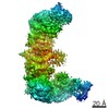
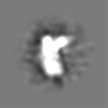
| 画像 | 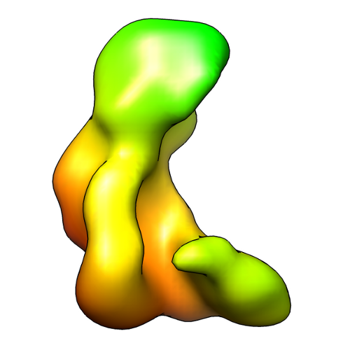 EMD-1646_image.png EMD-1646_image.png | 104 KB | ||
| アーカイブディレクトリ |  http://ftp.pdbj.org/pub/emdb/structures/EMD-1646ftp://ftp.pdbj.org/pub/emdb/structures/EMD-1646 http://ftp.pdbj.org/pub/emdb/structures/EMD-1646ftp://ftp.pdbj.org/pub/emdb/structures/EMD-1646 | HTTPS FTP |
-検証レポート
| 文書・要旨 | emd_1646_validation.pdf.gz | 186.4 KB | 表示 | EMDB検証レポート |
|---|---|---|---|---|
| 文書・詳細版 | emd_1646_full_validation.pdf.gz | 185.5 KB | 表示 | |
| XML形式データ | emd_1646_validation.xml.gz | 5.5 KB | 表示 | |
| アーカイブディレクトリ | https://ftp.pdbj.org/pub/emdb/validation_reports/EMD-1646ftp://ftp.pdbj.org/pub/emdb/validation_reports/EMD-1646 | HTTPS FTP |
-関連構造データ
-リンク
| EMDBのページ | EMDB (EBI/PDBe) / EMDataResource |
|---|
-マップ
| ファイル | ダウンロード / ファイル: emd_1646.map.gz / 形式: CCP4 / 大きさ: 6.4 MB / タイプ: IMAGE STORED AS FLOATING POINT NUMBER (4 BYTES) | ||||||||||||||||||||||||||||||||||||||||||||||||||||||||||||||||||||
|---|---|---|---|---|---|---|---|---|---|---|---|---|---|---|---|---|---|---|---|---|---|---|---|---|---|---|---|---|---|---|---|---|---|---|---|---|---|---|---|---|---|---|---|---|---|---|---|---|---|---|---|---|---|---|---|---|---|---|---|---|---|---|---|---|---|---|---|---|---|
| 注釈 | This is the 3D volume of human Dicer-TRBP complex | ||||||||||||||||||||||||||||||||||||||||||||||||||||||||||||||||||||
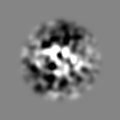
| 投影像・断面図 | 画像のコントロール
画像は Spider により作成 | ||||||||||||||||||||||||||||||||||||||||||||||||||||||||||||||||||||
| ボクセルのサイズ | X=Y=Z: 3.093 Å | ||||||||||||||||||||||||||||||||||||||||||||||||||||||||||||||||||||
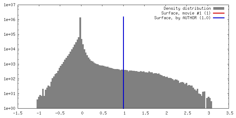
| 密度 |
| ||||||||||||||||||||||||||||||||||||||||||||||||||||||||||||||||||||
| 対称性 | 空間群: 1 | ||||||||||||||||||||||||||||||||||||||||||||||||||||||||||||||||||||
| 詳細 | EMDB XML:
CCP4マップ ヘッダ情報:
| ||||||||||||||||||||||||||||||||||||||||||||||||||||||||||||||||||||
 Z (Sec.)
 Y (Row.)
 X (Col.)


-添付データ
- 試料の構成要素
-全体 : Dicer-TRBP complex
| 全体 | 名称: Dicer-TRBP complex |
|---|---|
| 要素 |
|
-超分子 #1000: Dicer-TRBP complex
| 超分子 | 名称: Dicer-TRBP complex / タイプ: sample / ID: 1000 / 詳細: The sample was mostly monodisperse / 集合状態: One human Dicer binds to one TRBP molecule / Number unique components: 2 |
|---|---|
| 分子量 | 理論値: 250 KDa |
-分子 #1: Human Dicer
| 分子 | 名称: Human Dicer / タイプ: protein_or_peptide / ID: 1 / Name.synonym: hDicer / コピー数: 1 / 集合状態: Monomer / 組換発現: Yes |
|---|---|
| 由来(天然) | 生物種: Homo sapiens (ヒト) / 別称: Human |
| 分子量 | 理論値: 220 KDa |
| 組換発現 | 生物種:   Spodoptera frugiperda (ツマジロクサヨトウ) Spodoptera frugiperda (ツマジロクサヨトウ)組換プラスミド: Baculovirus |
-分子 #2: TAR RNA binding protein
| 分子 | 名称: TAR RNA binding protein / タイプ: protein_or_peptide / ID: 2 / Name.synonym: TRBP / コピー数: 1 / 集合状態: Monomer / 組換発現: Yes |
|---|---|
| 由来(天然) | 生物種: Homo sapiens (ヒト) / 別称: Human |
| 分子量 | 理論値: 30 KDa |
| 組換発現 | 生物種: Spodoptera frugiperda (ツマジロクサヨトウ) 組換プラスミド: Baculovirus |
-実験情報
-構造解析
| 手法 | ネガティブ染色法 |
|---|---|
 解析 解析 | 単粒子再構成法 |
| 試料の集合状態 | particle |
-試料調製
| 緩衝液 | pH: 7.4 / 詳細: 150mM KCl, 20mM HEPES |
|---|---|
| 染色 | タイプ: NEGATIVE 詳細: Grids with adsorbed protein floated on 2% w/v uranyl acetate for approximately 20 seconds. |
| 凍結 | 凍結剤: NONE / 装置: OTHER |
- 電子顕微鏡法
| 顕微鏡 | FEI TECNAI F20 |
|---|---|
| 日付 | 2009年5月31日 |
| 撮影 | カテゴリ: CCD フィルム・検出器のモデル: TVIPS TEMCAM-F415 (4k x 4k) 実像数: 947 / 平均電子線量: 20 e/Å2 |
| 電子線 | 加速電圧: 120 kV / 電子線源:  FIELD EMISSION GUN FIELD EMISSION GUN |
| 電子光学系 | 照射モード: FLOOD BEAM / 撮影モード: BRIGHT FIELD / Cs: 2.0 mm / 最小 デフォーカス(公称値): 0.15 µm / 倍率(公称値): 50000 |
| 試料ステージ | 試料ホルダー: Single tilt room temperature grid holder 試料ホルダーモデル: SIDE ENTRY, EUCENTRIC |
| 実験機器 |  モデル: Tecnai F20 / 画像提供: FEI Company |
-画像解析
| CTF補正 | 詳細: Whole image |
|---|---|
| 最終 再構成 | 想定した対称性 - 点群: C1 (非対称) / アルゴリズム: OTHER / 解像度のタイプ: BY AUTHOR / 解像度: 20.0 Å / 解像度の算出法: OTHER / ソフトウェア - 名称: EMAN, SPIDER / 使用した粒子像数: 129554 |
| 最終 2次元分類 | クラス数: 711 |
-原子モデル構築 1
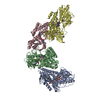
| 初期モデル | PDB ID: Chain - Chain ID: A |
|---|---|
| ソフトウェア | 名称: Chimera |
| 詳細 | PDBEntryID_givenInChain. Protocol: Rigid Body. Manual docking |
| 精密化 | 空間: REAL / プロトコル: RIGID BODY FIT |